. 2008 Aug 29;283(35):23774-81.
doi: 10.1074/jbc.M802542200. Epub 2008 Jun 20.

Revisiting the GroEL-GroES reaction cycle via the symmetric intermediate implied by novel aspects of the GroEL(D398A) mutant

Affiliations

Revisiting the GroEL-GroES reaction cycle via the symmetric intermediate implied by novel aspects of the GroEL(D398A) mutant

Ayumi Koike-Takeshita et al. J Biol Chem. .

Abstract

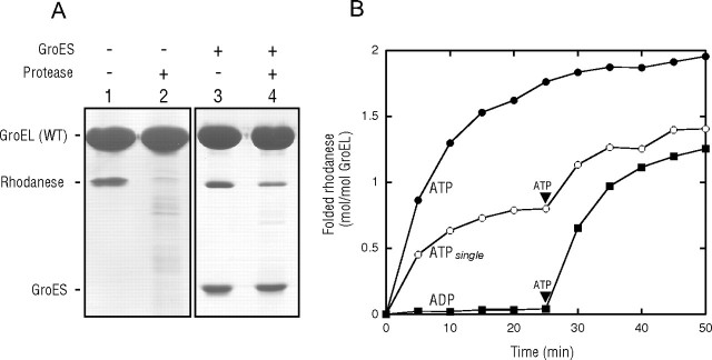
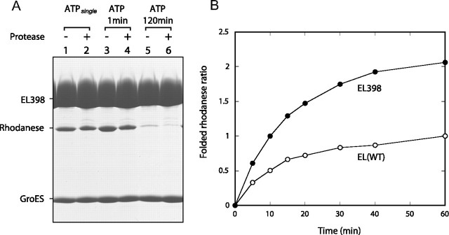
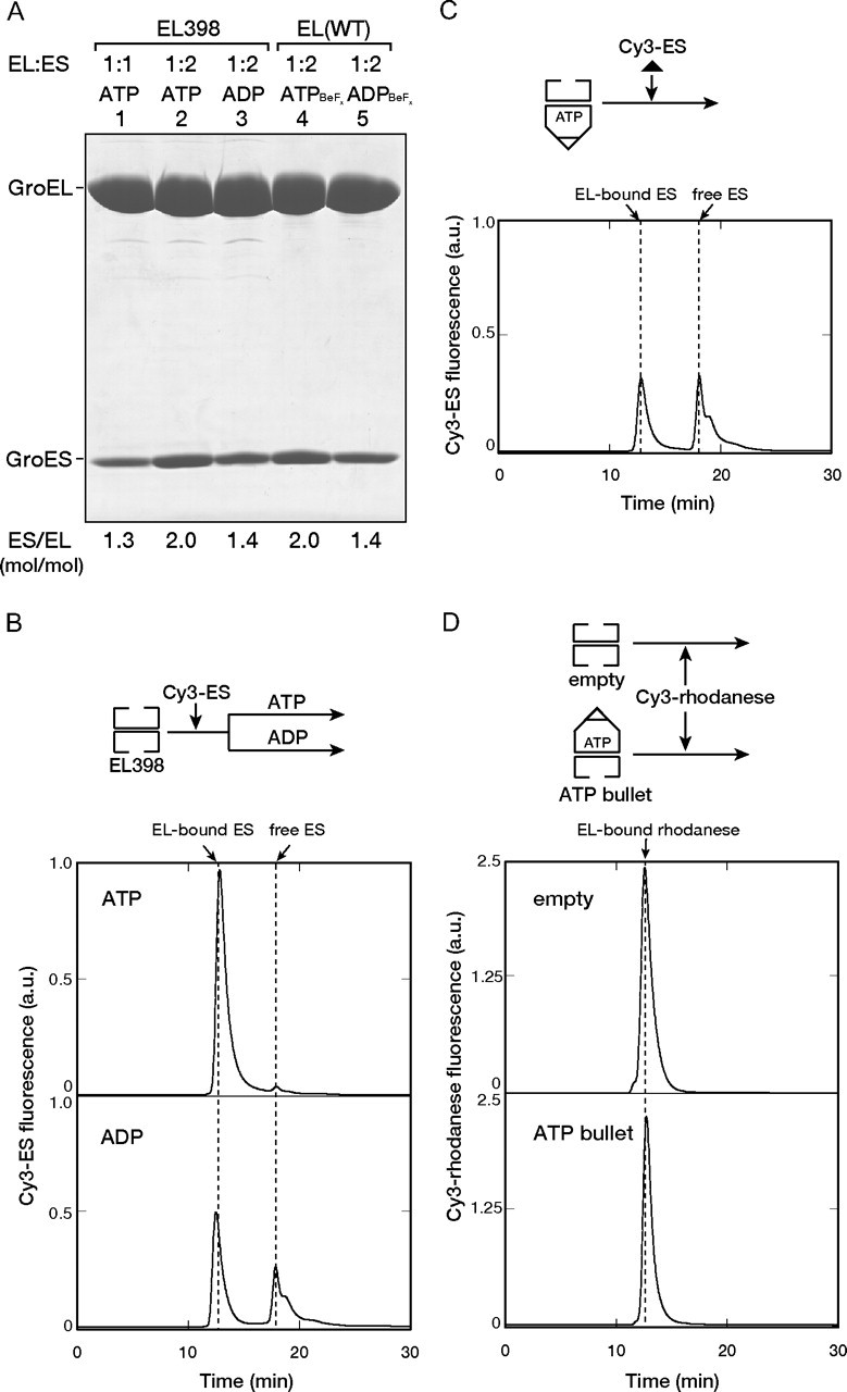
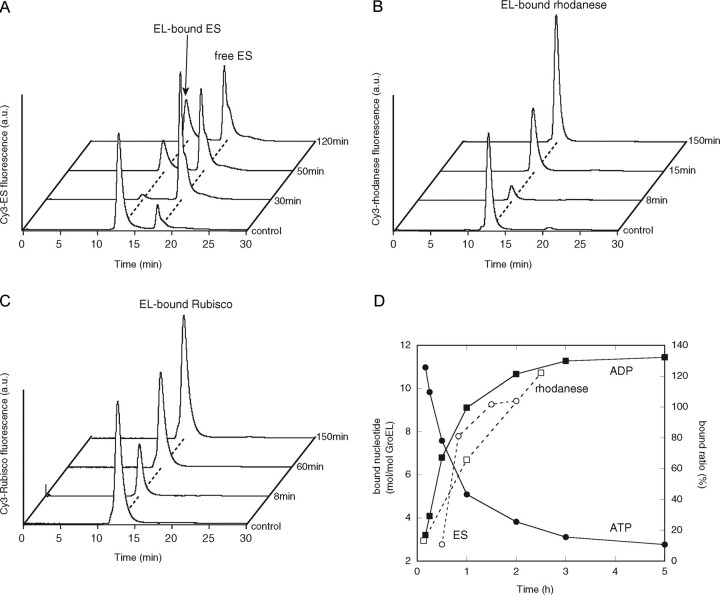
The Escherichia coli chaperonin GroEL is a double-ring chaperone that assists in protein folding with the aid of GroES and ATP. It is believed that GroEL alternates the folding-active rings and that the substrate protein (and GroES) can bind to the open trans-ring only after ATP in the cis-ring is hydrolyzed. However, we found that a substrate protein prebound to the trans-ring remained bound during the first ATP cycle, and this substrate was assisted by GroEL-GroES when the second cycle began. Moreover, a slow ATP-hydrolyzing GroEL mutant (D398A) in the ATP-bound form bound a substrate protein and GroES to the trans-ring. The apparent discrepancy with the results from an earlier study (Rye, H. S., Roseman, A. M., Chen, S., Furtak, K., Fenton, W. A., Saibil, H. R., and Horwich, A. L. (1999) Cell 97, 325-338) can be explained by the previously unnoticed fact that the ATP-bound form of the D398A mutant exists as a symmetric 1:2 GroEL-GroES complex (the "football"-shaped complex) and that the substrate protein (and GroES) in the medium is incorporated into the complex only after the slow turnover. In light of these results, the current model of the GroEL-GroES reaction cycle via the asymmetric 1:1 GroEL-GroES complex deserves reexamination.


Figures

FIGURE 1.
FIGURE 1.
Retention of rhodanese bound to the trans-ring of the GroEL-GroES complex during the ATPase cycle. A, sensitivity of rhodanese bound to the GroEL complex to protease digestion. ATP was added to the mixture containing 200 mm glucose, GroES, and GroEL saturated with heat-denatured rhodanese for a single reaction cycle. After 3 s, hexokinase was added to the solution to hydrolyze all free ATP. After 60 min, when the folding of rhodanese in the cis-ring was saturated during the single turnover conditions (22, 23), the GroEL-GroES complexes were isolated by ultrafiltration (100-kDa cutoff) and then subjected to SDS-PAGE analysis with (lane 4) or without (lane 3) proteinase K treatment. As a control experiment, the rhodanese-saturated GroEL was also subjected to SDS-PAGE analysis with (lane 2) or without (lane 1) proteinase K treatment. WT, wild-type. B, recoveries of rhodanese that had bound to the trans-ring during the single turnover experiment. Recovered yields are expressed as moles of recovered enzyme/mol of GroEL. At time 0, ATP or ADP was added to the mixtures containing the rhodanese-saturated GroEL and GroES. In the ATPsingle experiment, the mixture also contained 200 mm glucose, and then hexokinase was added 3 s after the addition of ATP. The rhodanese activity at a 2:1 molar ratio of native rhodanese to GroEL was taken as 2.0 mol/mol of GroEL.
FIGURE 2.
FIGURE 2.
Encapsulation of rhodanese bound to both rings of EL398 within the cavities. A, protection of encapsulated rhodanese from proteinase K. Mixtures of EL398 saturated with heat-denatured rhodanese, GroES, 200 mm glucose, and ATP were incubated for 3 s (lanes 1 and 2), 1 min (lanes 3 and 4), or 120 min (lanes 5 and 6). In the experiment denoted as ATPsingle (lanes 1 and 2), hexokinase was added to the aliquot after 3 s, and the reaction was left for 60 min longer as described for Fig. 1A. Aliquots underwent one of the two following treatments: ultrafiltration (100-kDa cutoff) and SDS-PAGE (lanes 1, 3, and 5) or ultrafiltration (100-kDa cutoff), proteinase K treatment, a second ultrafiltration (100-kDa cutoff), and SDS-PAGE (lanes 2, 4, and 6). Gels were stained with Coomassie Brilliant Blue. B, GroEL (wild-type (WT))- or EL398-assisted recovery of rhodanese activities for a single reaction cycle. A single round of folding was initiated by the addition of ATP to the mixtures containing 200 mm glucose, GroEL saturated with denatured rhodanese, and GroES. Hexokinase was then added after 3 s of the initiation. For a comparison, the ratio of the recovered activity to that at 60 min assisted by GroEL (wild-type) is shown. EL(WT), wild-type EL398.
FIGURE 3.
FIGURE 3.
Binding of GroES and the denatured substrate to the trans-ring of the EL398-GroES ATP bullet complex. A, SDS-PAGE analysis of the isolated EL398-GroES complex formed in the presence of ATP (lanes 1 and 2) or ADP (lane 3). EL398 and GroES were mixed at 1:1 (lane 1) and 1:2 (lanes 2 and 3) ratios. For comparison, the wild-type GroEL-GroES complexes formed in the presence of either ATP + BeFx (lane 4) or ADP + BeFx (lane 5) were also analyzed. The GroES/GroEL (ES/EL) molar ratios indicated at the bottom were determined by the band intensities. EL(WT), wild-type EL398. B and C, binding of Cy3-GroES to the empty EL398 (B) or the EL398-GroES ATP bullet formed using unlabeled GroES (C). Cy3-GroES was mixed with EL398 (B) or the EL398-GroES ATP bullet (C) at a 2:1 ratio in the presence of either ATP or ADP (B) or ATP (C). The mixtures were applied to a gel filtration HPLC column. Cy3 fluorescence was monitored in-line. The scale of the y axis is the same in B and C. a.u., arbitrary units. D, binding of denatured Cy3-rhodanese to the empty EL398 or the EL398-GroES ATP bullet. Denatured Cy3-rhodanese (0.15 μm) was diluted in buffer containing the empty EL398 or the EL398-GroES ATP bullet (0.3 μm) and was incubated for 5 min. The mixtures were subjected to gel filtration as described above. A schematic view of the experiments is shown above each graph.
FIGURE 4.
FIGURE 4.
Binding of GroES and the denatured substrate to the symmetric GroES-EL398-GroES ATP football complex. A, binding of Cy3-GroES to the EL398 ATP football complex formed using unlabeled GroES. The stable EL398 ATP football complex was generated by mixing EL398 (EL) with GroES (ES) at a 1:2 molar ratio in the presence of ATP (t = 0 min) and incubating the mixture for 5 min. The mixture was rapidly applied to a gel filtration column to isolate the ATP football complex. The isolated ATP football complex (0.5 μm) was mixed with Cy3-GroES (1 μm) and ATP (1 mm) at the indicated times and then separated by gel filtration HPLC with fluorescence detection. B and C, binding of denatured Cy3-labeled substrates to the EL398 ATP football complex. The ATP football complexes (0.5 μm) were mixed with either Cy3-rhodanese (0.25 μm)(B) or Cy3-Rubisco (0.25 μm)(C) at the indicated times and then separated by gel filtration with fluorescence detection. The trace labeled control represents the direct mixing of the empty EL398 (0.5 μm) with 1 μm Cy3-GroES and 1 mm ATP (A), 0.25 μm Cy3-rhodanese (B), or 0.25 μm Cy3-Rubisco (C). a.u., arbitrary units. D, time course of ATP hydrolysis by the isolated EL398 ATP football complex and quantification of the GroES and rhodanese bound to the EL398 ATP football complexes. The hydrolysis of ATP in the isolated ATP football complexes was determined as described under “Experimental Procedures.” The determination of the amounts of bound GroES and rhodanese was performed as follows. The integrated peak areas in the traces obtained by the binding assay of Cy3-GroES (A) and Cy3-rhodanese (B) were calculated. The amounts of GroES and rhodanese bound to unliganded EL398 (control in A and B) were taken as 200 and 100%, respectively.

References

    1. Hartl, F. U., and Hayer-Hartl, M. (2002) Science 295 1852–1858 - PubMed
    1. Young, J. C., Agashe, V. R., Siegers, K., and Hartl, F. U. (2004) Nat. Rev. Mol. Cell Biol. 5 781–791 - PubMed
    1. Thirumalai, D., and Lorimer, G. H. (2001) Annu. Rev. Biophys. Biomol. Struct. 30 245–269 - PubMed
    1. Taguchi, H. (2005) J. Biochem. (Tokyo) 137 543–549 - PubMed
    1. Horwich, A. L., Fenton, W. A., Chapman, E., and Farr, G. W. (2007) Annu. Rev. Cell Dev. Biol. 23 115–145 - PubMed

Publication types

MeSH terms

LinkOut - more resources