. 2008 Aug;190(16):5672-80.
doi: 10.1128/JB.01919-07. Epub 2008 Jun 20.

Direct visualization of the outer membrane of mycobacteria and corynebacteria in their native state

Affiliations

Direct visualization of the outer membrane of mycobacteria and corynebacteria in their native state

Benoît Zuber et al. J Bacteriol. 2008 Aug.

Abstract

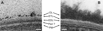
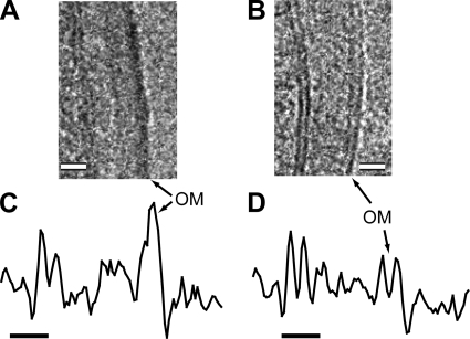
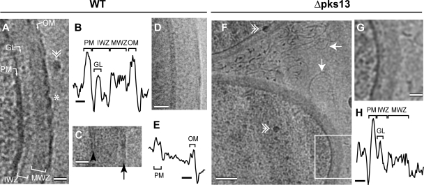
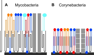
The cell envelope of mycobacteria, which include the causative agents of tuberculosis and leprosy, is crucial for their success as pathogens. Despite a continued strong emphasis on identifying the multiple chemical components of this envelope, it has proven difficult to combine its components into a comprehensive structural model, primarily because the available ultrastructural data rely on conventional electron microscopy embedding and sectioning, which are known to induce artifacts. The existence of an outer membrane bilayer has long been postulated but has never been directly observed by electron microscopy of ultrathin sections. Here we have used cryo-electron microscopy of vitreous sections (CEMOVIS) to perform a detailed ultrastructural analysis of three species belonging to the Corynebacterineae suborder, namely, Mycobacterium bovis BCG, Mycobacterium smegmatis, and Corynebacterium glutamicum, in their native state. We provide new information that accurately describes the different layers of the mycobacterial cell envelope and challenges current models of the organization of its components. We show a direct visualization of an outer membrane, analogous to that found in gram-negative bacteria, in the three bacterial species examined. Furthermore, we demonstrate that mycolic acids, the hallmark of mycobacteria and related genera, are essential for the formation of this outer membrane. In addition, a granular layer and a low-density zone typifying the periplasmic space of gram-positive bacteria are apparent in CEMOVIS images of mycobacteria and corynebacteria. Based on our observations, a model of the organization of the lipids in the outer membrane is proposed. The architecture we describe should serve as a reference for future studies to relate the structure of the mycobacterial cell envelope to its function.


Figures

FIG. 1.
FIG. 1.
Thin-section transmission electron microscopy of chemically fixed and dehydrated Corynebacterineae. (A) M. smegmatis mc2155. (B) C. glutamicum (CGL2020). The cell envelope of M. smegmatis is composed of a PM; a thick, electron-transparent layer (Peri ?; interpreted as the postulated periplasmic space); a thick, internal, electron-dense layer (EDL; considered a complex of peptidoglycan and arabinogalactan); a thin, electron-transparent layer (ETL; assumed to be composed of mycolic acids and other lipids); and an electron-dense OL (a complex protein-carbohydrate matrix with some lipids) (15, 16). The EDL and the mycolic acids of the ETL form the cell wall core. The cell envelope of C. glutamicum looks similar, but it does not possess an obvious low-density hypothetical periplasmic space and the OL is thicker than in M. smegmatis (43). In both cases, the use of ruthenium red stain resulted in enhanced staining of the OL. Bars, 20 nm.
FIG. 2.
FIG. 2.
CEMOVIS of a cross-sectioned M. smegmatis mc2155 cell. Arrows, knife marks. Bar, 500 nm.
FIG. 3.
FIG. 3.
Cell envelope of mycobacteria and gram-positive and gram-negative bacteria by CEMOVIS. (A, B, and C) M. smegmatis mc2155. (D, E, and F) M. bovis BCG. (G and H) S. gordonii. (I and J) E. coli. Images were acquired at 100 kV and defocused by −2.5 μm (A, D, and I), −5 μm (B), −3.7 μm (E), and −4.5 μm (G). They were denoised by Gaussian filtering in Adobe Photoshop (radius of 0.6 pixel for panels A and D and of 1 pixel for panels B, E, G, and I). The density profiles in panels C, F, H, and J were obtained from nondenoised images corresponding to panels A, E, G, and I, respectively. They were averaged over a width of 70 pixels (C) and 50 pixels (F, H, and J). Note that the GL is in IWZ. OWZ, outer wall zone; PG, peptidoglycan layer. Bars, 20 nm (A, B, D, E, G, and I) and 10 nm (C, F, H, and J).
FIG. 4.
FIG. 4.
Staining of M. smegmatis mc2155 with OsO4. (A) Glutaraldehyde-fixed and OsO4-postfixed cell. (B) Glutaraldehyde-fixed cell. (C) Density profile of the cell envelope in panel A. (D) Density profile of the cell envelope in panel B. Images A and B were acquired at 120 kV. They were denoised by Gaussian filtering in Adobe Photoshop (radius of 0.6 pixel). They have the same intensity scale. Images were defocused by −1.9 μm (A) and −2.1 μm (B) Density profiles were obtained from nondenoised images. They were averaged over a width of 35 pixels, and both profiles are shown at the same scale. The apparent difference in the distance between the PM and the OM in panels C and D is due to the fact that the measured cell envelopes had a different orientation in relation to the cutting direction. Bars, 20 nm (A and B) and 10 nm (C and D).
FIG. 5.
FIG. 5.
Cell envelope of corynebacteria visualized by CEMOVIS and whole-mount cryoEM. (A to E) Wild-type (WT) C. glutamicum. (F to G) C. glutamicum Δpks13. (A, C, F, and G) CEMOVIS. (D) Whole-mount cryoEM. (G) Higher magnification of the boxed area in panel F. Images A, C, F, and G were acquired at 100 kV, and image D was acquired at 200 kV. Images were defocused by −3.4 μm (A), −1.1 μm (C), −3 μm (D), and −5 μm (F and G). They were denoised by Gaussian filtering in Adobe Photoshop (radius of 1 pixel for panels A and D, 0.8 pixel for panel C, and 2.4 pixels for panels F and G). The density profiles in panels B and H were obtained from nondenoised images corresponding to panels A and G, respectively. The density profile in panel E was obtained from Gaussian filtered (radius of 1 pixel) image D. The density profiles were averaged over a width of 74 pixels (B), 65 pixels (E), and 70 pixels (H). The abbreviations are the same as those in Fig. 3. Double arrowhead, ice contamination; asterisk, cutting-induced defects in the OM; black arrowhead, PM; black arrow, OM; white arrow, filaments. Bars, 20 nm (A, C, D, and G), 10 nm (B, E, and H), and 100 nm (F).
FIG. 6.
FIG. 6.
Schematic representation, at scale, of the cell envelopes of E. coli, M. smegmatis, C. glutamicum, and S. gordonii as seen with CEMOVIS. The abbreviations are the same as those in Fig. 3. The GL is drawn as bound to the PM. This hypothesis is based on the conclusion of our previous work with gram-positive bacteria (58). The IWZ is attributed, by analogy to other bacteria, to a periplasmic space. The MWZ represents the peptidoglycan layer in E. coli and S. gordonii and would correspond to the peptidoglycan-arabinogalactan layer in M. smegmatis and C. glutamicum. The OL of M. smegmatis and C. glutamicum is not depicted (see Discussion).
FIG. 7.
FIG. 7.
Zipper model of the OM of Corynebacterineae. (A) Mycobacteria. (B) Corynebacteria. Hydrocarbon chains of the lipids are drawn to scale. Black, mycolic acid; dark blue, phospholipids (16- to 18-carbon-long chains); dark gray, peptidoglycan-arabinogalactan; light blue, GPL; light gray, porin; orange, trehalose dimycolate; red, trehalose monomycolate. Mycolic acids and trehalose mycolates are folded (54, 55). An unfolded mycolic acid is shown in panel A. It is too large to be accommodated in the OM. Porins are not drawn to scale. Of note, the porin of M. smegmatis MspA is expected to protrude out of the OM (27). The porin of corynebacteria has been proposed to be made by a stack of short proteins (6 kDa) (43). GPL are species-specific lipids found in M. smegmatis but not in M. bovis BCG (16). It has been suggested that the OM inner leaflet of corynebacteria contains a substantial amount of mycolic acids noncovalently bound to the peptidoglycan-arabinogalactan (43).

References

    1. Al-Amoudi, A., J. J. Chang, A. Leforestier, A. McDowall, L. M. Salamin, L. P. Norlen, K. Richter, N. S. Blanc, D. Studer, and J. Dubochet. 2004. Cryo-electron microscopy of vitreous sections. EMBO J. 233583-3588. - PMC - PubMed
    1. Al-Amoudi, A., D. Studer, and J. Dubochet. 2005. Cutting artefacts and cutting process in vitreous sections for cryo-electron microscopy. J. Struct. Biol. 150109-121. - PubMed
    1. Alderwick, L. J., E. Radmacher, M. Seidel, R. Gande, P. G. Hitchen, H. R. Morris, A. Dell, H. Sahm, L. Eggeling, and G. S. Besra. 2005. Deletion of Cg-emb in Corynebacterianeae leads to a novel truncated cell wall arabinogalactan, whereas inactivation of Cg-ubiA results in an arabinan-deficient mutant with a cell wall galactan core. J. Biol. Chem. 28032362-32371. - PubMed
    1. Bardou, F., A. Quemard, M. A. Dupont, C. Horn, G. Marchal, and M. Daffé. 1996. Effects of isoniazid on ultrastructure of Mycobacterium aurum and Mycobacterium tuberculosis and on production of secreted proteins. Antimicrob. Agents Chemother. 402459-2467. - PMC - PubMed
    1. Barksdale, L., and K. S. Kim. 1977. Mycobacterium. Bacteriol. Rev. 41217-372. - PMC - PubMed

Publication types

MeSH terms

LinkOut - more resources