. 2008 Aug;14(8):1671-80.
doi: 10.1261/rna.1039708. Epub 2008 Jun 20.

Monocistronic mRNAs containing defective hepatitis C virus-like picornavirus internal ribosome entry site elements in their 5' untranslated regions are efficiently translated in cells by a cap-dependent mechanism

Affiliations

Monocistronic mRNAs containing defective hepatitis C virus-like picornavirus internal ribosome entry site elements in their 5' untranslated regions are efficiently translated in cells by a cap-dependent mechanism

Graham J Belsham et al. RNA. 2008 Aug.

Abstract

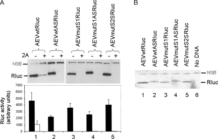
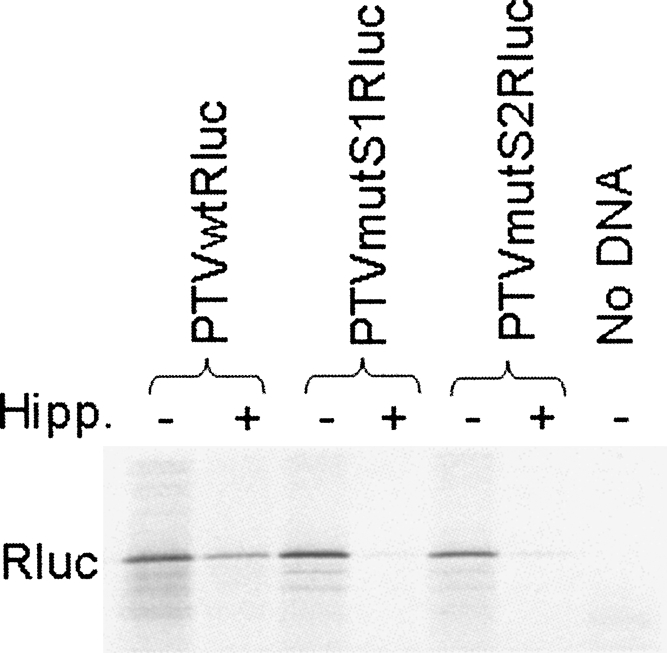
The initiation of protein synthesis on mRNAs within eukaryotic cells is achieved either by a 5' cap-dependent mechanism or through internal initiation directed by an internal ribosome entry site (IRES). Picornavirus IRES elements, located in the 5' untranslated region (5'UTR), contain extensive secondary structure and multiple upstream AUG codons. These features can be expected to inhibit cap-dependent initiation of translation. However, we have now shown that certain mutant hepatitis C virus-like picornavirus IRES elements (from porcine teschovirus-1 and avian encephalomyelitis virus), which are unable to direct internal initiation, are not significant barriers to efficient translation of capped monocistronic mRNAs that contain these defective elements within their 5'UTRs. Moreover, the translation of these mRNAs is highly sensitive to the expression of an enterovirus 2A protease (which induces cleavage of eIF4G) and is also inhibited by hippuristanol, a specific inhibitor of eIF4A function, in contrast to their parental wild-type IRES elements. These results provide a possible basis for the evolution of viral IRES elements within the context of functional mRNAs that are translated by a cap-dependent mechanism.


Figures

FIGURE 1.
FIGURE 1.
Cap-dependent and cap-independent translation within cells. (A) The structure of the basic pRBRluc reporter plasmid used in this study. Derivatives containing the wild-type or mutant EMCV IRES cDNA were made by insertion into the EcoRI (E) and BamHI (B) sites while the blunt-ended Fluc fragment (BamHI–XhoI fragment from pGEM-luc, see Materials and Methods) was inserted into the blunt-ended EcoRI site within pEMCVRluc to produce pFluc/EMCV/Rluc. The 5′UTR upstream of the Fluc coding region is 16 nt long. The SalI (S) site downstream from the Rluc coding sequence is indicated. It should be noted that the BamHI site located at the 3′ end of the EMCV cDNA was introduced at the position of AUG-10, modifying the ATG to ATC within this site (GGATCC); thus, AUG-10 is not present in these constructs. The line diagram of the EMCV IRES secondary structure shows the location of the GCGC sequence in place of the GNRA motif and the J domain as deleted in the ΔJ mutant. (B) Transient expression assays were performed by transfecting the indicated plasmids alone (−) or with (+) pGEM3Z/J1, which expresses the SVDV 2A protease, into vTF7-3 infected BHK cells. Cell extracts prepared after 20 h were analyzed by SDS-PAGE plus immunoblotting using anti-Rluc antibodies; detection was achieved using peroxidise labeled secondary antibodies plus chemiluminescence reagents. A nonspecific background band (labeled NSB) demonstrates the loading of cell extracts in each case. Expression levels within the cell extracts were quantified by Renilla luciferase assays. Results are presented as the mean (+S.D.) from four independent transfections. The inset shows the same data but on a different vertical scale for clarity. (C) The extracts from transfected cells were analyzed for Fluc expression by immunoblotting with anti-Fluc antibodies and by firefly luciferase enzyme assays.
FIGURE 2.
FIGURE 2.
Structure of monocistronic and dicistronic reporter plasmids containing wild-type and mutant forms of the PTV and AEV IRES elements. (A) Monocistronic derivatives of pRBRluc (Fig. 1) were prepared by inserting the indicated wild-type or mutant PTV-1 or AEV cDNA fragments into the BamHI (B) site. The wild-type and mutant sequences (in italics) for each derivative are indicated (the production of the mutants has been described previously, Chard et al. 2006a; Bakhshesh et al. 2008). Derivatives containing the IRES elements in the sense and antisense (AS) orientations were obtained by this procedure. (B) Dicistronic derivatives of pFluc were prepared by inserting EcoRI (E)–SalI (S) fragments from the monocistronic plasmids shown in A into the same sites of the vector downstream from the Fluc coding sequence. Note that the vector was prepared using a partial digestion with EcoRI and complete digestion with SalI. This construction therefore includes just nt 136–405 of the PTV cDNA, which clearly retains full IRES activity (as observed for nt 125–405). A line diagram of the secondary structure of the HCV-like IRES elements is shown to identify the locations of the individual IRES domains and the stems (S1 and S2), which are part of the pseudoknot (domain IIIf).
FIGURE 3.
FIGURE 3.
Cap-dependent translation of monocistronic mRNAs containing defective PTV IRES elements. Plasmids encoding monocistronic mRNAs containing the wild-type or mutant forms of the PTV IRES were transfected alone (−) or with (+) pGEM3Z/J1 into vTF7-3 infected BHK cells (as for Fig. 1B). Cell extracts were analyzed for Rluc expression by immunoblotting and by enzyme assay (results are the mean +S.D. for three or four independent transfections). The Rluc and a nonspecific background band (NSB) are indicated on the blots.
FIGURE 4.
FIGURE 4.
Comparison of translation from defective PTV IRES elements in mono- and dicistronic plasmids. The indicated monocistronic (Rluc alone) and dicistronic plasmids (Fluc/Rluc) containing the wild-type (A) or mutant (B) forms of the PTV IRES were assayed in parallel alone (−) or with (+) pGEM3Z/J1 (as for Fig. 1B). Cell extracts were prepared and analyzed for Fluc and Rluc expression as shown. Results are from one experiment but very similar results were observed in a second assay of the same set of plasmids.
FIGURE 5.
FIGURE 5.
Cap-dependent translation of monocistronic mRNAs containing defective AEV IRES elements. (A) Plasmids encoding monocistronic mRNAs containing the wild-type or mutant forms of the AEV IRES were transfected alone (−) or with (+) pGEM3Z/J1 into vTF7-3 infected BHK cells (as for Fig. 1B). Cell extracts were analyzed for Rluc expression by immunoblotting and by enzyme assay (results are the mean +S.D. for three or four independent transfections). (B) The indicated cell extracts were reanalyzed to facilitate comparison of the Rluc products expressed from plasmids containing the wild-type or mutant AEV IRES in the sense or antisense (AS) orientation. The Rluc and a nonspecific background band (NSB) are indicated on the blots.
FIGURE 6.
FIGURE 6.
Influence of the eIF4A inhibitor, hippuristanol, on the translation of monocistronic mRNAs containing wild-type and defective PTV IRES elements. The indicated plasmids were assayed in the presence or absence of hippuristanol (10 μM), as indicated, using an in vitro TNT system containing [35S]methionine, and the products were analyzed by SDS-PAGE and autoradiography.

References

    1. Ali I.K., McKendrick L., Morley S.J., Jackson R.J. Activity of the Hepatitis A virus IRES requires association between the cap-binding translation initiation factor (eIF4E) and eIF4G. J. Virol. 2001;75:7854–7863. - PMC - PubMed
    1. Baird S.D., Turcotte M., Korneluk R.G., Holcik M. Searching for IRES. RNA. 2006;12:1755–1785. - PMC - PubMed
    1. Bakhshesh M., Groppelli E., Willcocks M.M., Royall E., Belsham G.J., Roberts L.O. The picornavirus avian encephalomyelitis virus possesses a hepatitis C virus-like internal ribosome entry site (IRES) element. J. Virol. 2008;82:1993–2003. - PMC - PubMed
    1. Belsham G.J., Jackson R.J. Translation initiation on picornavirus RNA. In: Sonenberg N., editor. Translational Control of Gene Expression. Cold Spring Harbor Laboratory Press; Cold Spring Harbor, NY: 2000. pp. 869–900. Cold Spring Harbor Monograph 39,
    1. Bordeleau M.E., Mori A., Oberer M., Lindqvist L., Chard L.S., Higa T., Belsham G.J., Wagner G., Tanaka J., Pelletier J. Functional characterization of IRESes by an inhibitor of the RNA helicase eIF4A. Nature Chem. Biol. 2006;2:213–220. - PubMed

Publication types

MeSH terms

LinkOut - more resources