. 2008 Sep 1;112(5):1759-66.
doi: 10.1182/blood-2008-04-151068. Epub 2008 Jun 20.

Locally produced C5a binds to T cell-expressed C5aR to enhance effector T-cell expansion by limiting antigen-induced apoptosis

Affiliations

Locally produced C5a binds to T cell-expressed C5aR to enhance effector T-cell expansion by limiting antigen-induced apoptosis

Peter N Lalli et al. Blood. .

Abstract

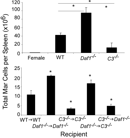
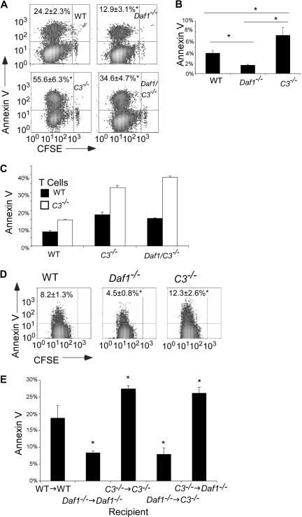
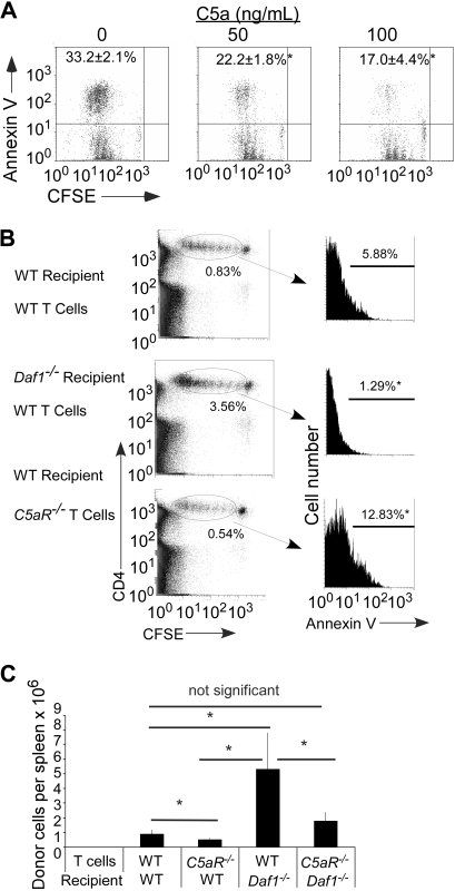
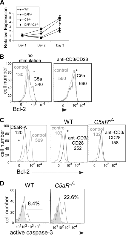
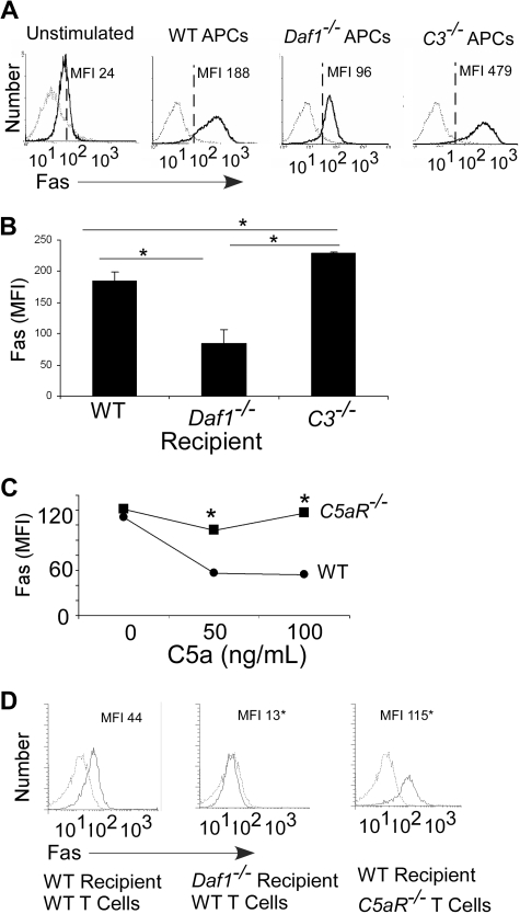
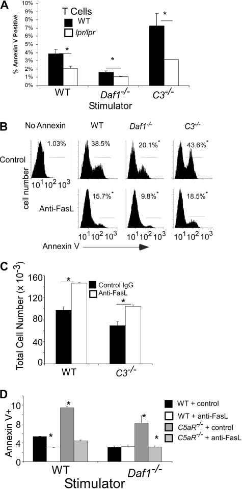
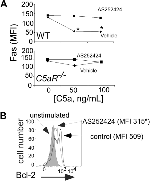
Our recent studies have shown that immune cell-produced complement provides costimulatory and survival signals to naive CD4(+) T cells. Whether these signals are similarly required during effector cell expansion and what molecular pathways link locally produced complement to T-cell survival were not clarified. To address this, we stimulated monoclonal and polyclonal T cells in vitro and in vivo with antigen-presenting cells (APCs) deficient in the complement regulatory protein, decay accelerating factor (DAF), and/or the complement component C3. We found that T-cell expansion induced by DAF-deficient APCs was augmented with diminished T-cell apoptosis, whereas T-cell expansion induced by C3(-/-) APCs was reduced because of enhanced T-cell apoptosis. These effects were traced to locally produced C5a, which through binding to T cell-expressed C5aR, enhanced expression of Bcl-2 and prevented Fas up-regulation. The results show that C5aR signal transduction in T cells is important to allow optimal T-cell expansion, as well as to maintain naive cell viability, and does so by suppressing programmed cell death.


Figures

Figure 1
Figure 1
APC-expressed C3 and DAF influence in vivo T-cell expansion. (Top) Absolute number of splenic GFP Mar T cells (percentage of GFP+CD4+ cells in the spleen multiplied by the total number of spleen cells) on day 4 after injection of 10 × 106 GFP+ Mar T cells into groups of male WT, Daf1−/−, or C3−/− or female control recipients (means ± SD of 3 or 4 animals/group). *P < .05. The total numbers of spleen cells in the male Daf1−/− mice (445 million) were significantly more than in the WT (252 million) or C3−/− (127 million) mice. (Bottom) Groups of male bone marrow chimeric mice (n = 3/group) were injected with 5 × 10⁶ CFSE-labeled Mar T cells, and total numbers of Mar cells in the spleen were quantified by flow cytometry (CFSE+/Vβ6+) on day 4. *P < .05 versus WT→WT control, n = 3 or 4 per group.
Figure 2
Figure 2
APC-expressed C3 and DAF influence T-cell apoptosis after antigen stimulation. (A) Flow cytometry plots showing annexin V staining of CFSE-labeled Mar T cells on day 3 after stimulation with WT, Daf1−/−, C3−/−, or Daf1/C3−/− macrophages plus HYDby peptide. Percentages of annexin V+ cells are given in the upper left. Data are representative of 4 individual experiments. *P < .05 versus WT APCs. Similar results were found when TUNEL staining was used as a readout (Figure S2). (B) Percentage of annexin V+ polyclonal H-2b T cells on day 4 of in vitro culture with allogeneic H-2d WT, Daf1−/−, or C3−/− macrophages. Values are means plus or minus SD of 3 individual experiments. *P < .05. (C) CFSE-labeled WT or C3−/− H-2d T cells were stimulated in vitro with WT, C3−/−, or Daf1/C3−/− allogeneic H-2b macrophages for 4 days and stained with annexin V. Values are means plus or minus SD of 3 individual experiments. *P < .05 versus WT T cells. (D) Flow cytometry plots depicting annexin V+ Mar T cells in the spleens of recipient mice on day 3 after adoptive transfer (10 × 106 Mar T cells per mouse) into groups of male WT, Daf1−/−, or C3−/− recipients (n = 3 per group). *P < .05 versus WT APCs. (E) Groups of male bone marrow chimeric mice (n = 3) were injected with 5 × 10⁶ CFSE-labeled Mar T cells and Vβ6+ splenic Mar cells were assayed for apoptosis by annexin V staining on day 3. *P < .05 versus WT→WT controls.
Figure 3
Figure 3
C5a binding to T cell–expressed C5aR regulates T-cell apoptosis and expansion. (A) Representative flow plots of annexin V+ Mar T cells after in vitro stimulation with H-2b bone marrow dendritic cells plus HYDby peptide+ recombinant mouse C5a. The experiment was repeated with similar results. Numbers in upper left quadrant represent percentage annexin V+ (n = 3 per group). *P < .05 versus no C5a control. (B) Left: Flow plots of polyclonal H-2b WT or C5aR−/− CD4 T cells (CFSE dilution) after injection into (B6xBalb/c) F1 WT or Daf1−/− recipients (day 3, n = 2 or 3/group). The percentages of injected cells per recipient spleen on day 3 are shown. Right: Annexin V expression on the injected cells within the gate. *P < .05 versus WT control. (C) Polyclonal H-2b WT or C5aR−/− CD4 T cells were injected into (B6xBalb/c) F1 WT or Daf1−/− recipients. Total numbers of donor T cells in recipient spleens were determined on day 4 after injection. Data are representative of 2 experiments. *P < .05.
Figure 4
Figure 4
T-cell Bcl-2 and caspase-3 expression are regulated by locally produced C5a binding to T cell–expressed C5aR. (A) Expression of message for Bcl-2 in responding Mar T cells after stimulation with Dby-loaded WT, Daf1−/−, C3−/−, or Daf1/C3−/− APCs (quantitative RT-PCR). Data are representative of 2 individual experiments. Error bars represent SD. (B) Flow cytometric analysis of intracellular Bcl-2 expression in unstimulated (left) or anti-CD3/CD28 stimulated (right) WT T cells plus recombinant C5a (50 ng/mL). Assays were performed at 72 hours. Data are representative replicates of 3 individual experiments. (C) Bcl-2 expression in anti-CD3/CD28-stimulated WT T cells plus or minus C5aR antagonist (C5aR-A, 0.1 μM, 72 hours) or vehicle control (left) and in anti-CD3/CD28–stimulated C5aR−/− T cells (right, 48 hours). Data are representative replicates of at least 2 or 3 individual experiments per group. Numbers in each panel depict the mean fluorescence intensity. *P < .05. (D) Caspase-3 expression in anti-CD3/CD28–stimulated WT or C5aR−/− T cells (120 hours). Analogous results were detected at 72 hours (2.1% caspase-3+ WT T cells vs 5.1% C5aR−/− T cells, not shown). Data are representative replicates of 3 individual experiments.
Figure 5
Figure 5
Local complement activation regulates T-cell Fas expression after antigen stimulation. (A) Flow cytometric plots of Fas expression on CFSE-labeled Mar T cells stimulated in vitro (3 days) with WT, Daf1−/−, or C3−/− macrophages plus HYDby peptide (MFI, isotype control-, anti-Fas-). Results are means of triplicate wells. Data are representative of 4 individual experiments. (B) Fas expression on Mar T cells in spleens (gated on Mar TCR Vβ6+ cells) of recipient male WT, Daf1−/−, or C3−/− mice 3 days after adoptive transfer (10 × 106 cells/mouse). Results are depicted as means plus or minus SD of n = 3 or 4 per group. (C) Mean fluorescent intensities of anti-CD3/CD28–stimulated WT or C5aR−/− T-cell surface Fas levels on addition of recombinant C5a after 72 hours. *P < .05 versus WT. Fas levels (MFI) on unstimulated WT was 25 and on C5aR−/− T cells was 28 (not shown). (D) Flow plots of polyclonal H-2b WT or C5aR−/− CD4 T cells after injection into B6xBalb/c F1 WT or Daf1−/− recipients (day 3) depicting surface Fas MFI. The experiment was repeated twice with similar results. *P < .05 versus WT.
Figure 6
Figure 6
Local complement activation regulates Fas-mediated T-cell apoptosis. (A) Polyclonal naive (> 90% CD62Lhi, CD44lo, not shown) CD4+ H-2b WT or B6.MRLlpr T cells were stimulated in vitro with allogeneic H-2d WT, Daf1−/−, or C3−/− macrophages and stained for annexin V on day 4. Results are means plus or minus SD of triplicate wells. The experiment was repeated with identical results. *P < .05. Data are representative of 3 individual experiments. (B) Representative flow plots of annexin V+ Mar T cells after stimulation for 3 days with WT, Daf1−/−, or C3−/− macrophages and HYDby peptide plus 10 μg/mL anti-FasL antibody or control. The percentage of cells within the gated region is listed in each panel. *P < .05 versus controls. (C) Total live (annexin V) Mar T cells after 3 days in culture with WT or C3−/− macrophages and HYDby peptide plus 10 μg/mL anti-FasL antibody or control. Results are means plus or minus SD of 2 experiments. *P < .05 versus control antibody. (D) Percentage annexin V+ polyclonal H-2b WT or C5aR−/− CD4 T cells after 4 days stimulation with allogeneic WT or Daf1−/− H-2d macrophages in the presence or absence of 10 μg/mL of anti-FasL antibody. Data are representative of 2 individual experiments. *P < .05 versus WT controls.
Figure 7
Figure 7
Complement-regulated Fas and Bcl-2 involves PI-3Kγ. (A) Mean fluorescent intensities for T-cell surface Fas expression on WT (top) or C5aR−/− (bottom) B6 T cells stimulated with anti-CD3/CD28 plus or minus recombinant C5a plus or minus 0.1 μM AS25242 (72 hours). *P < .05 versus control. The experiment was repeated with identical results. (B) Flow cytometry plot demonstrating Bcl-2 expression in anti-CD3/CD28-stimulated B6 T cells (gated on CD4+ cells) plus or minus 0.1 μM AS252424. Numbers in each panel depict the mean fluorescence intensity. Bcl-2 MFI in unstimulated cells was 130. Assay was performed at 72 hours. Data are representative of at least 2 independent experiments. P < .05.

Comment in

  • Complement dances with T cells.
    Ansari MJ, Sayegh MH. Ansari MJ, et al. Blood. 2008 Sep 1;112(5):1551-2. doi: 10.1182/blood-2008-07-168179. Blood. 2008. PMID: 18725570 No abstract available.

References

    1. Hildeman DA, Zhu Y, Mitchell TC, et al. Activated T cell death in vivo mediated by proapoptotic bcl-2 family member bim. Immunity. 2002;16:759–767. - PubMed
    1. Pellegrini M, Belz G, Bouillet P, Strasser A. Shutdown of an acute T cell immune response to viral infection is mediated by the proapoptotic Bcl-2 homology 3-only protein Bim. Proc Natl Acad Sci U S A. 2003;100:14175–14180. - PMC - PubMed
    1. Ju ST, Panka DJ, Cui H, et al. Fas(CD95)/FasL interactions required for programmed cell death after T-cell activation. Nature. 1995;373:444–448. - PubMed
    1. Stranges PB, Watson J, Cooper CJ, et al. Elimination of antigen-presenting cells and autoreactive T cells by Fas contributes to prevention of autoimmunity. Immunity. 2007;26:629–641. - PMC - PubMed
    1. Heeger PS, Lalli PN, Lin F, et al. Decay-accelerating factor modulates induction of T-cell immunity. J Exp Med. 2005;201:1523–1530. - PMC - PubMed

Publication types

MeSH terms