Review
. 2008 Jul;9(7):505-16.
doi: 10.1038/nrm2439.

Mediators of reprogramming: transcription factors and transitions through mitosis

Affiliations
Review

Mediators of reprogramming: transcription factors and transitions through mitosis

Dieter Egli et al. Nat Rev Mol Cell Biol. 2008 Jul.

Abstract

It is thought that most cell types of the human body share the same genetic information as that contained in the zygote from which they originate. Consistent with this view, animal cloning studies demonstrated that the intact genome of a differentiated cell can be reprogrammed to support the development of an entire organism and allow the production of pluripotent stem cells. Recent progress in reprogramming research now points to an important role for transcription factors in the establishment and the maintenance of cellular phenotypes, and to cell division as a mediator of transitions between different states of gene expression.


Figures

Figure 1 |
Figure 1 |. Cell-cycle synchronization after nuclear transfer.
a | Transfer of a G1 interphase nucleus into an oocyte in meiosis results in nuclear-envelope breakdown and chromosome condensation. Chromosome condensation converts the interphase nucleus into meiotic chromatin, thereby synchronizing the cell cycle of the donor cell with the recipient oocyte. Unfertilized oocytes are naturally arrested in meiosis. To enter the cell cycle and for development to occur, an artificial activation stimulus (intracellular calcium release) is required. Cytochalasin B (CytoB) is added to prevent cytokinesis and to prevent the extrusion of the second polar body (PB), which would result in two aneuploid cells. b | Transfer of chromosomes from a mitotic somatic cell to a mitotic zygote results in development. The cell cycle of the mitotic zygote and the mitotic genome of a somatic cell are synchronized from the moment of transfer. Cell-cycle progression and cleavage leads to the formation of a two-cell stage embryo. ICM, inner cell mass; TE, trophectoderm.
Figure 2 |
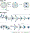
Figure 2 |. Genome exchange during cell division allows development of clones.
Meiotic progression of unfertilized oocytes and embryonic development (a). The germinal vesicle (GV) is the interphase nucleus of the oocyte before entry into meiotic division. Fertilization of the metaphase II oocyte by the sperm forms the fertilized zygote. Development is shown until the blastocyst stage at which embryonic stem (ES) cells can be derived. Removal of the condensed chromosomes from the unfertilized oocyte (b) or from the zygote (c) during mitosis, followed by the transfer of a new genome, results in the development of clones, from which ES cells can be derived. Nuclei or condensed chromosomes can be transferred into the unfertilized oocyte. Replacement of the GV nucleus with the interphase nucleus of a differentiated cell results in developmental arrest (d). Replacement of the two haploid interphase nuclei of the zygote with the interphase nuclei (nuclear transfer) or an M-phase genome (chromosome transfer) of a diploid cell results in developmental arrest in the next interphase (e). PB, polar body.
Figure 3 |
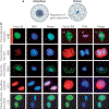
Figure 3 |. Regulators of gene expression dissociate from mitotic chromatin.
a | In a cell in interphase, regulators of gene expression (coloured dots) associate with the genetic information in the nucleus. During mitosis, the nuclear envelope breaks down, chromosomes condense and regulators of gene expression dissociate from the genetic information and localize to the cytoplasm. b | Gene expression is regulated at multiple levels and important components of these regulatory systems dissociate from the chromatin in mitosis. Interphase (I) and mitosis (M) stages are shown. Some of the main factors that dissociate from chromatin during mitosis include the transcriptional repressor heterochromatin protein-1β (HP1β; the red signal marks the phosphorylation of Ser10 on Histone H3 that causes the dissociation of HP1β from chromatin), BMI1, the chromatin remodelling protein BRG1, sister-chromatid cohesion-1 (SCC1) and the transcriptional activator heat-shock factor (HSF). All immunocytochemistry shown is in HeLa cells, except BRG1, which is shown in 2-cell-stage mouse embryos. Cells shown are at prometaphase of mitosis, with the exception of BMI1, which shows a cell at anaphase of mitosis with the separating two groups of chromatin. HP1β images adapted, with permission, from Nature REF. © (2005) Macmillan Publishers Limited. All rights reserved. BMI1 images adapted, with permission, from REF. © Company of Biologists. SCC1 images adapted, with permission, from Nature REF. © Macmillan Publishers Limited. All rights reserved. HSF images adapted, with permission, from REF. © (1995) Cell Press.
Figure 4 |
Figure 4 |. Model of reprogramming after transfer of a somatic cell genome into an oocyte or zygote in cell division.
a | A hypothetical locus in a repressed state, bound by polycomb repressive complex-1 (PRC1) and the heterochromatin protein-1 (HP1). The histone methyltransferase Suv39h methylates H3K9 to generate a binding site for HP1 and PRC2 methylates H3K27 to generate a binding site for PRC1. b | During mitosis, the phosphorylation of Ser10 on histone H3 by the mitotic kinase Aurora B results in the dissociation of HP1 from chromatin. Aurora B kinase also phosphorylates Ser28 on histone H3. PRC1 is phosphorylated during mitosis by 3pK (mitogen-activated protein kinase-activated protein kinase-3) and dissociates from methylated H3K27. An unknown kinase phosphorylates Suv39h, which also dissociates from chromatin. c | Following transfer of the somatic cell genome into a mitotic cell with an embryonic transcription factor environment, and following exit from mitosis, embryonic transcription factors bind to their target sites. Binding leads to the recruitment of chromatin-modifying activities and the establishment of a basal promoter complex. The recruitment of the histone demethylases jumonji domain-containing (JMJD) protein-2C (JMJD2C), JMJD-containing histone demethylation protein-2A (JHDM2A; not shown) and Lys-specific histone demethylase-1 (LSD1), which demethylate H3K9, can occur in a transcription-factor-dependent manner,. Unknown activities demethylate H3K27. The histone acetyl transferase general control of amino-acid-synthesis protein-5 (GCN5) acetylates Lys residues on the N-terminal tails of histone H3 and H4. d | The histone acetyl transferase CREB-binding protein (CBP)–RNA polymerase II (pol II) holoenzyme complex, is recruited to the promoter and nucleosome acetylation facilitates SWI/SNF recruitment to remodel chromatin and position nucleosomes. The transcription factor TFIID binds to the promoter and initiates transcriptional activation (see also ReF. 164). The active locus excludes the access of PRC1 or HP1 and prevents the formation of heterochromatin. CBX, chromobox homologue, a mammalian homologue of Drosophila melanogaster polycomb; E(z), enhancer of zeste; PHC, mammalian homologue of D. melanogaster polyhomeotic.
Figure 5 |
Figure 5 |. Different methods of reprogramming require transcriptional regulators and passage through cell division.
a | Removal of the condensed chromosomes from a zygote in mitosis (or an oocyte in meiosis) generates a karyoplast that lacks the factors that are required for the regulation of embryonic gene expression, and also generates a cytoplast that contains these factors. A new genome is transferred by microinjection. Following entry into interphase, nuclear factors localize to the new interphase nucleus and activate transcription, and thereby development occurs. b | Reprogramming by defined factors. Transgenes introduced with the help of viruses express the four transcription factors octamer-binding transcription factor-4 (Oct4), SRY-related high mobility group (HMG)-box protein-2 (Sox2), Kruppel-like factor 4 (Klf4) and c-Myc in a somatic cell. Following selection and multiple cell divisions, colonies arise that closely resemble embryonic stem (ES) cells. Reprogramming to induced-pluripotent stem (iPS) cells seems to be more efficient with rapidly dividing cell cultures. The persistence of somatic nuclear factors might counteract the reprogramming process. c | Fusion of a somatic cell (triangular shape) with an ES cell (round shape). A selection process results in the isolation of rare pluripotent stem cells with a tetraploid genome that contains both donor and recipient cell genomes.

Similar articles

Cited by

References

    1. Briggs R. & King TJ Transplantation of living nuclei from blastula cells into enucleated frogs’ eggs. Proc. Natl Acad. Sci. USA 38, 455–463 (1952). - PMC - PubMed
    1. Gurdon JB, Elsdale TR & Fischberg M. Sexually mature individuals of Xenopus laevis from the transplantation of single somatic nuclei. Nature 182, 64–65 (1958).

      Demonstrates that frogs can be cloned from somatic cells.

    1. Wilmut I, Schnieke AE, McWhir J, Kinnotd AJ & Campbell KH Viable offspring derived from fetal and adult mammalian cells. Nature 385, 810–813 (1997).

      Demonstrates the reversibility of mammalian differentiation.

    1. Eggan K. et al. Mice cloned from olfactory sensory neurons. Nature 428, 44–49 (2004). - PubMed
    1. Hochedlingoteer K. & Jaenisch R. Monoclonal mice generated by nuclear transfer from mature B and T donor cells. Nature 415, 1035–1038 (2002).

      Demonstrates that the genome of even the most differentiated cells can support development after nuclear transfer.

Publication types

Substances