. 2008 Jul;118(7):2438-47.
doi: 10.1172/JCI34694.

Proteinase 3 and neutrophil elastase enhance inflammation in mice by inactivating antiinflammatory progranulin

Affiliations

Proteinase 3 and neutrophil elastase enhance inflammation in mice by inactivating antiinflammatory progranulin

Kai Kessenbrock et al. J Clin Invest. 2008 Jul.

Abstract

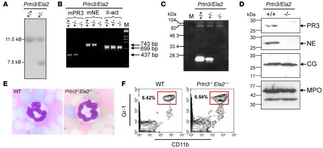
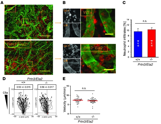
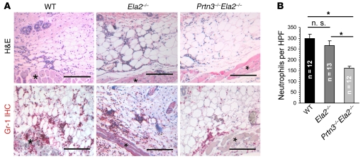
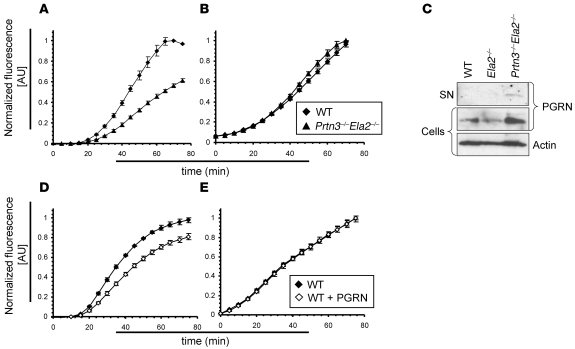
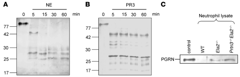
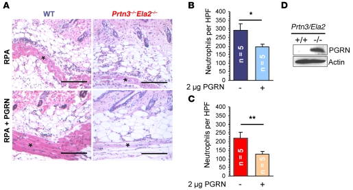
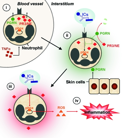
Neutrophil granulocytes form the body's first line of antibacterial defense, but they also contribute to tissue injury and noninfectious, chronic inflammation. Proteinase 3 (PR3) and neutrophil elastase (NE) are 2 abundant neutrophil serine proteases implicated in antimicrobial defense with overlapping and potentially redundant substrate specificity. Here, we unraveled a cooperative role for PR3 and NE in neutrophil activation and noninfectious inflammation in vivo, which we believe to be novel. Mice lacking both PR3 and NE demonstrated strongly diminished immune complex-mediated (IC-mediated) neutrophil infiltration in vivo as well as reduced activation of isolated neutrophils by ICs in vitro. In contrast, in mice lacking just NE, neutrophil recruitment to ICs was only marginally impaired. The defects in mice lacking both PR3 and NE were directly linked to the accumulation of antiinflammatory progranulin (PGRN). Both PR3 and NE cleaved PGRN in vitro and during neutrophil activation and inflammation in vivo. Local administration of recombinant PGRN potently inhibited neutrophilic inflammation in vivo, demonstrating that PGRN represents a crucial inflammation-suppressing mediator. We conclude that PR3 and NE enhance neutrophil-dependent inflammation by eliminating the local antiinflammatory activity of PGRN. Our results support the use of serine protease inhibitors as antiinflammatory agents.


Figures

Figure 1
Figure 1. Generation and characterization of Prtn3–/–Ela2–/– mice.
(A) Southern blot showing the WT (+/+) 11.5-kb band of the nontargeted allele and the presence of an additional 7.5-kb band in a heterozygous (+/–) PR3/NE-targeted embryonic stem cell clone. (B) RT-PCR revealed the complete lack of mouse PR3 (mPR3; 437 bp) and mouse NE (743 bp) transcripts in bone marrow cells from Prtn3–/–Ela2–/– mice (–/–), while expression of β-actin (699 bp) was normal. (C) Casein zymography showed prominent casein degradative activity at 27 kDa in WT neutrophil lysates, while intermediate degradation by heterozygous lines and no degradation by Prtn3–/–Ela2–/– lines was found at this size. M, marker. (D) Western blot analysis of granule enzyme expression in bone marrow–derived neutrophils. In Prtn3–/–Ela2–/– neutrophils, no signals for PR3 and NE were detected, while CG (~26 kDa), MPO (~59 kDa), and a smaller degradation product of MPO were detected at the same levels as in WT neutrophils. (E) Microscopic analysis of H&E-stained blood smears revealed normal granulocyte morphology in Prtn3–/–Ela2–/– mice, with a polymorphic nucleus (dark blue) identical to that of WT neutrophils. Original magnification, ×20. (F) Flow cytometry of peripheral blood with gating on Gr-1hiCD11b+ showed regular neutrophil populations (boxed regions) in Prtn3–/–Ela2–/– mice. Plots are representative of data obtained from 3 mice per group. Percentages denote percent cells in the boxed regions.
Figure 2
Figure 2. PR3 and NE are not principally required for neutrophil extravasation and interstitial migration.
(A and B) Phorbol ester–treated ear tissues of WT and Prtn3–/–Ela2–/– mice were immunostained for laminin (LN; green) to visualize EBM and Gr-1 (red) to identify neutrophils. Lesions were examined 4 h after stimulus application by fluorescence microscopy as described in Methods. (A) Representative images of tissue from WT and Prtn3–/–Ela2–/– mice. Both genotypes developed strong and widespread neutrophil infiltrations. (B) Higher-magnification images of boxed regions in A. Prtn3–/–Ela2–/– neutrophils showed no retention at the EBM. Scale bars: 200 μm (A); 25 μm (B). (C) Overall neutrophil infiltrates were quantified as the percentage of Gr-1–positive cells per microscopic field. Data are mean ± SEM. Intravascular cells were excluded. No significant difference between WT and Prtn3–/–Ela2–/– mice was found (P = 0.63). In vitro migration of WT and Prtn3–/–Ela2–/– neutrophils directed by C5a through 3-dimensional collagen matrices was analyzed by time-lapse video microscopy (see Supplemental Video 1). (D) The tracks of WT (n = 41) and Prtn3–/–Ela2–/– (n = 42) neutrophils are shown, and the factor for directionality ± SEM is indicated. No impairment was observed regarding chemotactic directionality of Prtn3–/–Ela2–/– versus WT neutrophils (P = 0.19). (E) Velocities of single cells (individual points) were calculated and averaged (red bar). Prtn3–/–Ela2–/– neutrophils showed no significant difference versus WT cells (P = 0.30).
Figure 3
Figure 3. Impaired inflammatory response to locally formed ICs in Prtn3–/–Ela2–/– mice.
(A) Representative photomicrographs of inflamed skin sections 4 h after IC formation. Neutrophils were identified morphologically (polymorphic nucleus) in H&E stainings and by Gr-1 staining (red). The cellular infiltrates were located to the adipose tissue next to the panniculus carnosus muscle (asterisks) and were primarily composed of neutrophil granulocytes. Scale bars: 200 μm. (B) Neutrophil infiltrates in lesions from Prtn3–/–Ela2–/– mice were significantly diminished compared with Ela2–/– mice and WT mice. Neutrophil influx in Ela2–/– mice was slightly, but not significantly, diminished compared with WT mice. Results are mean ± SEM infiltrated neutrophils per HPF. *P < 0.05.
Figure 4
Figure 4. Impaired oxidative burst and PGRN degradation by IC-activated Prtn3–/–Ela2–/– neutrophils.
Oxidative burst as the readout for neutrophil activation by ICs was measured over time. (A) While no difference was observed during the initial 20-min lag phase of the oxidative burst, Prtn3–/–Ela2–/– neutrophils exhibited diminished ROS production over time compared with WT neutrophils. (B) Bypassing receptor-mediated activation using 25 nM PMA restored the diminished oxidative burst of Prtn3–/–Ela2–/– neutrophils. Results are presented as normalized fluorescence in AU (relative to maximum fluorescence produced by WT cells). Data (mean ± SD) are representative of 3 independent experiments each conducted in triplicate. (C) Isolated mouse neutrophils were activated by ICs in vitro and tested for PGRN degradation by IB. In the cellular fraction, the PGRN (~80 kDa) signal was markedly increased in Prtn3–/–Ela2–/– cells compared with WT and Ela2–/– neutrophils. Intact PGRN was present in the supernatant (SN) of IC-activated Prtn3–/–Ela2–/– neutrophils only, not of WT or Ela2–/– cells. (D and E) Exogenous administration of 100 nM PGRN significantly reduced ROS production of neutrophils activated by ICs (D), but not when activated by PMA (E). Data (mean ± SD) are representative of 3 independent experiments each conducted in triplicate.
Figure 5
Figure 5. PR3 and NE are major PGRN processing enzymes of neutrophils.
(A and B) Silver-stained SDS-PAGE analysis of recombinant human PGRN incubated at a 1:10 enzyme/substrate ratio with purified human NE (A) and recombinant mouse PR3 (B). Both NE and PR3 completely cleaved ~80-kDa PGRN to smaller molecular fragments within 5 min of incubation. (C) Recombinant mouse PGRN was incubated with neutrophil lysates from WT, Ela2–/–, and Prtn3–/–Ela2–/– mice for 1 h at 37°C and analyzed by anti-mouse PGRN Western blot. Compared with untreated PGRN (control), WT neutrophils completely degraded PGRN. In Ela2–/– neutrophils, a faint band of intact PGRN was detected, while in Prtn3–/–Ela2–/– neutrophils, a distinct PGRN band remained, comparable to control.
Figure 6
Figure 6. PGRN is a potent inhibitor of IC-stimulated inflammation in vivo.
Recombinant mouse PGRN (2 μg) was intradermally applied with anti-OVA IgG, and the RPA was started in WT and Prtn3–/–Ela2–/– mice (n = 5 per group). (A) After 4 h, the effect of PGRN application was evaluated by histological analyses. Representative images show neutrophil infiltrates at the panniculus carnosus muscle (asterisks). Scale bars: 200 μm. (B and C) Effect of PGRN administration on neutrophil influx. In both WT (B) and Prtn3–/–Ela2–/– (C) mice, neutrophil infiltration was significantly diminished at PGRN-treated sites compared with untreated sites. This effect appeared to be more pronounced in the protease-deficient mice. Data are mean ± SEM infiltrated neutrophils per HPF. *P < 0.05; **P < 0.01. (D) Neutrophils isolated ex vivo from inflamed peritoneum of WT and Prtn3–/–Ela2–/– mice were analyzed by anti-mouse PGRN Western blot of concentrated neutrophil lysates. Intact PGRN was found abundantly in Prtn3–/–Ela2–/– but not WT neutrophils. Loading was controlled using anti-actin Western blot.
Figure 7
Figure 7. Proposed function of PR3 and NE in IC-mediated inflammation.
TNF-α–primed neutrophils extravasate from blood vessels, translocate PR3/NE to the cellular surface, and discharge PGRN to the pericellular environment (i). During transmigration of interstitial tissues (ii), neutrophil activation is initially suppressed by relatively high pericellular levels of antiinflammatory PGRN (green shading), which is also produced locally by keratinocytes and epithelial cells of the skin. Until IC depots are reached, neutrophil activation is inhibited by PGRN. Surface receptors (e.g., Mac-1) recognize ICs, which results in signal transduction (black dotted arrow) and activation of the phox. The molecular pathway of PGRN-mediated inhibition is not completely understood, but it may interfere with integrin signaling after IC encounter (green dotted line inside the cell). Adherence of neutrophils to ICs (iii) further increases pericellular PR3 and NE activity. PR3 and NE cooperatively degrade PGRN in the early stage of neutrophilic activation to facilitate optimal neutrophil activation (red shading), resulting in sustained integrin signaling (red arrow) and robust production of ROS by the phox system. Subsequently, neutrophils release ROS together with other proinflammatory mediators and chemotactic agents, thereby enhancing the recruitment of further neutrophils and establishing inflammation (iv). In the absence of PR3/NE, the switch from inflammation-suppressing (ii) to inflammation-enhancing (iii) conditions is substantially delayed, resulting in diminished inflammation in response to ICs (iv).

References

    1. Nathan C. Neutrophils and immunity: challenges and opportunities. Nat. Rev. Immunol. 2006;6:173–182. doi: 10.1038/nri1785. - DOI - PubMed
    1. Jancar S., Sanchez C.M. Immune complex-mediated tissue injury: a multistep paradigm. Trends Immunol. 2005;26:48–55. doi: 10.1016/j.it.2004.11.007. - DOI - PubMed
    1. Ravetch J.V. Fc receptors: rubor redux. Cell. 1994;78:553–560. doi: 10.1016/0092-8674(94)90521-5. - DOI - PubMed
    1. Belaaouaj A., et al. Mice lacking neutrophil elastase reveal impaired host defense against gram negative bacterial sepsis. Nat. Med. 1998;4:615–618. doi: 10.1038/nm0598-615. - DOI - PubMed
    1. Belaaouaj A., Kim K.S., Shapiro S.D. Degradation of outer membrane protein A in Escherichia coli killing by neutrophil elastase. Science. 2000;289:1185–1188. doi: 10.1126/science.289.5482.1185. - DOI - PubMed

Publication types

MeSH terms