Review
. 2008 May;33(5):405-20.
doi: 10.1080/02713680802018419.

Understanding and analyzing meibomian lipids--a review

Affiliations
Review

Understanding and analyzing meibomian lipids--a review

Igor A Butovich et al. Curr Eye Res. 2008 May.

Abstract

Purpose: This review is intended to bring to the informed reader the current state of knowledge about meibomian lipids and the art for analyzing them.

Methods: At the forefront of any endeavor, there are controversies, and these, along with future directions in the field, are brought to the reader's attention.

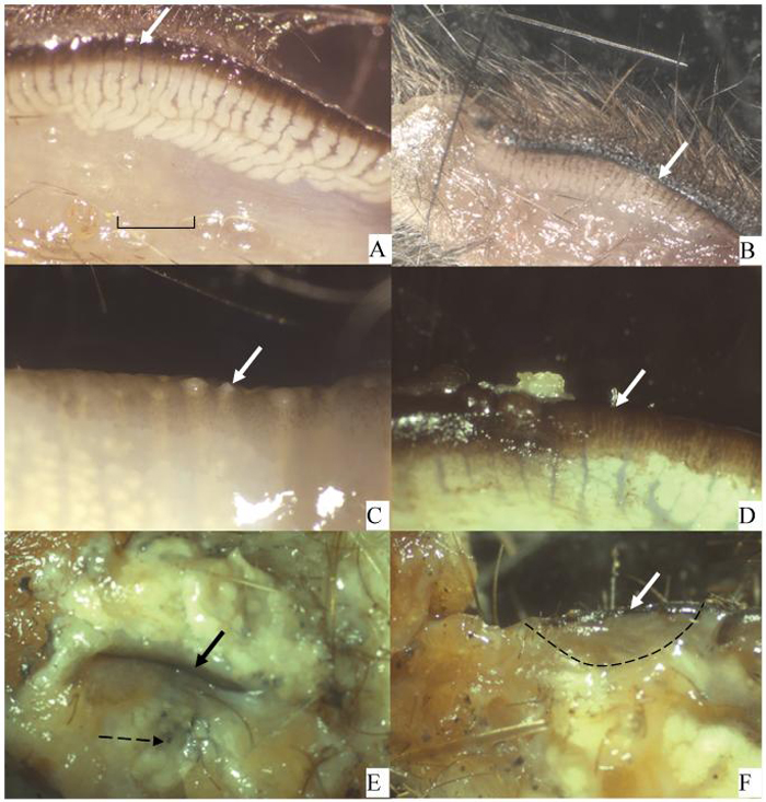
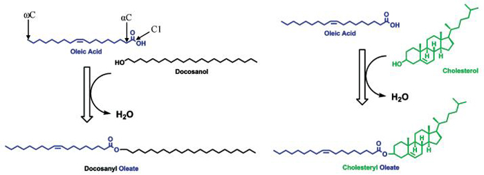
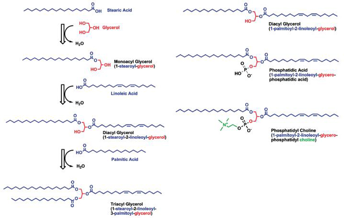
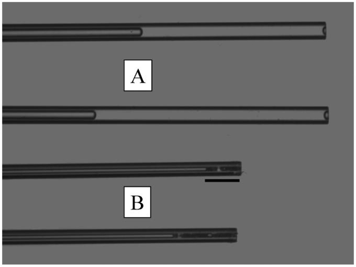
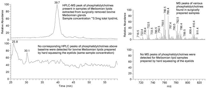
Results: Function and anatomy of meibomian glands are briefly covered, giving insight into possible mechanisms for secretory controls. Anatomically, some anomalies in meibomian gland distribution of different species, such as whales versus dolphins, are presented, and, for the first time, the structure of the meibomian glands in a selection of marsupials is presented. In attempting to make the literature more accessible, lipid structure and nomenclature are described, and these structures are related to their possible effects on the physicochemical properties of meibomian lipids. The advantages and disadvantages of various collection and storage techniques are described, as well as how gas chromatography and combined HPLC and mass spectrometry coupled with fragmentation are currently enabling us to determine the nature of the lipids in very small samples.

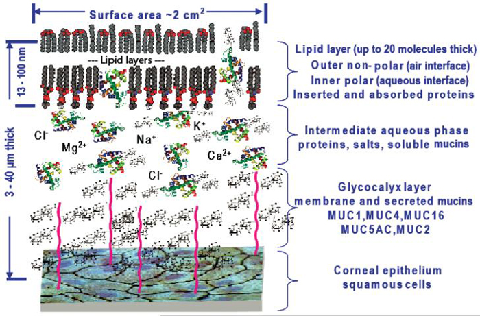
Conclusions: This review extends to discussing the lipids in tears (as opposed to meibomian gland lipids) and briefly highlights new thoughts about the interactions between proteins of the tear film and meibomian lipids. A model that includes proteins in the outer layer of the tear film is also presented. This model is currently being critically analyzed by the ocular community. It concludes briefly by highlighting possible further areas of research in this area.


Figures

FIGURE 1
FIGURE 1
View of inner tarsal plate from various marsupials and an echidna. Bold arrow indicates eyelid margin. The meibomian glands are visible below this. (A) Kultarr (Antechinmys laniger). An endangered desert dwelling species. (B) Antechinus (Antechinus stuartii). Coastal forest dweller. (C) Koala (Phascolarctos cinereus). Tree dweller living in coastal forests. Just left of the arrow are large meibomian gland openings. (D) Red necked wallaby (Macropus rufogriseus). A coastal plain dweller. Just left of the arrow is a meibomian gland opening with a small amount of meibomian oil extruded. (E) Echidna (Tachyglossus aculeatus). Both eyelids are present surrounding the ocular slit. This region has a massive fat pad consisting of sebaceous glands. The broken arrow points to the end of a hair shaft that is surrounded by the sebaceous gland. (F) Echidna. The edge of one eyelid has been dissected (from lid margin to dotted line) revealing no meibomian glands. Scale in (A) to be used for all figures according to this legend: A = 0.5 mm; B = 2.5 mm; C = 2.0 mm; D = 2.0 mm; E = 2.5 mm; F = 2.5 mm.
FIGURE 2
FIGURE 2
Wax and sterol esters. Oleic acid C18:1:9Z is an 18-carbon fatty acid with a cis double bond 9 bonds away from C1. The same double bond is 9 bonds from the omega or last carbon (ωC), so it is an omega-9 fatty acid. It condenses with a long-chain alcohol, which results in an ester bond to form a wax ester. Similarly, a condensation with a sterol (in this case, cholesterol) will form a sterol ester. These are families of molecules because the fatty acid, alcohol, or sterol may vary.
FIGURE 3
FIGURE 3
Glycerolipids are formed by a condensation of various fatty acids to glycerol. For monoacyl- and diacylglycerols, the free hydroxyl groups make this part of the molecule water soluble, while triacylglycerols, with no free hydroxyl, are very hydrophobic. Phospholipids have a phosphate condensed to C3 of glycerol, and this head group is very hydrophilic. An alcohol can further condense onto the phosphate group to form phospholipids. If the acid group were removed at C2 (common) or C1 (uncommon), it would be called a lysophospholipid.
FIGURE 4
FIGURE 4
Comparative appearance and volumes of tear samples (A) and meibomian lipid samples (B) collected with a micropipette at room temperature. Scale = 1.46 mm.
FIGURE 5
FIGURE 5
Comparisons of HPLC and MS spectra obtained from homogenized bovine glands versus extruded lipids.
FIGURE 6
FIGURE 6
Revised tear film illustrating the closer interaction of the lipid-binding proteins of the tear and the outer lipid layer.

References

    1. McDonald JE. Surface phenomena of tear films. Trans Am Ophthalmol Soc. 1968;66:905–939. - PMC - PubMed
    1. McDonald JE. Surface phenomena of tear films. Am J Ophthalmol. 1969;67:56–64. - PubMed
    1. Guillon J-P. Tear film photography and contact lens wear. J Br Contact Lens Assoc. 1982;5:84–87.
    1. Bron AJ, Tiffany JM, Gouveia SM, et al. Functional aspects of the tear film lipid layer. Exp Eye Res. 2004;78:347–360. - PubMed
    1. Tiffany JM. Advances in Lipid Research. Academic Press; New York/London: 1987. The lipid secretion of the meibomian glands; pp. 1–62. - PubMed

Publication types

LinkOut - more resources