. 2008 Jun 20:8:65.
doi: 10.1186/1471-213X-8-65.

Endothelial-specific ablation of serum response factor causes hemorrhaging, yolk sac vascular failure, and embryonic lethality

Affiliations

Endothelial-specific ablation of serum response factor causes hemorrhaging, yolk sac vascular failure, and embryonic lethality

Mary L Holtz et al. BMC Dev Biol. .

Abstract

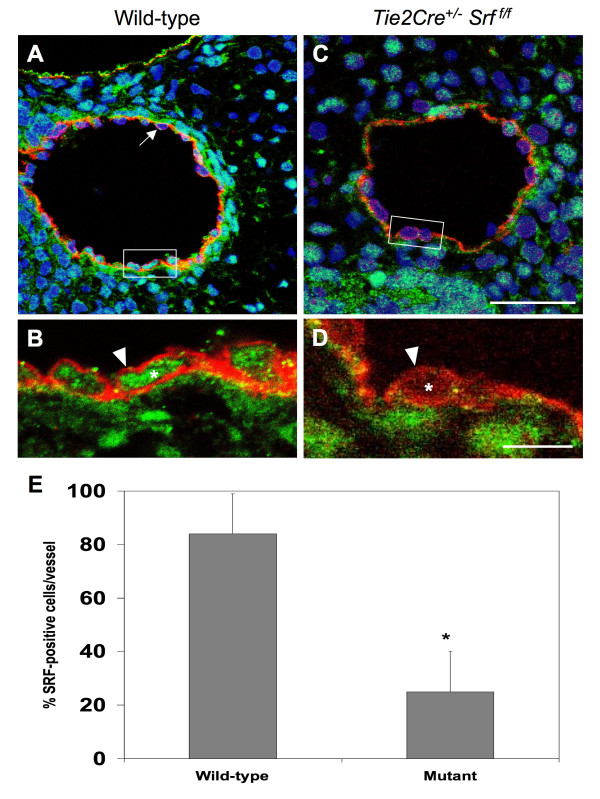
Background: Serum response factor (SRF), a member of the MADS box family of nuclear transcription factors, plays an important role in cardiovascular development and function. Numerous studies demonstrate a central role for SRF in regulating smooth and cardiac muscle cell gene expression. Consistent with this, loss of SRF function blocks differentiation of coronary vascular smooth muscle cells from proepicardial precursors, indicating SRF is necessary for coronary vasculogenesis. The role of SRF in endothelial cell contribution during early vascular development, however, has not been addressed. To investigate this, we generated transgenic mice lacking expression of SRF in endothelial cells. Mice expressing Cre recombinase (Tie2Cre+) under Tie2 promoter control were bred to mice homozygous for Srf alleles containing loxP recombination sites within the Srf gene (Srff/f). Tie2 is a tyrosine kinase receptor expressed predominantly on endothelial cells that mediates signalling during different stages of blood vessel remodelling. Resulting embryos were harvested at specific ages for observation of physical condition and analysis of genotype.

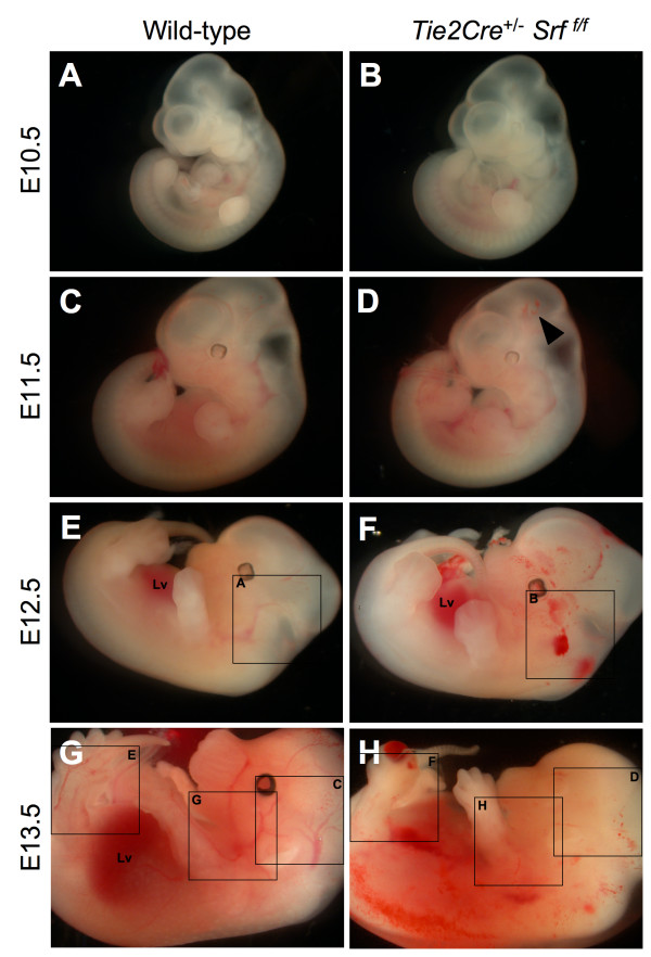
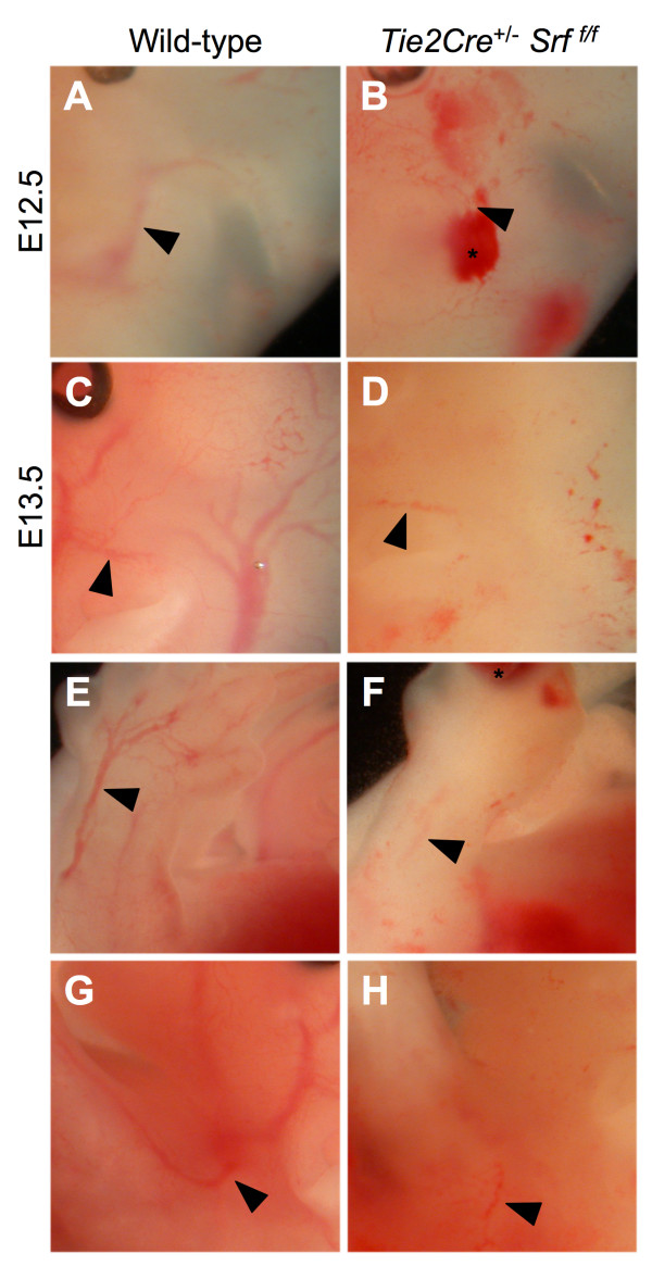
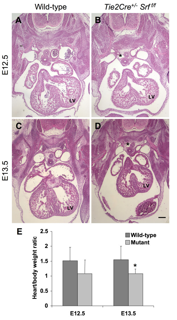
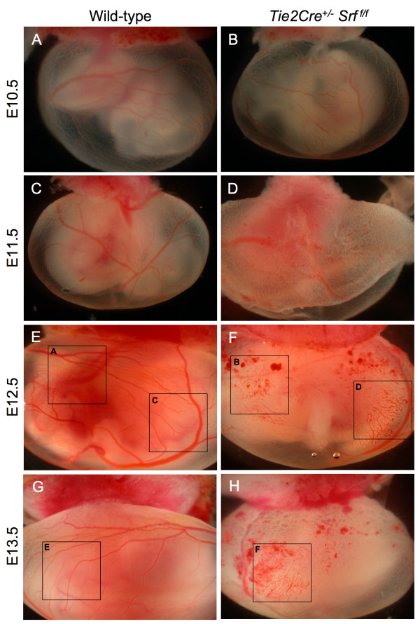
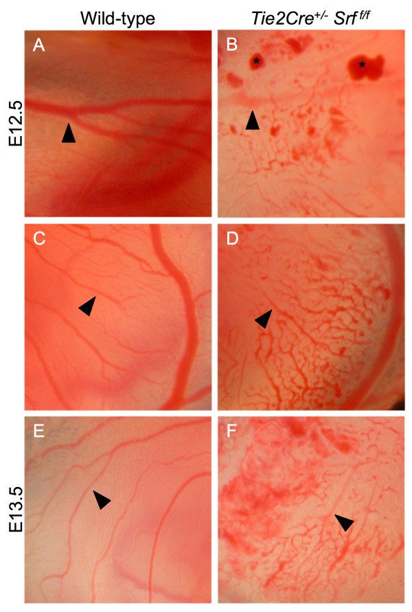
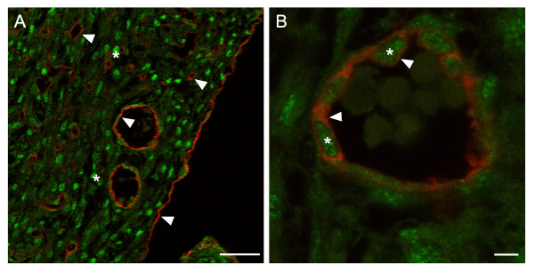
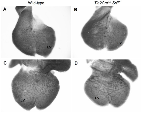
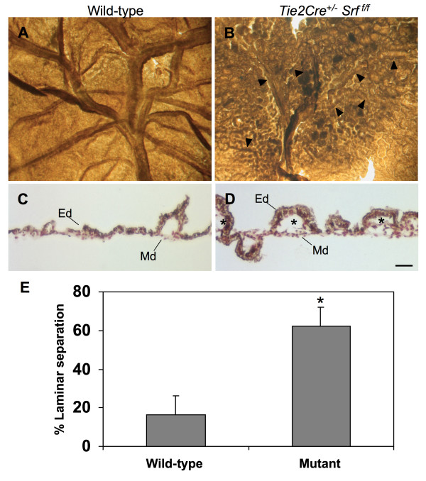
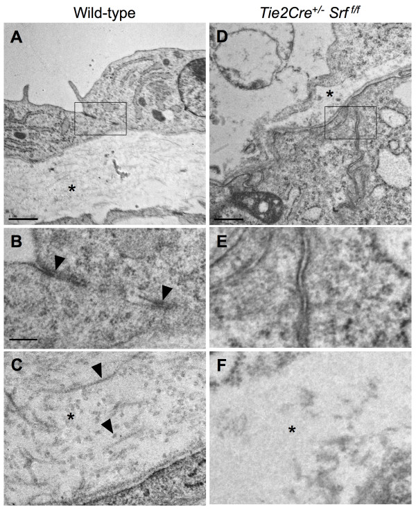
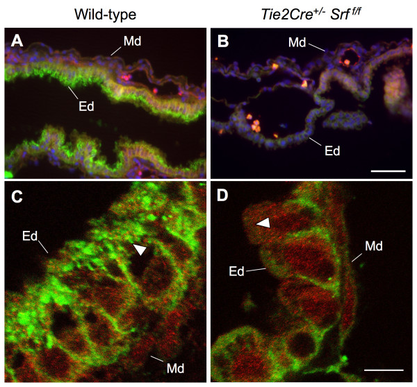
Results: Tie2Cre+/-Srff/f embryos appeared to develop normally compared to wild-type littermates until embryonic day 10.5 (E10.5). Beginning at E11.5, Tie2Cre+/-Srff/f embryos exhibited cerebrovascular hemorrhaging and severely disrupted vascular networks within the yolk sac. Hemorrhaging in mutant embryos became more generalized with age, and by E14.5, most Tie2Cre+/-Srff/f embryos observed were nonviable and grossly necrotic. Hearts of mutant embryos were smaller relative to overall body weight compared to wild-type littermates. Immunohistochemical analysis revealed the presence of vascular endothelial cells; however, vessels failed to undergo appropriate remodelling. Initial analysis by electron microscopy suggested a lack of appropriate cell-cell contacts between endothelial cells. Consistent with this, disrupted E-cadherin staining patterns were observed in mutant embryos.

Conclusion: These results provide the first in vivo evidence in support of a role for SRF in endothelial cell function and strongly suggest SRF is required for appropriate vascular remodelling.


Figures

Figure 1
Figure 1
Tie2Cre+/-Srff/f embryos exhibit internal hemorrhaging. Photographs of whole unfixed, unstained mouse embryos at E10.5 (A, B), E11.5 (C, D), E12.5 (E, F) and E13.5 (G, H). Tie2Cre+/-Srff/f mutant embryos (B, D, F, H) exhibited internal hemorrhaging compared to wild-type littermates (A, C, E, G). This effect progressed with increasing gestational age. E14.5 mutant embryos were grossly necrotic and unsuitable for analysis. Labelled boxes in Fig. 1 correspond to images in Fig. 2.; Lv = liver. Arrowhead marks minimal hemorrhaging observed in E11.5 embryos.
Figure 2
Figure 2
Vascular fragmentation and internal hemorrhaging in Tie2Cre+/-Srff/f embryos. Higher magnification photographs of whole unfixed, unstained mouse embryos as in Figure 1; embryos were examined at E12.5 (A, B) and E13.5 (C-H). Tie2Cre+/-Srff/f mutant embryos (B, D, F, H) exhibited blood pooling (asterisks) and loss of visible vascular structures (arrowheads mark similar vessels; compare A & B, C & D, E & F, and G & H).
Figure 3
Figure 3
Hearts in mutant embryos are smaller and dysmorphic. Photomicrographs (40×) of transverse sections of mouse embryos at E12.5 (A, B) and E13.5 (C, D) stained with hematoxylin and eosin. Tie2Cre+/-Srff/f embryos exhibited reduced heart size by E12.5 (B, compare to A), which became more pronounced and consistent at E13.5 (D, compare to C). (E) Quantitation of heart size normalized to overall body weight revealed smaller hearts in both E12.5 (1.5 ± 0.4 vs. 1.08 ± 0.5, wild-type vs. mutant respectively; p = 0.054) and E13.5 embryos (1.4 ± 0.39% vs. 1.04 ± 0.17%, wild-type vs. mutant respectively; p = 0.021). Enlarged vessels apparently caused by edema due to vascular insufficiency were also noted in mutant embryos (asterisks). LV = left ventricle. Scale bar: 200 μm.
Figure 4
Figure 4
Tie2Cre+/-Srff/f embryos exhibit yolk sac vascular failure. Photographs of whole unfixed, unstained intact yolk sacs containing mouse embryos at E10.5 (A, B), E11.5 (C, D), E12.5 (E, F) and E13.5 (G, H). Embryos with Tie2Cre+/-Srff/f genotype exhibited severely disrupted vascular networks at E12.5 (F) and E13.5 (H) as compared to wild-type littermates (E and G respectively). Labelled boxes in Fig. 4 correspond to images in Fig. 5.
Figure 5
Figure 5
Yolk sac tissues of Tie2Cre+/-Srff/f embryos display vascular disintegration and blood pooling. Photographs of whole unfixed, unstained intact yolk sacs containing mouse embryos at E12.5 (A, B, C, D) and E13.5 (E, F). Yolk sac tissues from mutant embryos showed disorganized vascular networks at E12.5 (B) that become more pronounced at E13.5 (D, F arrowheads). Blood pools (B, asterisks) were also observed in mutant yolk sac tissues.
Figure 6
Figure 6
Detection of SRF protein within vascular endothelial cells. Single-plane confocal laser photomicrographs (A-630×, B-1,000×) of ventricular tissue from wild-type E16.5 mouse heart immunolabelled with the VEC marker PECAM-1 (red) and SRF (green). (A) PECAM-1-stained cells surround vessels as well as form the epicardium (arrowheads). Cardiomyocytes within ventricular tissue show strong SRF staining (asterisks). (B) Endothelial cell-specific PECAM-1 (red; arrowheads) encircles the nuclear-localized SRF protein (green; asterisks). Scale bars: A = 40 μm; B = 4 μm.
Figure 7
Figure 7
SRF protein is decreased in endothelial tissues of Tie2Cre+/-Srff/f embryos. Photomicrographs (A, C:1,000×; B, D:4,000×) of cross-sections of blood vessels from E10.5 wild-type (A, B) and mutant (C, D) embryos were used to count the total number of VEC and the number of VEC exhibiting SRF (boxes in A, C correspond to images in C, D respectively). Tissues were labelled with antibodies against SRF (green) and the VEC-marker PECAM-1 (red) and analyzed using single-plane confocal laser microscopy (blue = Topro3 nuclear staining). Only those cells showing SRF signal completely encircled by PECAM-1 staining were scored as SRF-positive VEC (B, D arrowheads). Not all VEC displayed detectable SRF protein (A, arrow). Final numbers were expressed as percent SRF-positive VEC of the total counted for that vessel (E): 84 ± 14.3 vs. 25 ± 15.7, wild-type vs. mutant respectively; *p = 0.0000004, student's t test. Scale bars: A, C = 30 μm; B, D = 10 μm.
Figure 8
Figure 8
Early coronary vessel development in Tie2Cre+/-Srff/f embryos appears normal compared to wild-type littermates. Photographs of whole-mount immunostained wild-type (A, C) and Tie2Cre+/-Srff/f (B, D) hearts. Samples were stained with the endothelial cell marker PECAM-1 to detect vascular structures. Vessels on ventral (A, B) and dorsal (C, D) heart surfaces were clearly visible, indicating apparently normal assembly of initial coronary vasculature. LV = left ventricle.
Figure 9
Figure 9
Tie2Cre+/-Srff/f yolk sac tissues show loss of blood vessel integrity and separation of dermal layers. Yolk sac tissues immunostained with VEC-specific marker PECAM-1 to label endothelial cells and vascular structures. Photographs of wild-type (A) and Tie2Cre+/-Srff/f (B) whole yolk sac at E13.5; arrowheads (B) mark apparent blood vessel remnants in mutant yolk sac tissue. Photomicrographs (200×) of cross-sections of wild-type (C) and Tie2Cre+/-Srff/f (D and E) yolk sac at E13.5 immunolabelled for PECAM-1 (brown-gray = PECAM-1, red = neutral red counterstain). Normal cellular layers of endoderm (Ed) and extraembryonic mesoderm (Md) are tightly associated in wild-type as compared to mutant yolk sac tissue. Asterisks mark areas of inappropriate tissue layer separation in Tie2Cre+/-Srff/f yolk sac (D). (E) Quantitation of laminar separation in cross-sections of yolk sac tissues as in C and D revealed increased delamination in mutant animals (16.3 ± 10.8% vs. 62.0 ± 10.4%, wild-type vs. mutant respectively; p = 0.0031). Scale bar: C, D = 50 μm.
Figure 10
Figure 10
Lack of junctional complexes and collagen deposition revealed by electron microscopy analysis of yolk sac tissues. Yolk sac tissues from wild-type (A, B, C) and Tie2Cre+/-Srff/f (D, E, F) embryos were subjected to transmission electron microscopy to assess ultrastructural detail. Desmosomal-type junctional complexes are observed in wild-type tissue (B arrowheads) but not in mutant tissue (E). Intralaminar collagen deposition occurs normally between endodermal and mesodermal cell layers in wild-type (A, C) but not mutant (D, F) tissues. Arrowheads in C denote longitudinal and cross-sectional views of individual collagen fibrils. Boxes in A and D correspond to images in B and E, respectively; asterisks in A and D correspond to images in C and F, respectively; scale bars: A, D = 1 μm; B, C, E, F = 0.2 μm.
Figure 11
Figure 11
E-Cadherin localization is disrupted in yolk sac tissues of Tie2Cre+/-Srff/f embryos. Photomicrographs of wild-type (A, C; 400×) and mutant (B, D; 2,000×) yolk sac tissues stained for PECAM (red) and E-cadherin (green). Immunofluorescent analysis revealed a decrease in detectable E-cadherin protein in mutant samples. Robust E-cadherin staining is observed in the endodermal (Ed) but not mesodermal (Md) layer of wild-type yolk sac (A). In contrast, very little E-cadherin staining can be detected in yolk sac from Tie2Cre+/-Srff/f embryos (B). Single-plane confocal microscopy (C, D; 4,000×) confirms the lack of E-cadherin immunostaining at the apical brush-border surface of mutant yolk sac endodermal cells (compare A and B, arrowheads). Blue stain in A and B is DAPI nuclear stain. Scale bars: A, B = 50 μm; C, D = 7.5 μm.

References

    1. Miano JM. Serum response factor: toggling between disparate programs of gene expression. Journal of Molecular and Cellular Cardiology. 2003;35:577–593. doi: 10.1016/S0022-2828(03)00110-X. - DOI - PubMed
    1. Treisman R. The serum response element. Trends in Biochemical Sciences. 1992;17:423–426. doi: 10.1016/0968-0004(92)90013-Y. - DOI - PubMed
    1. Arsenian S, Weinhold B, Oelgeschlager M, Ruther U, Nordheim A. Serum response factor is essential for mesoderm formation during mouse embryogenesis. EMBO Journal. 1998;17:6289–6299. doi: 10.1093/emboj/17.21.6289. - DOI - PMC - PubMed
    1. Posern G, Treisman R. Actin' together: serum response factor, its cofactors and the link to signal transduction. . Trends in Cell Biology. 2006;16:588–596. doi: 10.1016/j.tcb.2006.09.008. - DOI - PubMed
    1. Belaguli NS, Schildmeyer LA, Schwartz RJ. Organization and myogenic restricted expressin of the murine serum response factor gene. A role for autoregulation. . Journal of Biological Chemistry. 1997;272:18222–18231. doi: 10.1074/jbc.272.29.18222. - DOI - PubMed

Publication types

Substances