. 2008 Jul 8;18(13):933-42.
doi: 10.1016/j.cub.2008.05.045. Epub 2008 Jun 19.

Aven-dependent activation of ATM following DNA damage

Affiliations

Aven-dependent activation of ATM following DNA damage

Jessie Yanxiang Guo et al. Curr Biol. .

Abstract

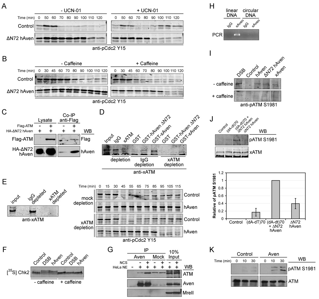
Background: In response to DNA damage, cells undergo either cell-cycle arrest or apoptosis, depending on the extent of damage and the cell's capacity for DNA repair. Cell-cycle arrest induced by double-stranded DNA breaks depends on activation of the ataxia-telangiectasia (ATM) protein kinase, which phosphorylates cell-cycle effectors such as Chk2 and p53 to inhibit cell-cycle progression. ATM is recruited to double-stranded DNA breaks by a complex of sensor proteins, including Mre11/Rad50/Nbs1, resulting in autophosphorylation, monomerization, and activation of ATM kinase.

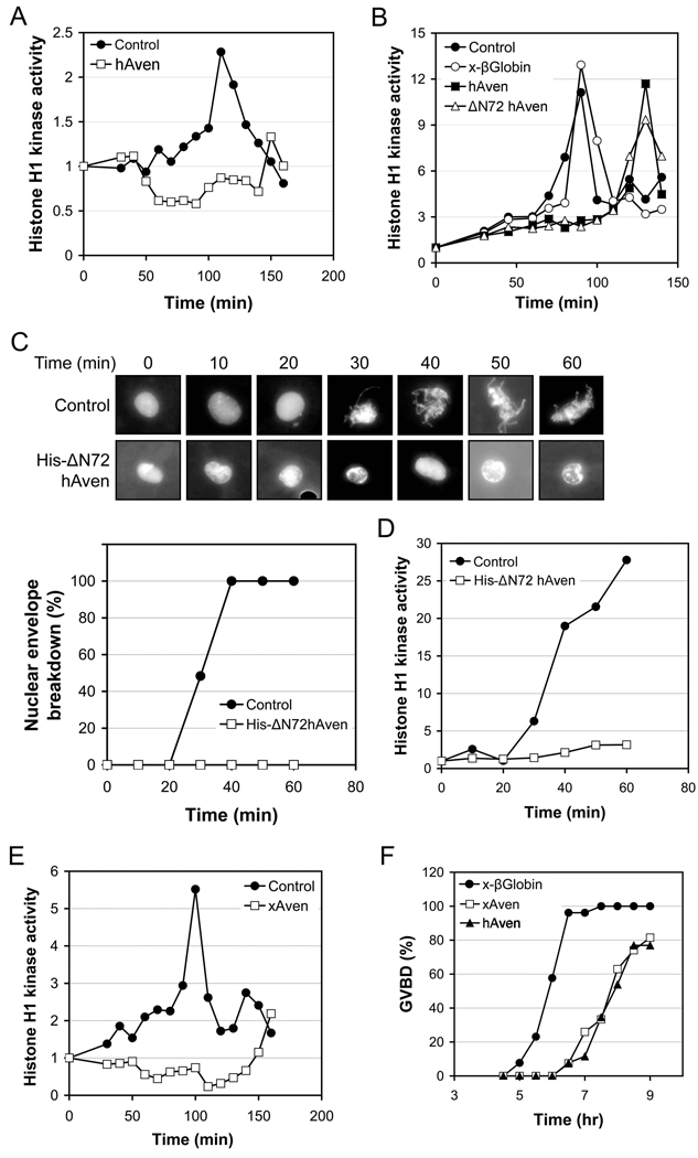
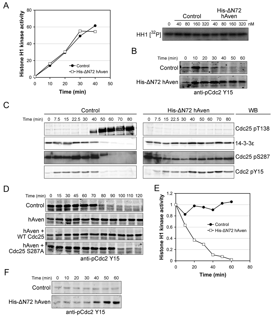
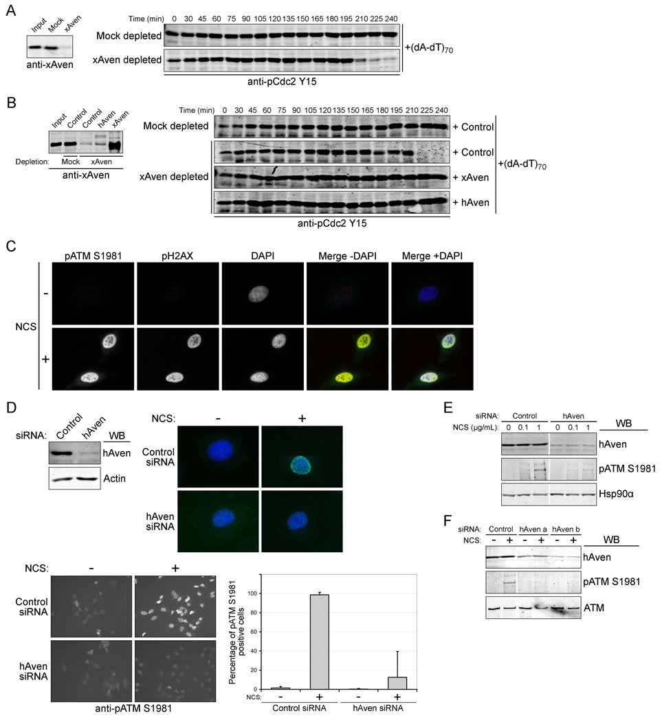
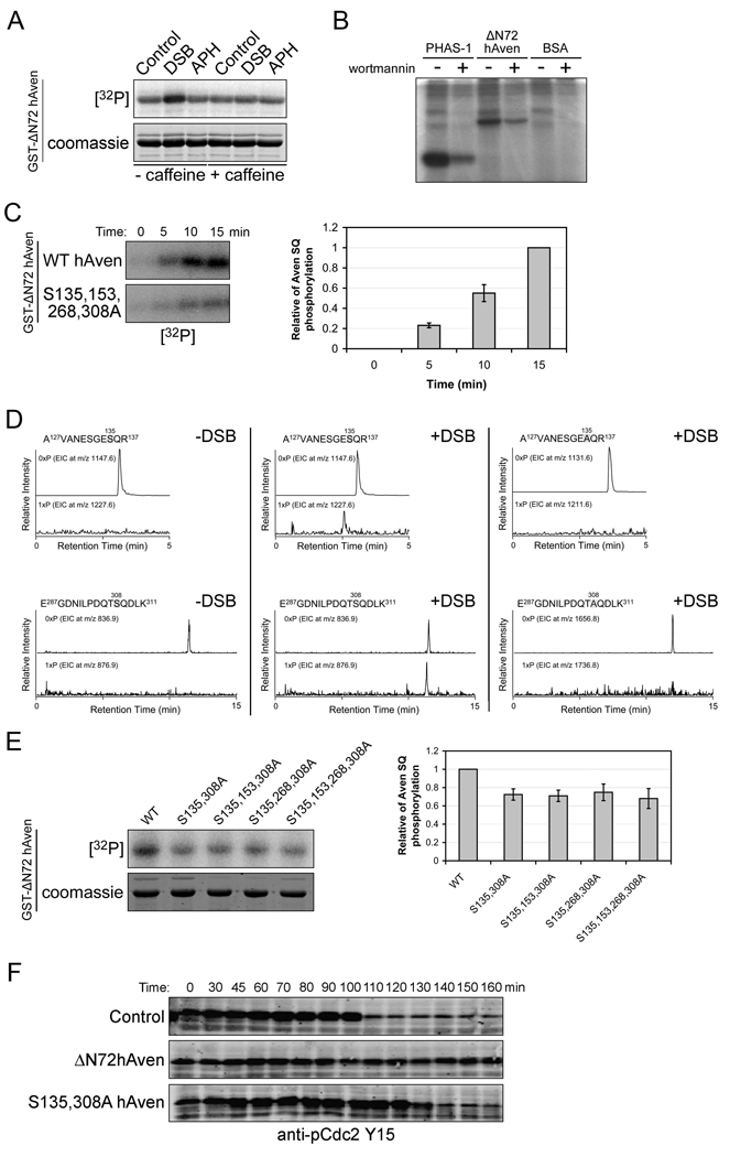
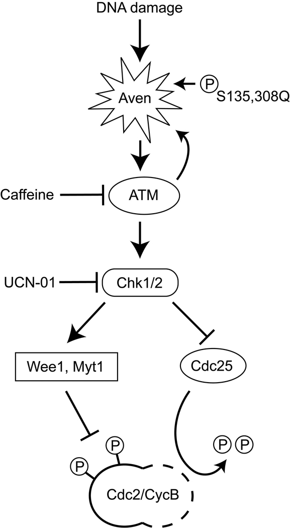
Results: In characterizing Aven protein, a previously reported apoptotic inhibitor, we have found that Aven can function as an ATM activator to inhibit G2/M progression. Aven bound to ATM and Aven overexpressed in cycling Xenopus egg extracts prevented mitotic entry and induced phosphorylation of ATM and its substrates. Immunodepletion of endogenous Aven allowed mitotic entry even in the presence of damaged DNA, and RNAi-mediated knockdown of Aven in human cells prevented autophosphorylation of ATM at an activating site (S1981) in response to DNA damage. Interestingly, Aven is also a substrate of the ATM kinase. Mutation of ATM-mediated phosphorylation sites on Aven reduced its ability to activate ATM, suggesting that Aven activation of ATM after DNA damage is enhanced by ATM-mediated Aven phosphorylation.

Conclusions: These results identify Aven as a new ATM activator and describe a positive feedback loop operating between Aven and ATM. In aggregate, these findings place Aven, a known apoptotic inhibitor, as a critical transducer of the DNA-damage signal.


Figures

Figure 1
Figure 1. Aven overexpression delays mitotic entry
(A) Cycling Xenopus egg extracts were supplemented with mRNA encoding full-length human Aven (hAven), or an equal volume buffer (control). Samples were withdrawn at the indicated times and assayed for their ability to phosphorylate histone H1 in the presence of [γ-32P] ATP. (B) mRNA encoding hAven, ΔN72hAven or Xenopus β-globin (x- βGlobin), or buffer control were added to cycling Xenopus egg extracts. Aliquots of extracts were withdrawn at the indicated times and assayed as (A). (C and D) His-ΔN72hAven protein or GST control was added to interphase extracts and incubated for 30 minutes. Following the addition of non-degradable CyclinB1, aliquots were taken at the indicated times (C) to visualize nuclear envelope breakdown and chromatin condensation by fluorescence microscopy (Hoechst DNA stain), and (D) to assay histone H1 phosphorylation. (C) Bottom: represents the average of three replicates. (E) Cycling extracts were incubated with mRNA encoding Xenopus Aven (xAven) or buffer, and aliquots were taken at the indicated times and assayed as (A). (F) 40 stage VI oocytes were injected with mRNAs encoding x-βGlobin, xAven, or hAven. After 12 hr, oocytes were treated with progesterone and scored for germinal vesicle breakdown (GVBD). Data shown in this and all subsequent figures are the result of at least three repetitions.
Figure 2
Figure 2. Aven indirectly inhibits Cdc2/CyclinB kinase activity by modulating Cdc2 phosphorylation status
(A) Left panel: 1 µl (4 unit) of purified Cdc2/Cyclin B kinase was incubated with 80 nm His-ΔN72hAven protein or GST control, radiolabeled ATP and Histone H1 substrate. Samples were taken at the indicated time points and assayed for Histone H1-directed kinase activity and quantified. Right panel: 1 µl (4 unit) of purified Cdc2/Cyclin B kinase was incubated with His-ΔN72hAven protein or His-GST control at different concentrations for 30 min, and assayed for Histone H1-directed kinase activity. (B) Interphase extracts were incubated with His-ΔN72hAven protein or His-GST control for 30 minutes. Non-degradable cyclinB1 was then added to the extracts and aliquots were taken at the indicated times for immunoblotting with anti- pCdc2Y15. (C) His-ΔN72hAven protein together with GST-Cdc25 coupled to glutathione beads was incubated in interphase egg extract for 30 minutes. Following the addition of non-degradable CyclinB1, GST-Cdc25 beads were retrieved and washed at the indicated times and immunoblotted with anti-14-3-3ε antibody. In addition, aliquots were taken at the same time points and immunoblotting was performed with anti-pCdc25T138, anti-pCdc25S287, or anti-pCdc2Y15 antibody. (D) Cycling extracts were incubated with mRNA encoding hAven, or hAven together with wild type Cdc25 or Cdc25S287A. Samples were taken at the indicated times and immunoblotted with anti-pCdc2Y15 antibody. (E and F) His-ΔN72hAven protein or GST control protein was added to mitotic extracts, aliquots were taken at the indicated times and assayed for their ability to phosphorylate histone H1 or immunoblotted with anti-pCdc2Y15 antibody.
Figure 3
Figure 3. DNA damage checkpoint pathway components are involved in Aven-induced delay of mitotic entry
(A and B) mRNA encoding ΔN72hAven or control buffer was added to cycling extracts +/− UCN-01(5ng/ul) or caffeine (5mM). Aliquots were taken at the indicated times and immunoblotted with anti-pCdc2Y15 antibody. (C) ΔN72hAven was transfected into 293T cells with or without Flag -tagged full-length ATM. Cell lysates and anti-Flag immunoprecipitates were separated by SDS-PAGE and analyzed by immunoblotting with the indicated antibodies. (D) Glutathione beads coupled to GST, GST-ΔN72hAven or GST-xAven were incubated in interphase extracts depleted with xATM antibody or control IgG for 1 hr at room temperature, then retrieved, washed, and immunoblotted with anti-xATM antibody. (E) Left panel: cycling extracts were immunodepleted with anti-xATM or control IgG and aliquots were immunoblotted with anti-xATM antibody. Right panel: hAven mRNA was incubated in cycling extracts immunodepleted of ATM or mock-depleted with IgG. Samples were taken at the indicated times and immunoblotted with anti-pCdc2Y15 antibody. (F) Cycling extracts were supplemented with either buffer, hAven mRNA or DNA bearing double stranded breaks (DSB) in the presence of 35S-labeled Chk2 +/− caffeine (5mM) for 1 hr. Aliquots were resolved by SDS-PAGE and Chk2 electrophoretic mobility was examined by phosphoimager. (G) Hela cells treated with or without NCS (200 ng/ml) for 1hr were harvested to make nuclear fractions. Rabbit anti-Aven antibody or control IgG immunoprecipitates were immunoblotted with indicated antibodies. (H) Circular pGEX-KG-Emi2 or Not-I cut linear pGEX-KG-Emi2 plasmid was incubated in the interphase extracts for 30min. Anti-Xenopus Aven antibody or control IgG was used for immunoprecipitation and washed immunoprecipitates were used as PCR templates to detect DNA binding to Xenopus Aven. (I) Cycling extracts were supplemented with buffer control, DSB DNA, or mRNA encoding full-length hAven, ΔN72hAven, or full-length xAven +/− caffeine (5mM) for 1hr. Samples were taken and immunoblotted with anti-pATM S1981 antibody. (J) Top panel: buffer control, 10 ng of (dA-dT)70, 10 ng of (dA-dT)70 with mRNA encoding ΔN72hAven, or mRNA encoding ΔN72hAven was added to cycling extracts. After 1hr, samples were immunoblotted with the indicated antibodies. Bottom panel: the amount of pATM S1981 was quantified, normalized, and plotted. Error bars represent the standard deviation of three replicate experiments. (K) Hela cell transfected with vector control or Aven were treated with NCS (200ng/ml) for 10 or 30 min. Cell lysates were resolved by SDS-PAGE and immunoblotted with the indicated antibodies.
Figure 4
Figure 4. Aven depletion overrides a DNA damage checkpoint
(A) Left panel: Anti-xAven immunoblotting of cycling extracts before and after depletion of Xenopus Aven by three sequential incubations with IgG or anti-xAven antibody at 4°C. Right panel: immunoblot of Aven- or mock-depleted extracts with anti-pCdc2Y15 antibody following addition of 10ng/µl (dA-dT)70; aliquots were removed at the indicated times. (B) Left panel: immunoblot for xAven in depleted extracts (as in A) with subsequent addition of buffer control, mRNA for human or Xenopus Aven. Note that anti-xAven antibody weakly recognizes hAven in cycling extracts. Right Panel: immunoblotting of the same extracts with anti-pCdc2Y15 antibody at the indicated times following addition of 10ng/µl (dA-dT)70. (C) Hela cells treated with NCS (200 ng/ml) for 30 minutes were stained with anti-pATM S1981(green), anti-pH2AX (red) or with DAPI (blue) to stain DNA. Yellow coloring indicates co-localization of pATM S1981 and pH2AX. (D) Top left panel: immunoblotting of human Aven in HeLa cells transfected with control siRNA or pooled hAven siRNAs with anti-hAven antibody. Actin serves as a loading control. Top right and bottom left panels: control or hAven siRNA-transfected Hela cells were incubated with NCS (200 ng/ml) for 30 minutes and stained with anti-pATM S1981 (green) or with DAPI to stain DNA (blue). Top right panel shows a single high magnification nucleus; pATM S1981 staining of a larger field of cells is shown on the bottom left. Bottom right: Columns represent the average of pATM S1981 positive cells from three replicate experiments and error bars represent the standard deviation. (E) anti-pATM S1981 immunoblots of HeLa cells transfected with control siRNA, or pooled hAven siRNAs and treated +/− 100 or 1000 ng/ml of NCS. Hsp90α was used as a loading control. (F) Immunoblots of HeLa cells transfected with single hAven siRNAs (a or b) or control siRNA and treated +/− NCS (200 ng/ml) with the indicated antibodies.
Figure 5
Figure 5. Positive feedback activation of Aven by phosphorylation
(A) GST-ΔN72hAven coupled to glutathione beads were dipped into interphase egg extracts pre-treated with DSB or sperm chromatin and Aphidicolin (APH) in the presence of [γ-32P] ATP +/− caffeine (5mM) for 15 min, then retrieved, washed, and resolved on 10% SDS-PAGE. Phosphorylated hAven was detected using a phosphoimager. (B) ATM kinase was immunoprecipitated from 293T cells treated with NCS +/− wortmannin using anti-ATM antibody coupled to protein A sepharose beads. Purified His-ΔN72hAven protein was then incubated with the immunoprecipitates in kinase buffer for 30 min and resolved by SDS-PAGE. PHAS-1, a known ATM substrate, and BSA were included as positive and negative controls, respectively. Phosphorylation was detected by phosphoimager. (C) Left panel: GST-ΔN72hAven wild type (WT) or S135,153,268,308A mutant protein conjugated to glutathione beads were dipped into interphase extracts in the presence of [γ-32P] ATP. Extracts were then treated with DSB and aliquots were taken at the indicated times and assayed as (A). Right panel: the amount of Aven SQ phosphorylation was quantified, normalized, and plotted. Error bars represent standard deviation of three replicate experiments. (D) GST-ΔN72hAven wild type or S135, 153, 268, 308A protein was incubated in interphase extracts - (left) or + (middle and right) DSB DNA. After incubation, the protein was retrieved on glutathione Sepharose beads and resolved by SDS-PAGE, and the relevant bands were excised from the gel, trypsin digested, and analyzed by LC/MS mass spectrometry. LC/MS-extracted ion chromatograms (EIC) of unphosphorylated (0×P) and presumed monophosphorylated (1×P) tryptic peptides were examined, and EIC peaks (middle; at 2.6 and 11.5 min in the 1×P traces) indicate the phosphorylation of the tryptic peptides upon DSB DNA treatment. Such peaks were not detected in the tryptic peptides derived from the GST-AN72hAven S135, 153, 268, 308A protein (right), indicating a lack of phosphorylation. (E) Left panel: xATM kinase was immunoprecipitated from interphase extracts pre-treatment with DSB. Purified GST-ΔN72hAven WT or AQ mutant proteins were incubated with the immunoprecipitates in the kinase buffer for 30 min and assayed as (A). Right panel: the amount of Aven SQ phosphorylation was quantified, normalized, and plotted. Error bars represent standard deviation of three replicate experiments (T test, P<0.05). (F) mRNA-encoding ΔN72hAven or ΔN72hAvenS135, 308A was added to the cycling extracts, and aliquots were taken at the indicated times and immunoblotted with anti-pCdc2Y15 antibody.
Figure 6
Figure 6. Aven’s role in DNA damage-induced ATM activation and G2/M arrest
During DNA damage, Aven serves as a signal transducer in the ATM activation pathway. Activated ATM in turn phosphorylates Aven on S135 and S308, and causes full activation of Aven. Fully activated Aven then further enhances ATM activation, leading to activation of downstream pathway components to inhibit Cdc25 activation and enhance Wee1/Myt kinase activity, leading to Cdc2/CyclinB inactivation and inhibition of mitotic entry.

References

    1. Lew DJ, Kornbluth S. Regulatory roles of cyclin dependent kinase phosphorylation in cell cycle control. Current opinion in cell biology. 1996;8:795–804. - PubMed
    1. Coleman TR, Dunphy WG. Cdc2 regulatory factors. Current opinion in cell biology. 1994;6:877–882. - PubMed
    1. Abraham RT. Cell cycle checkpoint signaling through the ATM and ATR kinases. Genes & development. 2001;15:2177–2196. - PubMed
    1. Raleigh JM, O'Connell MJ. The G(2) DNA damage checkpoint targets both Wee1 and Cdc25. Journal of cell science. 2000;113(Pt 10):1727–1736. - PubMed
    1. Furnari B, Blasina A, Boddy MN, McGowan CH, Russell P. Cdc25 inhibited in vivo and in vitro by checkpoint kinases Cds1 and Chk1. Molecular biology of the cell. 1999;10:833–845. - PMC - PubMed

Publication types

MeSH terms