Comparative Study
. 2008 Aug 15;56(11):1250-62.
doi: 10.1002/glia.20694.

Molecular basis of oncostatin M-induced SOCS-3 expression in astrocytes

Affiliations
Comparative Study

Molecular basis of oncostatin M-induced SOCS-3 expression in astrocytes

Brandi J Baker et al. Glia. .

Abstract

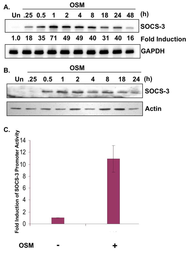
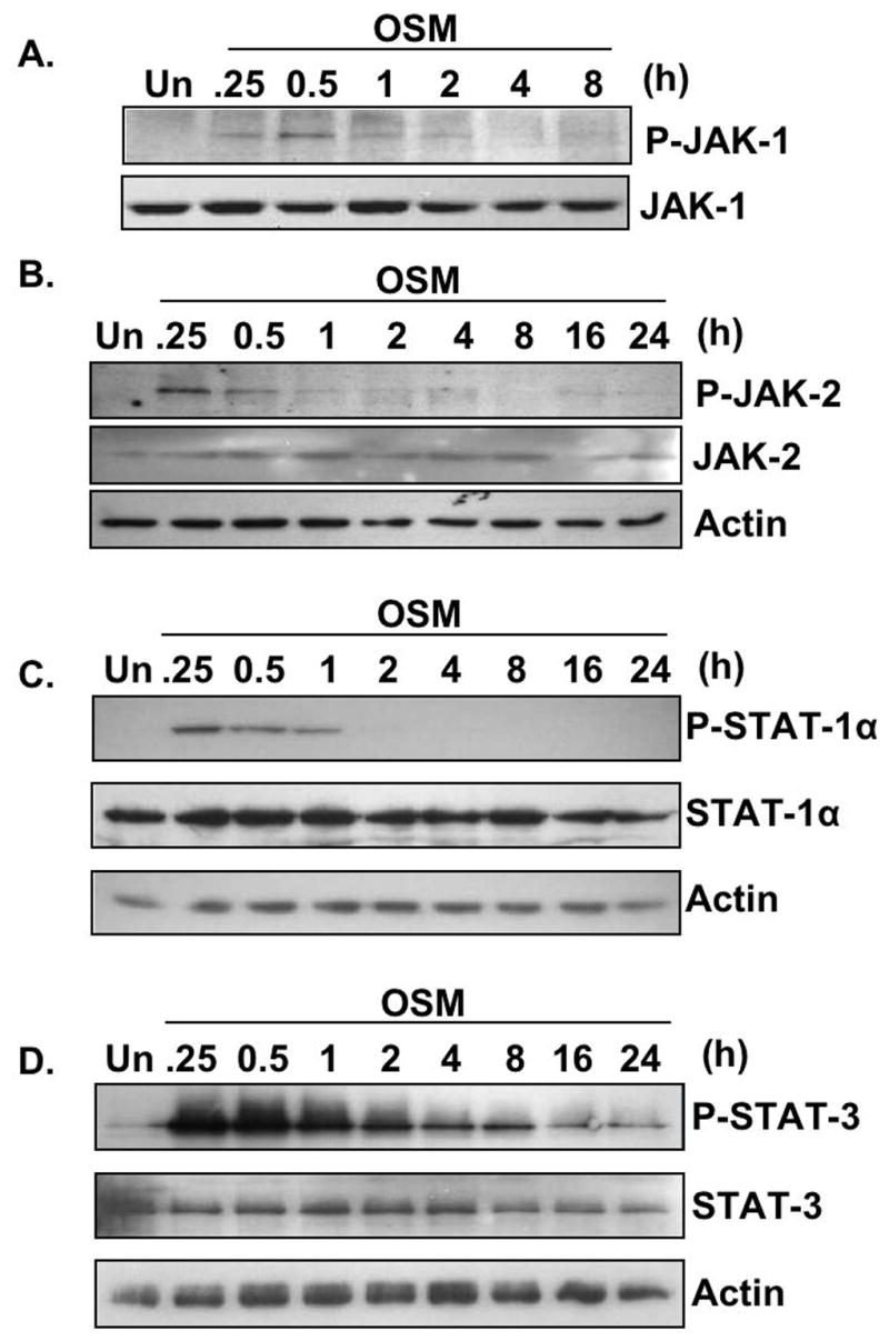
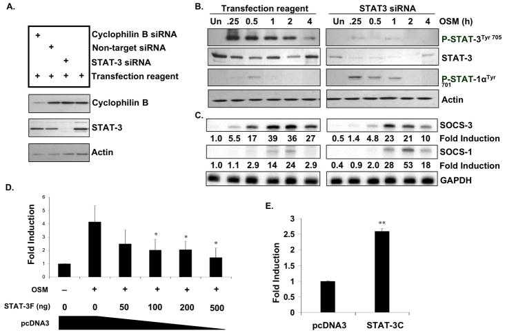
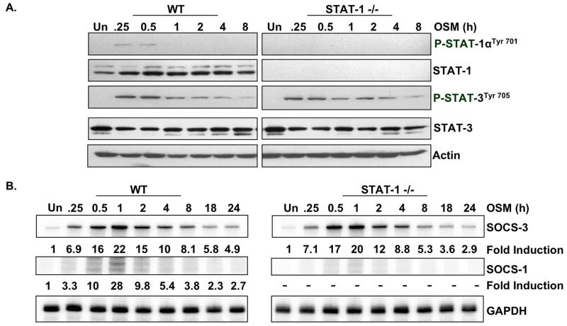
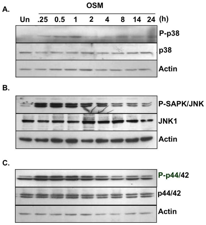
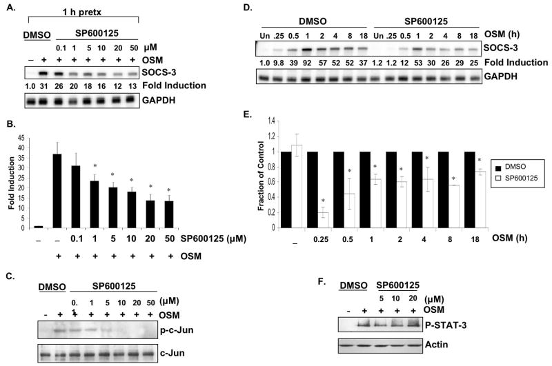
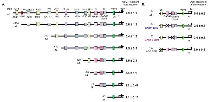
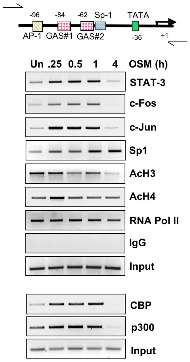
Under neuropathological conditions, reactive astrocytes release cytokines and chemokines, which act in an autocrine and/or paracrine fashion to modulate production of immunoregulatory factors from cells including microglia, astrocytes, and neurons. In this way, astrocytes play an important role in orchestrating immune responses within the central nervous system (CNS). Suppressor of cytokine signaling (SOCS) proteins are endogenous, negative regulators of the JAK/STAT signaling pathway and function as attenuators of the immune and inflammatory responses. As such, SOCS proteins may have critical roles in the CNS under neuroinflammatory conditions. In the inflamed CNS, expression of IL-6 cytokine family member oncostatin M (OSM) is elevated; however, its functional effects are not well understood. We demonstrate that OSM is a potent inducer of SOCS-3 in astrocytes. Analysis of the SOCS-3 promoter revealed that an AP-1 element, two IFN-gamma activation sequence (GAS) elements, and a GC-rich region are crucial for SOCS-3 gene expression. Using small interfering RNA against STAT-3, as well as a STAT-3 dominant-negative construct, we demonstrate that STAT-3 activation is critical for OSM induction of SOCS-3 expression. The ERK1/2 and JNK pathways also contribute to OSM-induced SOCS-3 gene expression. OSM stimulation led to a time-dependent recruitment of the transcription factors STAT-3, c-Fos, c-Jun, and Sp1 and the coactivators CREB-binding protein (CBP) and p300 to the endogenous SOCS-3 promoter. These data indicate that OSM-induced activation of STAT-3 and the ERK1/2 and JNK pathways are critical for astrocytic expression of SOCS-3, which provides for feedback inhibition of cytokine-induced inflammatory responses in the CNS.


Figures

Figure 1
Figure 1. Induction of SOCS-3 Expression by OSM
A, B. Primary astrocytes were treated with OSM (5 ng/ml) for 0–48 h. A. 10 μg of RNA extracts was analyzed for SOCS-3 mRNA expression by RPA. GAPDH mRNA expression serves as a control. B. Protein lysates were collected and separated by SDS-PAGE, immunoblotted for SOCS-3 protein, and for actin as a loading control. C. Primary astrocytes were transiently transfected with the pGL3 firefly luciferase-expression vector containing the murine SOCS-3 promoter. Cells recovered overnight and were then treated with OSM for 16 h. Lysates were then analyzed for luciferase activity. Values were normalized to total protein, and fold induction calculated by dividing the OSM treatment values by untreated levels. Data represent the mean +/− S.D. of three experiments.
Figure 2
Figure 2. OSM-induced Activation of the JAK/STAT Pathway in Astrocytes
A. Primary astrocytes were treated with OSM (5 ng/ml) for 0–8 h. 500 μg of protein lysates were immunoprecipitated with an anti-JAK-1 antibody, separated by SDS-PAGE, and immunoblotted for phosphorylated JAK-1Tyr1022/1023. The membrane was stripped and reprobed with an antibody against total JAK-1. Representative of three experiments. B–D. Primary astrocytes were treated with OSM (5 ng/ml) for 0–24 h. Protein lysates were prepared and subjected to immunoblot analysis with antibodies against phosphorylated JAK-2Tyr1007/1008, STAT-1Tyr701, or STAT-3Tyr705. Membranes were stripped and reprobed for total JAK-2, STAT-1, and STAT-3, respectively, and actin as a loading control. Representative of four experiments.
Figure 3
Figure 3. Inhibition of STAT-3 Expression or Activation Inhibits OSM-induced SOCS-3 Expression and Promoter Activity
A. Astrocytes were transiently transfected with 100 nM siRNA against STAT-3, cyclophilin B as a positive control, or non-target siRNA as a negative control, and incubated for 72 h. Specific STAT-3 knock-down was confirmed by immunobloting for total STAT-3 protein, cyclophilin B and actin. B, C. Primary astrocytes were incubated with transfection reagent alone or with transfection reagent plus 100 nM siRNA against STAT-3, and incubated for 72 h. Cells were then treated with OSM (5 ng/ml) for the indicated times (0–4 h). B. Protein lysates were prepared and subjected to immunoblot analysis with antibodies against phosphorylated STAT-3Tyr705 or STAT-1Tyr701. Membranes were stripped and reprobed for total STAT-3 and actin as loading controls. Representative of three independent experiments. C. 10 μg of RNA was analyzed for SOCS-3 and SOCS-1 mRNA expression by RPA. GAPDH mRNA expression serves as a loading control. Representative of three independent experiments. D. Primary astrocytes were transiently co-transfected with increasing concentrations of a STAT-3 dominant-negative vector (STAT-3F), along with the mSOCS-3 promoter (200 ng). The total amount of cDNA was kept constant using an empty pcDNA3 vector. Following transfection, cells recovered for 48 h to allow for expression of the STAT-3F construct. The cells were then treated with OSM for 24 h, and analyzed for luciferase expression as a measure of SOCS-3 promoter activity. Mean +/− S.D. of three independent experiments. *, p ≤ 0.05. E. Astrocytes were transiently co-transfected with the mSOCS-3 promoter (200 ng) and either 200 ng of pcDNA3 or 200 ng of STAT-3C. Cells recovered for 48 h and then were analyzed for luciferase expression. Mean +/− S.D. of three experiments. **, p ≤ 0.001.
Figure 4
Figure 4. OSM-induced Expression of SOCS-1, but not SOCS-3, Depends on STAT-1
A, B. Astrocytes isolated from STAT-1+/+ (WT) or STAT-1/ mice were incubated with OSM (5 ng/ml) for the indicated times. A. Protein lysates were subjected to immunoblotting for phospho-STAT-1αTyr701 and phospho-STAT-3Tyr705. Blots were stripped and reprobed for total STAT-1, total STAT-3 and actin. B. 10 μg of RNA was analyzed for SOCS-3 and SOCS-1 mRNA expression by RPA. GAPDH mRNA expression serves as a loading control. Representative of three independent experiments.
Figure 5
Figure 5. OSM-induced Activation of the MAPK Pathways in Primary Astrocytes
A–C. Primary astrocytes were treated with OSM (5 ng/ml) for 0–24 h. Protein lysates were prepared, separated by SDS-PAGE, and subjected to immunoblot analysis with antibodies against (A) phosphorylated p38Thr180/Tyr182, (B) SAPK/JNKThr183/Tyr182 or (C) p44/42 ERKThr202/Tyr204. Membranes were stripped and reprobed for total p38, SAPK/JNK, and p44/42 ERK, respectively, and actin as a loading control. Representative of three experiments.
Figure 6
Figure 6. OSM-induced SOCS-3 Expression Partially Depends on Activation of the ERK Pathway
A, B. Primary astrocytes were pretreated with 0.2% DMSO or with increasing concentrations (0–20 μM) of U0126, a specific inhibitor of MEK1 and MEK2, or U0124, a negative control, for 1 h. Cells were then incubated with OSM (5 ng/ml) for 30 min. A. 10 μg of RNA was analyzed for SOCS-3 mRNA expression by RPA. GAPDH mRNA expression serves as a control. Representative of three independent experiments. B. Protein lystes were prepared, separated by SDS-PAGE and immunoblotted for phosphorylated p44/42 ERKThr202/Tyr204. Lysates were also probed for total p44/42 ERK as loading control. Representative of three independent experiments. C. Primary astrocytes were transiently transfected with the SOCS-3 promoter (200 ng) and allowed to recover for 24 h. The cells were then pretreated with 0.2% DMSO or with increasing concentrations of U0126 or U0124 for 1 h. Cells were then incubated in the absence or presence of OSM (5 ng/ml) for 8 h, and analyzed for luciferase expression as a measure of SOCS-3 promoter activity. Mean +/− S.D. of three independent experiments. *, p ≤ 0.05; **, p ≤ 0.001 compared to DMSO condition. D. Primary astrocytes were pretreated with 0.2% DMSO or with 20 μM of U0126 for 1 h. Cells were then incubated with OSM (5 ng/ml) for the indicated times. 10 μg of RNA was analyzed for SOCS-3 mRNA expression by RPA. GAPDH mRNA expression serves as a control. Representative of three independent experiments. E. Average fraction of SOCS-3 mRNA expression induced by OSM in the presence of 20 μM U0126. Data represent the mean +/− S.D. of three experiments. *, p ≤ 0.05 compared to DMSO condition. F. Primary astrocytes were pretreated with 0.2% DMSO or with increasing concentrations (0–20 μM) of U0126 for 1 h. Cells were then incubated with OSM (5 ng/ml) for 30 min. Protein lysates were subjected to immunoblotting for phospho-STAT-3Tyr705 and actin.
Figure 7
Figure 7. OSM-induced SOCS-3 Expression Involves the JNK Pathway
A–C. Primary astrocytes were incubated with 0.5% DMSO or with increasing concentrations (0–50 μM) of SP600125, a specific JNK inhibitor, for 1 h. Cells were then stimulated with OSM (5 ng/ml) for 30 min. A. 10 μg of RNA was analyzed for SOCS-3 mRNA expression by RPA. GAPDH mRNA expression serves as a control. Representative of three independent experiments. B. Average SOCS-3 mRNA fold inductions were calculated from three independent experiments. Data represent the mean +/− S.D. of three experiments. *, p ≤ 0.05 compared to DMSO condition. C. Protein lystes were prepared, separated by SDS-PAGE and immunoblotted for phosphorylated c-Jun (Ser 63). Lysates were also probed for total c-Jun as loading control. Representative of three independent experiments. D. Primary astrocytes were pretreated with 0.2% DMSO or with 20 μM of SP600125 for 1 h. Cells were then incubated with OSM (5 ng/ml) for the indicated times. 10 μg of RNA was analyzed for SOCS-3 and GAPDH mRNA expression by RPA. Representative of three independent experiments. E. Average fraction of SOCS-3 mRNA expression was calculated from three independent experiments. Data represent the mean +/− S.D. of three experiments. *, p ≤ 0.05 compared to DMSO condition. F. Primary astrocytes were pretreated with 0.2% DMSO or with increasing concentrations (0–20 μM) of SP600125 for 1 h. Cells were then incubated with OSM (5 ng/ml) for 30 min. Protein lysates were subjected to immunoblotting for phospho-STAT-3Tyr705 and actin.
Figure 8
Figure 8. The Proximal AP-1 Site and the Two GAS Elements are Critical for OSM-induced SOCS-3 Promoter Activity
A. Primary astrocytes were transiently transfected with 200 ng of the WT or Δ1–Δ7 SOCS-3 promoter constructs. Cells recovered for 24 h, and were treated with OSM for 18 h. Cell lysates were analyzed for luciferase expression. Mean +/− S.D. of three independent experiments. B. Site-directed mutagenesis was performed on the proximal AP-1 site and on each of the two GAS elements in the Δ4 SOCS-3 promoter. Primary astrocytes were transfected with either the Δ4, Δ4 GAS #1 SDM, Δ4 GAS #2 SDM, or Δ4 AP-1 SDM construct (200 ng). After recovering for 24 h, cells were treated with OSM for 18 h and cell lysates were analyzed for luciferase expression. Mean +/− S.D. of three independent experiments.
Figure 9
Figure 9. Characterization of OSM-induced Effects at the SOCS-3 Promoter In Vivo
Primary astrocytes were treated with OSM (5 ng/ml) for the indicated times, and subjected to chromatin immunoprecipitation (ChIP) assay. Briefly, formaldehyde was used to cross-link proteins to DNA and the soluble chromatin was subjected to immunoprecipitation with IgG control serum or 5 μg of antibody against STAT-3, c-Fos, c-Jun, Sp1, acetylated histone H3, acetylated histone H4 and RNA Polymerase II. In a separate experiment, immunoprecipitation was performed with 5 μg of antibody against CBP and p300. Primers specific for the indicated region of the SOCS-3 promoter were used to amplify the DNA bound to particular proteins at different times. Input serves as a control for total DNA. Representative of three independent experiments.

References

    1. Alexander WS, Hilton DJ. The role of suppressors of cytokine signaling (SOCS) proteins in regulation of the immune response. Annu Rev Immunol. 2004;22:503–529. - PubMed
    1. Auernhammer CJ, Bousquet C, Melmed S. Autoregulation of pituitary corticotroph SOCS-3 expression: characterization of the murine SOCS-3 promoter. Proc Natl Acad Sci U S A. 1999;96:6964–6969. - PMC - PubMed
    1. Baetz A, Frey M, Heeg K, Dalpke AH. Suppressor of cytokine signaling (SOCS) proteins indirectly regulate toll-like receptor signaling in innate immune cells. J Biol Chem. 2004;279:54708–54715. - PubMed
    1. Barclay JL, Anderson ST, Waters MJ, Curlewis JD. Characterization of the SOCS3 promoter response to prostaglandin E2 in T47D cells. Mol Endocrinol. 2007a;21:2516–2528. - PubMed
    1. Barclay JL, Anderson ST, Waters MJ, Curlewis JD. Regulation of suppressor of cytokine signaling 3 (SOC3) by growth hormone in pro-B cells. Mol Endocrinol. 2007b;21:2503–2515. - PubMed

Publication types

MeSH terms

Substances