

PELP1--a novel estrogen receptor-interacting protein
- PMID: 18571832
- PMCID: PMC2578818
- DOI: 10.1016/j.mce.2008.04.019
PELP1--a novel estrogen receptor-interacting protein
Abstract
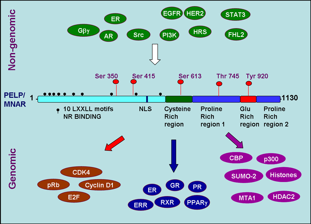
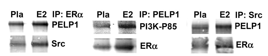
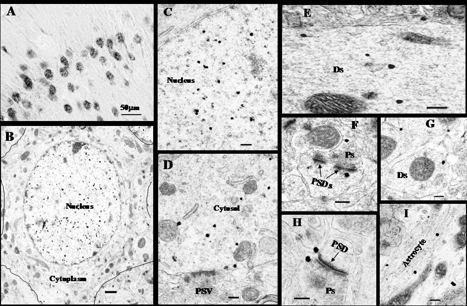
PELP1 (proline-, glutamic acid-, and leucine-rich protein-1) is a novel estrogen receptor (ER)-interacting protein that has been implicated to be important for mediation of both the genomic and nongenomic signaling of 17beta-estradiol (E2). PELP1 contains ten nuclear receptor-interacting boxes (LXXLL motifs), which allow it to interact with ER and other nuclear hormone receptors, a zinc finger, a glutamic acid-rich domain, and two proline-rich domains. The proline-rich regions contain several consensus PXXP motifs, which allow PELP1 to couple the ER with SH3 domain-containing kinase signaling proteins, such as Src and PI3K P85 regulatory subunit. PELP1 is expressed in many different brain regions, including the hippocampus, hypothalamus, and cerebral cortex. Further work has demonstrated that PELP1 is colocalized with ER-alpha in neurons in various brain regions. PELP1 is primarily expressed in neurons, with some expression also observed in glia. Subcellular localization studies revealed that PELP1 is highly localized in the cell nucleus of neurons, with some cytoplasm localization as well, and PELP1 is also localized at synaptic sites. Work in other tissues has demonstrated that PELP1 is critical for nongenomic and genomic signaling by E2, as PELP1 knockdown studies significantly attenuates E2-induced activation of ERK and Akt signaling pathways, and inhibits E2 genomic transcriptional effects on gene expression in breast cancer cells. Preliminary studies in the brain, suggests that similar roles may exist for PELP1 in the brain, but this remains to be established, and further work to characterize the precise roles and functions of PELP1 in the brain are needed.
Figures
Similar articles
-
Molecular cloning and characterization of PELP1, a novel human coregulator of estrogen receptor alpha.J Biol Chem. 2001 Oct 12;276(41):38272-9. doi: 10.1074/jbc.M103783200. Epub 2001 Jul 31. J Biol Chem. 2001. PMID: 11481323
-
Cloning, expression, and localization of MNAR/PELP1 in rodent brain: colocalization in estrogen receptor-alpha- but not in gonadotropin-releasing hormone-positive neurons.Endocrinology. 2005 Dec;146(12):5215-27. doi: 10.1210/en.2005-0276. Epub 2005 Sep 1. Endocrinology. 2005. PMID: 16141397
-
Extranuclear functions of ER impact invasive migration and metastasis by breast cancer cells.Cancer Res. 2010 May 15;70(10):4092-101. doi: 10.1158/0008-5472.CAN-09-3834. Epub 2010 May 11. Cancer Res. 2010. PMID: 20460518 Free PMC article.
-
PELP1: a review of PELP1 interactions, signaling, and biology.Mol Cell Endocrinol. 2014 Jan 25;382(1):642-651. doi: 10.1016/j.mce.2013.07.031. Epub 2013 Aug 8. Mol Cell Endocrinol. 2014. PMID: 23933151 Free PMC article. Review.
-
Emerging significance of ER-coregulator PELP1/MNAR in cancer.Histol Histopathol. 2007 Jan;22(1):91-6. doi: 10.14670/HH-22.91. Histol Histopathol. 2007. PMID: 17128415 Free PMC article. Review.
Cited by
-
Expression of estrogen receptors, PELP1, and SRC in human spermatozoa and their associations with semen quality.Hum Cell. 2023 Mar;36(2):554-567. doi: 10.1007/s13577-022-00847-6. Epub 2022 Dec 28. Hum Cell. 2023. PMID: 36577884 Free PMC article.
-
Hirsutism, virilism, polycystic ovarian disease, and the steroid-gonadotropin-feedback system: a career retrospective.Am J Physiol Endocrinol Metab. 2012 Jan 1;302(1):E4-E18. doi: 10.1152/ajpendo.00488.2011. Epub 2011 Oct 25. Am J Physiol Endocrinol Metab. 2012. PMID: 22028409 Free PMC article. Review.
-
Proteomic analysis of native hepatocyte nuclear factor-4α (HNF4α) isoforms, phosphorylation status, and interactive cofactors.J Biol Chem. 2011 Jan 7;286(1):674-86. doi: 10.1074/jbc.M110.154732. Epub 2010 Nov 3. J Biol Chem. 2011. PMID: 21047794 Free PMC article.
-
Extranuclear coactivator signaling confers insensitivity to tamoxifen.Clin Cancer Res. 2009 Jun 15;15(12):4123-30. doi: 10.1158/1078-0432.CCR-08-2347. Epub 2009 May 26. Clin Cancer Res. 2009. PMID: 19470742 Free PMC article.
-
Integration of ERα-PELP1-HER2 signaling by LSD1 (KDM1A/AOF2) offers combinatorial therapeutic opportunities to circumventing hormone resistance in breast cancer.Breast Cancer Res. 2012 Sep 17;14(5):112. doi: 10.1186/bcr3249. Breast Cancer Res. 2012. PMID: 22992372 Free PMC article.
References
-
- Alkayed NJ, Harukuni I, Kimes AS, London ED, Traystman RJ, Hurn PD. Gender-linked brain injury in experimental stroke. Stroke. 1998;29:159–165. discussion 166. - PubMed
-
- Ballif BA, Villen J, Beausoleil SA, Schwartz D, Gygi SP. Phosphoproteomic analysis of the developing mouse brain. Mol Cell Proteomics. 2004;3:1093–1101. - PubMed
-
- Barletta F, Wong CW, McNally C, Komm BS, Katzenellenbogen B, Cheskis BJ. Characterization of the interactions of estrogen receptor and MNAR in the activation of cSrc. Mol Endocrinol. 2004;18:1096–1108. - PubMed
-
- Bjornstrom L, Sjoberg M. Mechanisms of estrogen receptor signaling: convergence of genomic and nongenomic actions on target genes. Mol Endocrinol. 2005;19:833–842. - PubMed
Publication types
MeSH terms
Substances
Grants and funding
LinkOut - more resources
Full Text Sources
Other Literature Sources
Research Materials
Miscellaneous
