

Polymersomes: a new multi-functional tool for cancer diagnosis and therapy
- PMID: 18572025
- PMCID: PMC2714227
- DOI: 10.1016/j.ymeth.2008.05.006
Polymersomes: a new multi-functional tool for cancer diagnosis and therapy
Abstract
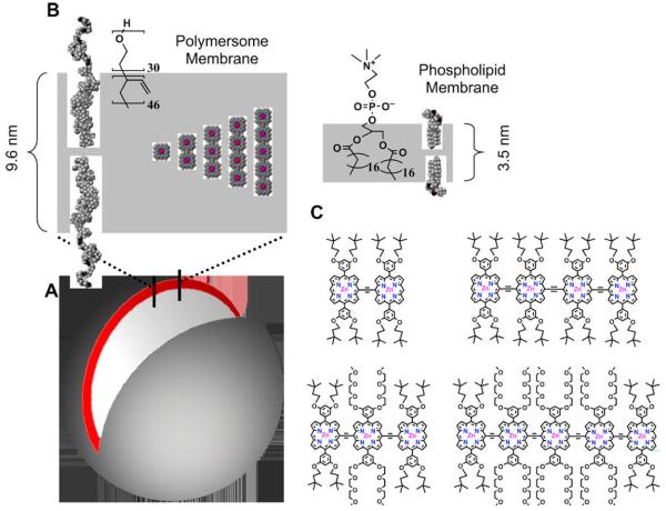
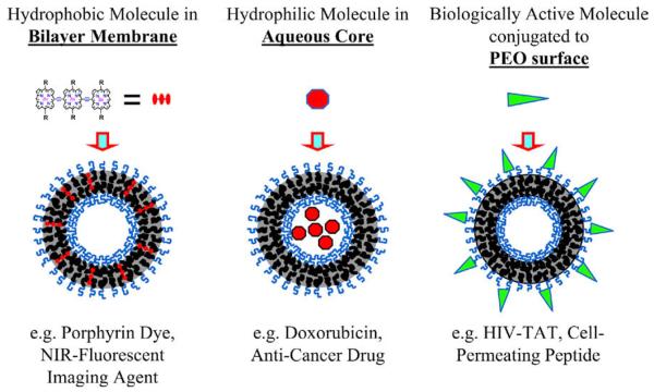
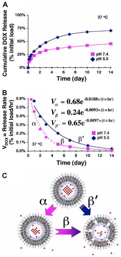
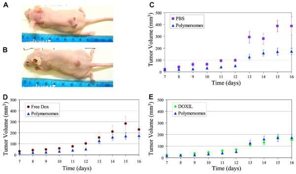
Nanoparticles are being developed as delivery vehicles for therapeutic pharmaceuticals and contrast imaging agents. Polymersomes (mesoscopic polymer vesicles) possess a number of attractive biomaterial properties that make them ideal for these applications. Synthetic control over block copolymer chemistry enables tunable design of polymersome material properties. The polymersome architecture, with its large hydrophilic reservoir and its thick hydrophobic lamellar membrane, provides significant storage capacity for both water soluble and insoluble substances (such as drugs and imaging probes). Further, the brush-like architecture of the polymersome outer shell can potentially increase biocompatibility and blood circulation times. A further recent advance is the development of multi-functional polymersomes that carry pharmaceuticals and imaging agents simultaneously. The ability to conjugate biologically active ligands to the brush surface provides a further means for targeted therapy and imaging. Hence, polymersomes hold enormous potential as nanostructured biomaterials for future in vivo drug delivery and diagnostic imaging applications.
Figures
Similar articles
-
Engineering Polymersomes for Diagnostics and Therapy.Adv Healthc Mater. 2018 Apr;7(8):e1701276. doi: 10.1002/adhm.201701276. Epub 2018 Jan 15. Adv Healthc Mater. 2018. PMID: 29334183 Free PMC article. Review.
-
Constructing aptamer anchored nanovesicles for enhanced tumor penetration and cellular uptake of water soluble chemotherapeutics.Acta Biomater. 2016 Apr 15;35:269-79. doi: 10.1016/j.actbio.2016.02.012. Epub 2016 Feb 9. Acta Biomater. 2016. PMID: 26873366
-
Stimuli-responsive polymersomes for cancer therapy.Biomater Sci. 2016 Jan;4(1):55-69. doi: 10.1039/c5bm00268k. Biomater Sci. 2016. PMID: 26456625 Review.
-
Biocompatible polymersomes-based cancer theranostics: Towards multifunctional nanomedicine.Int J Pharm. 2017 Mar 15;519(1-2):287-303. doi: 10.1016/j.ijpharm.2017.01.037. Epub 2017 Jan 20. Int J Pharm. 2017. PMID: 28115259 Review.
-
Polymersomes from Hybrids -Polypeptides for Drug Delivery Applications.Methods Mol Biol. 2021;2207:139-150. doi: 10.1007/978-1-0716-0920-0_11. Methods Mol Biol. 2021. PMID: 33113133
Cited by
-
Synthesis of Multifunctional Polymersomes Prepared by Polymerization-Induced Self-Assembly.Polymers (Basel). 2023 Jul 17;15(14):3070. doi: 10.3390/polym15143070. Polymers (Basel). 2023. PMID: 37514459 Free PMC article.
-
Delivery of size-controlled long-circulating polymersomes in solid tumours, visualized by quantum dots and optical imaging in vivo.Biotechnol Biotechnol Equip. 2015 Jan 2;29(1):175-180. doi: 10.1080/13102818.2014.984894. Epub 2014 Nov 26. Biotechnol Biotechnol Equip. 2015. PMID: 26019630 Free PMC article.
-
Biodistribution and bioimaging studies of hybrid paclitaxel nanocrystals: lessons learned of the EPR effect and image-guided drug delivery.J Control Release. 2013 Nov 28;172(1):12-21. doi: 10.1016/j.jconrel.2013.06.039. Epub 2013 Aug 3. J Control Release. 2013. PMID: 23920039 Free PMC article.
-
Strategies to Obtain Encapsulation and Controlled Release of Small Hydrophilic Molecules.Front Bioeng Biotechnol. 2020 May 13;8:437. doi: 10.3389/fbioe.2020.00437. eCollection 2020. Front Bioeng Biotechnol. 2020. PMID: 32478055 Free PMC article. Review.
-
Progressive Saturation Improves the Encapsulation of Functional Proteins in Nanoscale Polymer Vesicles.Pharm Res. 2016 Mar;33(3):573-89. doi: 10.1007/s11095-015-1809-9. Epub 2015 Oct 27. Pharm Res. 2016. PMID: 26508477
References
-
- Cegnar M, Kristl J, Kos J. Expert Opinion on Biological Therapy. 2005;5:1557–1569. - PubMed
-
- Antonietti M, Forster S. Advanced Materials. 2003;15:1323–1333.
-
- Discher BM, Won YY, Ege DS, Lee JCM, Bates FS, Discher DE, Hammer DA. Science. 1999;284:1143–1146. - PubMed
-
- Discher DE, Eisenberg A. Science. 2002;297:967–973. - PubMed
-
- Lee JCM, Bermudez H, Discher BM, Sheehan MA, Won YY, Bates FS, Discher DE. Biotechnology and Bioengineering. 2001;73:135–145. - PubMed
Publication types
MeSH terms
Substances
Grants and funding
LinkOut - more resources
Full Text Sources
Other Literature Sources
