. 2008 Aug 1;320(1):72-8.
doi: 10.1016/j.ydbio.2008.04.018. Epub 2008 Apr 23.

Ascl3 expression marks a progenitor population of both acinar and ductal cells in mouse salivary glands

Affiliations

Ascl3 expression marks a progenitor population of both acinar and ductal cells in mouse salivary glands

Tara Bullard et al. Dev Biol. .

Abstract

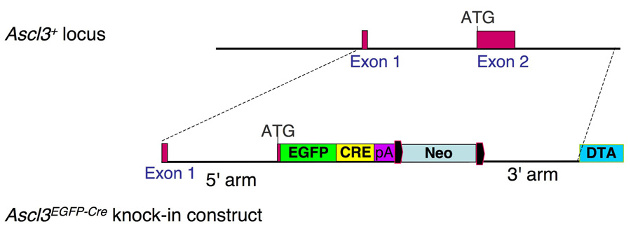
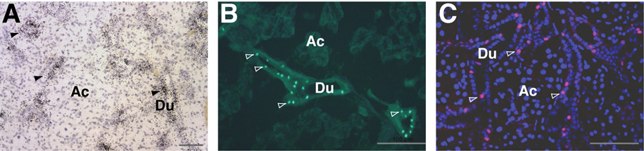
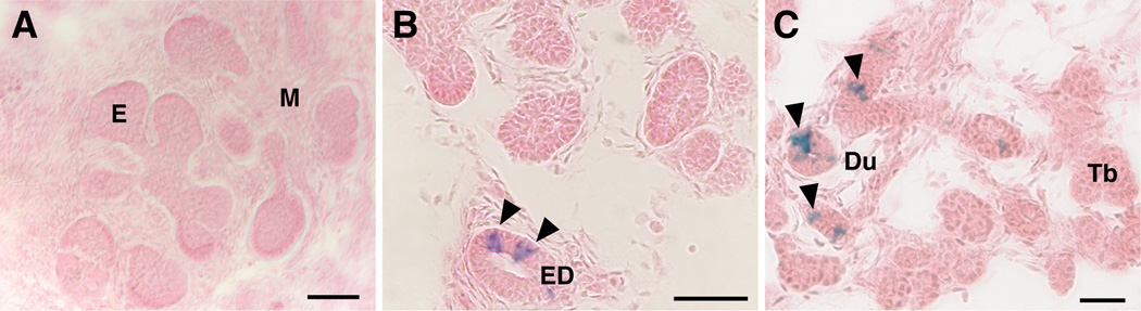
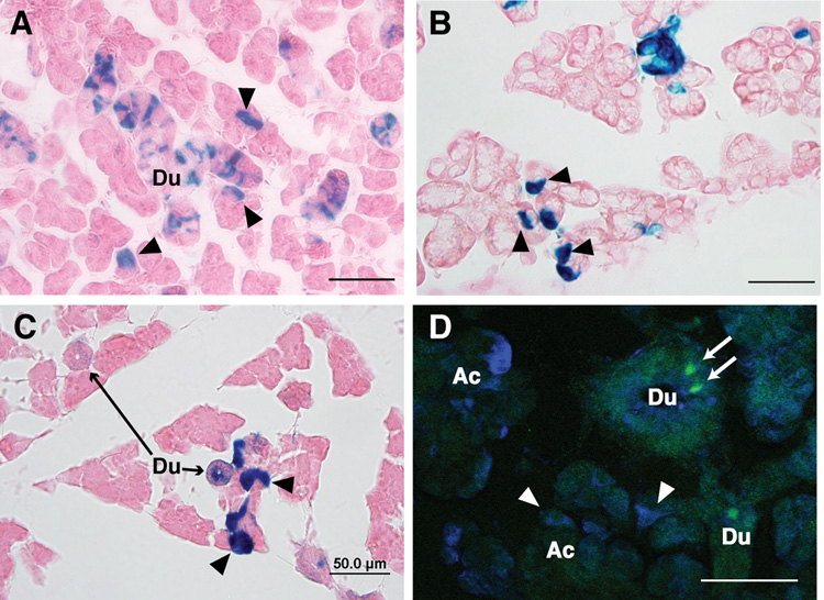
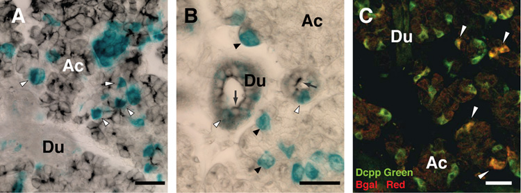
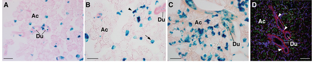
Ascl3, also know as Sgn1, is a member of the mammalian achaete scute (Mash) gene family of transcription factors, which have been implicated in cell fate specification and differentiation. In the mouse salivary gland, expression of Ascl3 is restricted to a subset of duct cells. Salivary gland function depends on the secretory acinar cells, which are responsible for saliva formation, and duct cells, which modify the saliva and conduct it to the oral cavity. The salivary gland ducts are also the putative site of progenitor cells in the adult gland. Using a Cre recombinase-mediated reporter system, we followed the fate of Ascl3-expressing cells after the introduction of an EGFP-Cre expression cassette into the Ascl3 locus by homologous recombination. Lineage tracing shows that these cells are progenitors of both acinar and ductal cell types in all three major salivary glands. In the differentiated progeny, expression of Ascl3 is down-regulated. These data directly demonstrate a progenitor-progeny relationship between duct cells and the acinar cell compartment, and identify a population of multipotent progenitor cells, marked by expression of Ascl3, which is capable of generating both gland cell types. We conclude that Ascl3-expressing cells contribute to the maintenance of the adult salivary glands.


Figures

Figure 1
Figure 1
Introduction of the EGFP-Cre expression cassette into the Ascl3 locus. The Ascl3 gene locus is comprised of only two exons. A knock-in construct was generated which replaces the second exon with an expression cassette encoding EGFP and Cre recombinase as a fusion protein. This construct was targeted to the Ascl3 locus by homologous recombination, placing EGFP-Cre under control of the Ascl3 promoter. The neomycin gene used for ES cell targeting is flanked by LoxP sites (black) and was subsequently removed by Cre recombination. The diphtheria toxin A (DTA) gene was used for negative selection, and is not retained in the recombined Ascl3EGFP-Cre allele.
Figure 2
Figure 2
EGFP-Cre recombinase expression recapitulates endogenous Ascl3 expression. AIn situ hybridization on a paraffin section of submandibular gland from Ascl3EGFP-Cre/+ female using a radioactively labeled antisense probe to Ascl3 coding sequence. Positive signal from endogenous Ascl3 mRNA expression (black grains) is concentrated in the ducts. Hematoxylin staining was used to stain all cells. Arrowheads indicate labeled duct structures. Ac, acinar cells; Du, duct cells. Scale bar is 50 µm. B. Fluorescent image of a frozen section from Ascl3EGFP-Cre/+ female submandibular gland. EGFP expression driven by the Ascl3 promoter is detectable in the nuclei of a subset of ductal cells (arrowheads). Ac, acinar cells; Du, duct cells. Scale bar is 100 µm. C. Immunohistochemical staining of paraffin section from Ascl3EGFP-Cre/+ submandibular gland examined using antibodies to Cre recombinase and aquaporin 5. Ascl3-expressing Cre+ cells are labeled red. Antibody to aquaporin 5 labels apical membranes of acinar cells and is shown in green. All nuclei are stained with ToPro3 (blue). Large round nuclei are acinar cells; smaller aligned nuclei are from duct cells. Arrowheads indicate duct cells in which Ascl3-driven Cre recombinase expression is detected. Ac, acinar cells; Du, duct cells. Scale bar is 50 µm.
Figure 3
Figure 3
Ascl3 expression is initiated during embryogenesis and is confined to the presumptive duct cells of the developing salivary gland. Ascl3EGFP-Cre/+/Rosa embryos taken at A) embryonic (E) day 13.5, B) E15.5 and C) E17.5 were fixed, sectioned and stained for LacZ expression in order to determine the Ascl3 expression pattern during salivary gland development. A. At the pseudoglandular stage, there is no detectable expression of Ascl3 in the embryonic submandibular gland. B. In the canalicular stage, at E15.5, limited expression of Ascl3 is observed only in the large excretory duct (arrowheads), based on the detection of LacZ activation. C. At the terminal bud stage Ascl3-expressing cells are limited to the cells of the presumptive ducts (arrowheads). E, epithelial buds; M, mesenchyme; ED, excretory duct; Du, presumptive ducts; Tb, terminal end buds. Scale bars are 50 µm.
Figure 4
Figure 4
Lineage tracing of progeny from Ascl3-expressing cells in salivary glands. Frozen sections of salivary glands from 3-week-old female Ascl3EGFP-Cre/+/Rosa26R mice, stained for LacZ expression, which marks the progeny of cells in which Cre recombinase, driven by the Ascl3 promoter, has been expressed. A. Submandibular B. Sublingual C. Parotid gland. Arrowheads point to acinar cells in which the LacZ gene has been activated. D. Immunohistochemistry of Ascl3EGFP-Cre/+ submandibular gland section probed with antibody to beta-galactosidase (blue). Expression of the EGFP-Cre fusion protein, as detected by EGFP (green, arrows), is limited to cells in the duct and is not ectopically activated in LacZ-positive cells in the acinar compartment (arrowheads). Scale bars are 50 µm.
Figure 5
Figure 5
Ascl3-expressing cells generate both duct and acinar cell progeny. A. Co-localization of LacZ expression with the acinar cell-specific protein aquaporin 5. Submandibular gland isolated from 6-week-old female Ascl3EGFP-Cre+/Rosa26 mouse was fixed and stained for LacZ in whole mount (blue). Following post-fixation, the tissue was embedded in paraffin, sectioned and stained with antibody to aquaporin 5, which labels apical membranes of acinar cells (black). Arrows indicate cells in which aquaporin 5 protein is co-localized with LacZ, demonstrating that differentiated acinar cells are among the descendants of Ascl3-expressing cells. Duct cells (Du) are not positive for aquaporin 5. Ac, acinar cells. B. Co-localization of LacZ-positive progeny with duct cell-specific marker, cytokeratin 19. Submandibular gland tissue stained for LacZ expression (blue), was embedded, sectioned and probed with antibody to cytokeratin 19 (black), which is localized to the apical membrane of duct cells (arrows). LacZ-positive cells in the ducts show co-localization with cytokeratin 19 (white arrowheads); LacZ-positive cells in the acinar (Ac) compartment (black arrowheads) do not react with duct cell-specific antibodies. C. Co-localization of LacZ expression with Dcpp, a secretory protein expressed in serous demilune cells of the sublingual gland. Dcpp antibody localizes to serous demilune cells (green). Antibody to beta-galactosidase is shown in red. Co-localization of the two antibodies (yellow), is indicated by arrowheads, and demonstrates that Ascl3-expressing cells are precursors of demilune cells in the sublingual gland. Du, duct cells; Ac, acinar cells. Scale bars are 50 µm.
Figure 6
Figure 6
Lineage tracing of Ascl3-expressing cells reveals continued contribution to the sublingual acinar cell population with age. Sublingual glands were removed from females of the Ascl3EGFP-Cre/+/Rosa26R strain at 3 weeks (A), 3 months (B), and 1 year of age (C), embedded, sectioned and analyzed for LacZ expression. With increasing age, there is a growing contribution of Ascl3-expressing progeny to the acinar cell compartment of the gland. The arrow in (B) indicates LacZ expression in an acinar cell, while the arrowhead points to a LacZ-positive demilune cell. D. Immunohistochemical staining using antibodies to Cre recombinase and aquaporin 5 on paraffin section of 5-month-old female Ascl3EGFP-Cre/+/Rosa26R mouse reconfirms that the endogenous Ascl3 expression in sublingual gland is restricted to the ducts, and is not ectopically activated in LacZ-positive acinar cells. Cre-expressing cells are red (arrows). Antibody to aquaporin 5 stains the apical membranes of acinar cells green. All nuclei are stained with ToPro. Du, ducts; Ac, acinar cells. Scale bars are 50 µm.

References

    1. Ball W, et al. Secretory proteins as markers for cellular phenotypes in rat salivary glands. Dev. Biol. 1988;125:265–279. - PubMed
    1. Battiste J, et al. Ascl1 defines sequentially generated lineage-restricted neuronal and oligodendrocyte precursor cells in the spinal cord. Development. 2007;134:285–293. - PubMed
    1. Bekhor I, et al. cDNA cloning, sequencing and in situ localization of a transcript specific to both sublingual demilune cells and parotid intercalated duct cells in mouse salivary glands. Arch. Oral Biol. 1994;39:1011–1022. - PubMed
    1. Cau E, et al. Mash1 activates a cascade of bHLH regulators in olfactory neuron progenitors. Development. 1997;124:1611–1621. - PubMed
    1. Chai Y, et al. Proliferative and structural differences between male and female mouse submandibular glands. Anat. Rec. 1993;235:303–311. - PubMed

Publication types

Substances