

VEP indices of cortical lateral interactions in epilepsy treatment
- PMID: 18572221
- PMCID: PMC2873633
- DOI: 10.1016/j.visres.2008.04.030
VEP indices of cortical lateral interactions in epilepsy treatment
Abstract
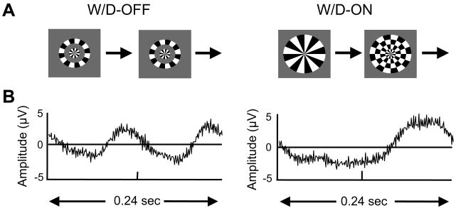
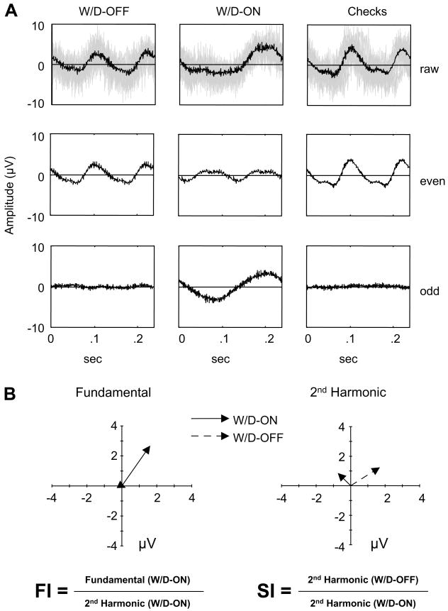
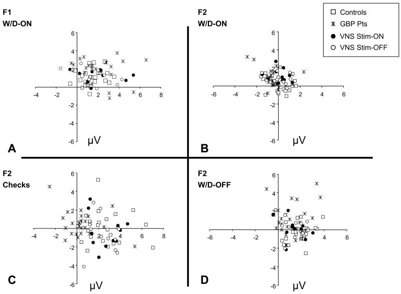
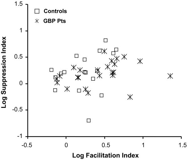
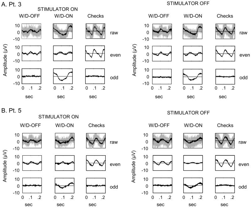
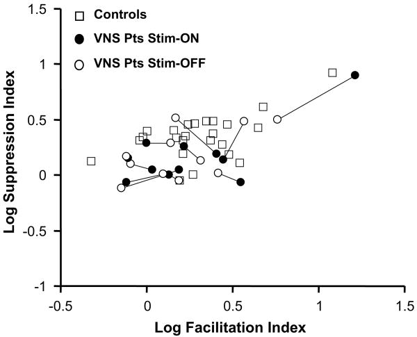
We extend Spekreijse's strategy for analyzing lateral interactions in visual evoked potentials (VEPs) to clinical neurophysiologic testing of patients with epilepsy. Stimuli consisted of the radial windmill/dartboard pattern [Ratliff, F., & Zemon, V. (1982). Some new methods for the analysis of lateral interactions that influence the visual evoked potential. In: Bodis-Wollner (Ed.), Evoked potentials, Vol. 388. (pp. 113-124). New York: Annals of the New York Academy of Sciences.] and conventional checkerboards. The fundamental and 2nd-harmonic components of the steady-state responses were used to calculate indices reflecting facilitatory (FI) and suppressive (SI) cortical interactions. We carried out two studies. In the first, VEPs in 38 patients receiving antiepileptic drug (AED) therapy were compared to those of age-matched controls. For three AEDs (tiagabine, topiramate, and felbamate), addition of the drug did not change the FI and SI compared to baseline values or those of normal controls. However, the addition of gabapentin was associated with an increase of the FI, and this change was reversed when the medication was withdrawn. This suggested a medication-specific change in cortical lateral interactions. The second study focused on the effects of neurostimulation therapy. Eleven epilepsy patients receiving chronic vagus nerve stimulation (VNS) treatment were tested. By comparing VEPs recorded with the stimulator on (Stim-ON) and turned off (Stim-OFF) in the same session, we determined that VNS did not have a short-acting effect on lateral interactions. However, when compared with normal controls, the VNS patients had a significantly smaller SI (p<.05), but no difference in the FI, demonstrating the presence of a chronic effect. We conclude that with the appropriate stimuli, VEPs can be used as a measure of cortical lateral interactions in normals and epileptic patients, and demonstrate specific changes in these interactions associated with certain treatment modalities.
Figures
References
-
- Bandini F, Pierantozzi M, Bodis-Wollner I. Parkinson's disease changes the balance of onset and offset visual responses: An evoked potential study. Clinical Neurophysiology. 2001;112:976–983. - PubMed
-
- Ben-Menachem BE, Hambergerm JA, Hedner T, Hammond HE, Uthman RB, Slater J, et al. Effects of vagus nerve stimulation on amino acids and other metabolites in the CSF of patients with partial seizures. Epilepsy Research. 20(3):221–227. - PubMed
-
- Ben-Menachem E, Hellstroem K, Waldton C, Augustinsson LE. Evaluation of refractory epilepsy treated with vagus nerve stimulation for up to 5 years. Neurology. 1999;52:1731–1735. - PubMed
-
- Bodis-Wollner I, Hendley CD, Mylin LH, Thornton J. Visual evoked potentials and the visuogram in multiple sclerosis. Annals of Neurology. 1979;5:40–47. - PubMed
-
- Bodis-Wollner I, Yahr MD. Measurement of visual evoked potentials in Parkinson's disease. Brain. 1978;101:661–671. - PubMed
Publication types
MeSH terms
Substances
Grants and funding
LinkOut - more resources
Full Text Sources
Other Literature Sources
Medical
Miscellaneous
