. 2008;32(11):1385-97.
doi: 10.1016/j.dci.2008.05.008. Epub 2008 Jun 10.

B cell receptor accessory molecules in the channel catfish, Ictalurus punctatus

Affiliations

B cell receptor accessory molecules in the channel catfish, Ictalurus punctatus

Manoranjan Sahoo et al. Dev Comp Immunol. 2008.

Abstract

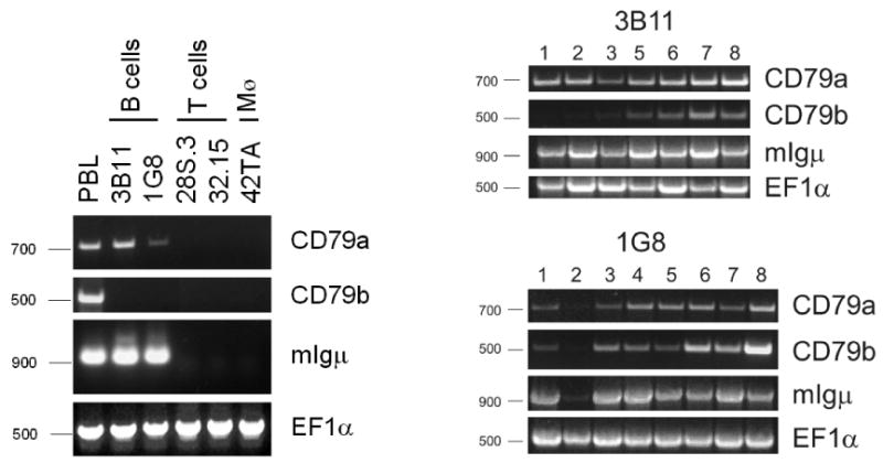
B cell receptor (BCR) accessory molecules CD79a and CD79b homologs were identified in the channel catfish, Ictalurus punctatus. Both are found as single copy genes that encode proteins containing a signal peptide, an extracellular immunoglobulin domain, a transmembrane region and a cytoplasmic tail containing an immune-receptor tyrosine-dased activation motif (ITAM). IpCD79a and IpCD79b transcripts correlate well with IgM message expression. They are highly expressed in peripheral blood leukocytes (PBL) enriched in membrane (m) IgM+ cells and catfish clonal B cell lines, but not in catfish clonal T cells, indicating that IpCD79a and IpCD79b expression is B cell restricted. Studies using catfish clonal B cells (3B11) transfected with constructs encoding epitope-tagged IpCD79a and IpCD79b revealed that IpCD79a was expressed as a 45 kDa protein and IpCD79b was expressed as a 32 kDa protein. Furthermore, co-immunoprecipitations of epitope-tagged CD79 proteins demonstrate that these molecules are non-covalently associated with mIgM. These data correlate with some of the previous immunoprecipitation data demonstrating that catfish mIgM associates with proteins of 45 and 32 kDa.


Figures

Fig. 1
Fig. 1
Catfish IpCD79a and IpCD79b. Nucleotide and predicted amino acid sequences of IpCD79a and IpCD79b. The predicted signal peptide (SP), Ig domains, TM and CYT are labeled above the sequence. Potential N-linked glycosylation sites are underlined and the conserved charged/polar amino acid residue in the TM is in bold. The ITAM motifs are gray shaded and in bold, the J-like sequences are double-underlined and the stop (TGA) codons are marked with (*). In IpCD79a, Tyr 204 is also double-underlined. Nucleotide and amino acid numbers are at left. GenBank accession numbers are IpCD79a (EF173897) and IpCD79b (EF173898).
Fig. 2
Fig. 2
CD79 amino acid comparisons. Amino acid alignment of IpCD79a with fish, mouse and human CD79a sequences and IpCD79b with fish, chicken, mouse and human CD79b sequences. The predicted extracellullar, TM and CYT boundaries are labeled, conserved cysteine residues that form the intrachain disulfide bond are marked with (▼) and the cysteines that form the interchain disulfide bond between CD79a and CD79b in mammals are marked (↑). Other conserved cysteine residues in the fish CD79 Ig domains and TMs are marked with (↓). The ITAM motifs are gray shaded, tyrosine residue 204 in CD79a is marked by ‡, and (.) represent amino acid identities and (-) represent gaps in the alignment. The pufferfish (Tr) and rainbow trout (Om) CD79 sequences are from Guselnikov et al 2003 [34]. Abbreviations and GenBank accession numbers for the other CD79 sequences are: chicken (Gg CD79b, AB062512), mouse (Mm CD79a, NP_031681; CD79b, CAJ18566), human (Hs CD79a1, NP-001774; CD79a2, NP-067612; CD79b P40259). There is no available chicken CD79a sequence. Number of amino acids is listed at the right.
Fig. 3
Fig. 3
Phylogenetic analyses of CD79 molecules. (A) Neighbor-Joining trees with pairwise gap deletions were drawn using MEGA3.1 [48] with 5000 bootstrap replications. (B) Comparison of CD79 percent amino acid identity values from pairwise alignments of full length sequences, Ig domains, TM and CYT regions. GenBank accession numbers and sequence references used are as in Fig. 2.
Fig. 4
Fig. 4
Southern blot analysis of IpCD79a and IpCD79b. Genomic DNA from two gynogenetic (labeled G) and three out bred catfish was hybridized with either an IpCD79a or IpCD79b specific probe. Ten μg of genomic DNA was digested to completion with either EcoR I or Pst I. Kb Size markers are at left.
Fig. 5
Fig. 5
RT-PCR analysis of IpCD79 message expression in catfish PBL. Total RNA was obtained from catfish PBL and B cell enriched (IgM+) and depleted (IgM-) fractions from the same fish. RT-PCR was performed using primers specific for catfish membrane (m) IgM, mIgD, IpCD79a, IpCD79b and EF1α (see supplementary Table 1). Base pair markers are at left.
Fig. 6
Fig. 6
RT-PCR analyses of IpCD79 message in catfish clonal cell lines. (A) Total RNA was obtained from catfish PBL and log phase 3B11 and 1G8 B cells, 28S.3 and 32.15 T cells and 42TA macrophages. RT-PCR was performed using the same primers as described for fig. 4. (B) Time course of IpCD79 message expression in catfish 1G8 and 3B11 B cells. Days after culture initiation are labeled numerically. Base pair markers are at left.
Fig. 7
Fig. 7
Predicted sizes of catfish recombinant CD79a, CD79b and Igμ2-cyt using Western blots. Catfish recombinant proteins were expressed in 3B11 B cells and cell lysates (2.5 × 10⁶ cells/lane) were analyzed by SDS-PAGE at 36 hrs post transfection. Proteins were transferred to nitrocellulose membranes and visualized using an anti-HA or anti-FLAG mAbs. (A) Recombinant HA-tagged CD79A and FLAG-tagged CD79B were expressed in 3B11 cells and cell lysates were electrophoresed under reducing conditions. (B) Recombinant HA-tagged Igμ2-cyt (containing Igμ domains 2-3, TM and CYT) was expressed in 3B11 cells and cell lystates electrophoresed using non-reducing and reducing conditions. Under non-reducing conditions, Igμ2-cyt dimers can be observed. In each panel, lysates from untransfected cells are used as controls. Arrows mark the identified proteins and molecular weight size markers are at left.
Fig. 8
Fig. 8
Recombinant catfish HA-tagged Igμ2-cyt, FLAG-tagged CD79a and HA-tagged CD79b are expressed on the surface of 3B11 B cells. (A) Schematic showing the expressed tagged proteins on the surface of 3B11 B cells (B) Surface expression of IgM, recombinant Igμ2-cyt, CD79a and CD79b molecules were analyzed by flow cytometry using anti-IgM (9E1), anti-FLAG, and anti-HA mAbs, followed by PE-conjugated goat anti-mouse secondary antibodies. 3B11 B cells transfected with all three constructs (shaded histograms) showed increased anti-IgM staining as compared to untransfected B cells (open histograms). Percentage of stained transfected cells are indicated on the individual histograms. (C) The transfected cells shown in A were lysed in 2% octyl-β-glucoside and the resulting lysates were immunoselected using anti-trout IgM 1.14 (negative control), anti-HA, or anti-FLAG mAbs coated Protein G Sepharose beads. The immunoselected proteins were separated by 10% SDS-PAGE under reducing conditions and visualized by Western blot using anti-HA-conjugated to horse radish peroxidase (HRP) and (D) anti-FLAG-HRP mAbs. Arrows mark identified proteins, IP indicates mAbs used for immunoselection and WB indicates mAb used in Western blotting. The anti-HA-HRP mAb was reactive with the IgH chain of the immunoselecting mAb and is indicated by (*). Molecular weight size markers are at left.

References

    1. Hombach J, Leclercq L, Radbruch A, Rajewsky K, Reth M. A novel 34-kd protein co-isolated with the IgM molecule in surface IgM-expressing cells. Embo J. 1988;7:3451–6. - PMC - PubMed
    1. Campbell KS, Cambier JC. B lymphocyte antigen receptors (mIg) are non-covalently associated with a disulfide linked, inducibly phosphorylated glycoprotein complex. Embo J. 1990;9:441–8. - PMC - PubMed
    1. Hombach J, Tsubata T, Leclercq L, Stappert H, Reth M. Molecular components of the B-cell antigen receptor complex of the IgM class. Nature. 1990;343:760–2. - PubMed
    1. Matsuo T, Kimoto M, Sakaguchi N. Direct identification of the putative surface IgM receptor-associated molecule encoded by murine B cell-specific mb-1 gene. J Immunol. 1991;146:1584–90. - PubMed
    1. Campbell KS, Hager EJ, Friedrich RJ, Cambier JC. IgM antigen receptor complex contains phosphoprotein products of B29 and mb-1 genes. Proc Natl Acad Sci U S A. 1991;88:3982–6. - PMC - PubMed

Publication types

Associated data