

Peptides containing membrane-interacting motifs inhibit herpes simplex virus type 1 infectivity
- PMID: 18572274
- PMCID: PMC7172891
- DOI: 10.1016/j.peptides.2008.04.022
Peptides containing membrane-interacting motifs inhibit herpes simplex virus type 1 infectivity
Abstract
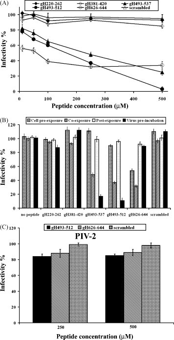
Herpes simplex virus (HSV) membrane fusion represents an attractive target for anti-HSV therapy. To investigate the structural basis of HSV membrane fusion and identify new targets for inhibition, we have investigated the different membranotropic domains of HSV-1 gH envelope glycoprotein. We observed that fusion peptides when added exogenously are able to inhibit viral fusion likely by intercalating with viral fusion peptides upon adopting functional structure in membranes. Interestingly, peptides analogous to the predicted HSV-1 gH loop region inhibited viral plaque formation more significantly. Their inhibitory effect appears to be a consequence of their ability to partition into membranes and aggregate within them. Circular dichroism spectra showed that peptides self-associate in aqueous and lipidic solutions, therefore the inhibition of viral entry may occur via peptides association with their counterpart on wild-type gH. The antiviral activity of HSV-1 peptides tested provides an attractive basis for the development of new fusion peptide inhibitors corresponding to regions outside the fusion protein heptad repeat regions.
Figures


Similar articles
-
Heptad repeat 2 in herpes simplex virus 1 gH interacts with heptad repeat 1 and is critical for virus entry and fusion.J Virol. 2006 Mar;80(5):2216-24. doi: 10.1128/JVI.80.5.2216-2224.2006. J Virol. 2006. PMID: 16474129 Free PMC article.
-
Analysis of synthetic peptides from heptad-repeat domains of herpes simplex virus type 1 glycoproteins H and B.J Gen Virol. 2006 May;87(Pt 5):1085-1097. doi: 10.1099/vir.0.81794-0. J Gen Virol. 2006. PMID: 16603508
-
Dendrimers functionalized with membrane-interacting peptides for viral inhibition.Int J Nanomedicine. 2013;8:521-34. doi: 10.2147/IJN.S37739. Epub 2013 Feb 5. Int J Nanomedicine. 2013. PMID: 23429490 Free PMC article.
-
The identification and characterization of fusogenic domains in herpes virus glycoprotein B molecules.Chembiochem. 2008 Mar 25;9(5):758-67. doi: 10.1002/cbic.200700457. Chembiochem. 2008. PMID: 18311743
-
gH625: a milestone in understanding the many roles of membranotropic peptides.Biochim Biophys Acta. 2015 Jan;1848(1 Pt A):16-25. doi: 10.1016/j.bbamem.2014.10.006. Epub 2014 Oct 12. Biochim Biophys Acta. 2015. PMID: 25305339 Free PMC article. Review.
Cited by
-
Analysis of a membrane interacting region of herpes simplex virus type 1 glycoprotein H.J Biol Chem. 2008 Oct 31;283(44):29993-30009. doi: 10.1074/jbc.M803092200. Epub 2008 Aug 4. J Biol Chem. 2008. PMID: 18678872 Free PMC article.
-
Update on emerging antivirals for the management of herpes simplex virus infections: a patenting perspective.Recent Pat Antiinfect Drug Discov. 2013 Apr;8(1):55-67. doi: 10.2174/1574891x11308010011. Recent Pat Antiinfect Drug Discov. 2013. PMID: 23331181 Free PMC article. Review.
-
Antiviral activity of mycosynthesized silver nanoparticles against herpes simplex virus and human parainfluenza virus type 3.Int J Nanomedicine. 2013;8:4303-14. doi: 10.2147/IJN.S50070. Epub 2013 Nov 6. Int J Nanomedicine. 2013. PMID: 24235828 Free PMC article.
-
Daphnia magna and Xenopus laevis as in vivo models to probe toxicity and uptake of quantum dots functionalized with gH625.Int J Nanomedicine. 2017 Apr 4;12:2717-2731. doi: 10.2147/IJN.S127226. eCollection 2017. Int J Nanomedicine. 2017. PMID: 28435254 Free PMC article.
-
Liposome armed with herpes virus-derived gH625 peptide to overcome doxorubicin resistance in lung adenocarcinoma cell lines.Oncotarget. 2016 Jan 26;7(4):4077-92. doi: 10.18632/oncotarget.6013. Oncotarget. 2016. PMID: 26554306 Free PMC article.
References
-
- Carfi A., Willis S.H., Whitbeck J.C., Krummenacher C., Cohen G.H., Eisenberg R.J., Wiley D.C. Herpes simplex virus glycoprotein D bound to the human receptor HveA. Mol Cell. 2001;8:169–179. - PubMed
-
- Chan D.C., Kim P.S. HIV entry and its inhibition. Cell. 1998;93:681–684. - PubMed
-
- Chetal P., Chauhan V.S., Sahal D. A meccano set approach of joining trpzip a water soluble beta-hairpin peptide with a didehydrophenylalanine containing hydrophobic helical peptide. J Peptide Res. 2005;65:475–484. - PubMed
MeSH terms
Substances
LinkOut - more resources
Full Text Sources
Other Literature Sources
