

Antemortem MRI based STructural Abnormality iNDex (STAND)-scores correlate with postmortem Braak neurofibrillary tangle stage
- PMID: 18572417
- PMCID: PMC3097053
- DOI: 10.1016/j.neuroimage.2008.05.012
Antemortem MRI based STructural Abnormality iNDex (STAND)-scores correlate with postmortem Braak neurofibrillary tangle stage
Abstract
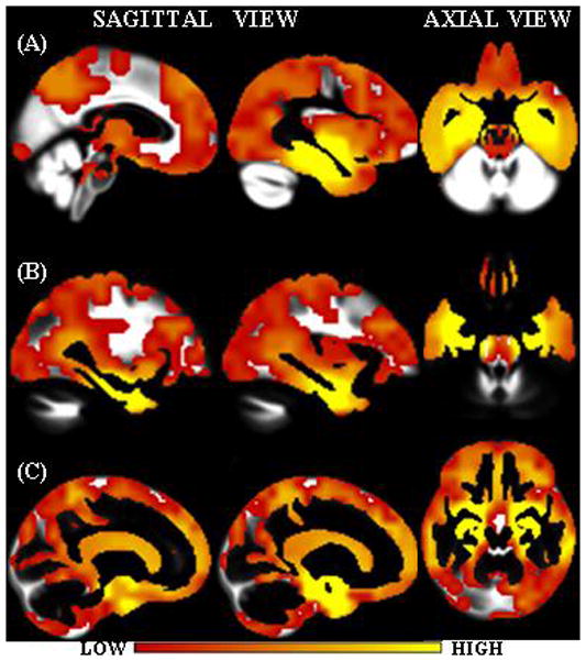
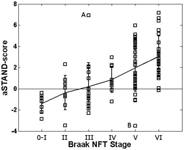
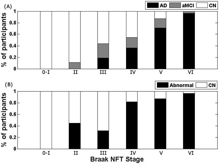
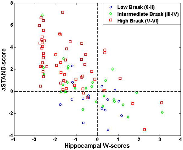
The clinical diagnosis of Alzheimer's disease (AD) does not exactly match the pathological findings at autopsy in every subject. Therefore, in-vivo imaging measures, such as Magnetic Resonance Imaging (MRI) that reflect underlying pathology, would be clinically useful independent supplementary measures of disease stage. We have developed an algorithm that extracts atrophy information from individual patient's 3D MRI scans and assigns a STructural Abnormality iNDex (STAND)-score to the scan based on the degree of atrophy in comparison to patterns extracted from a large library of clinically well characterized AD and CN (cognitively normal) subject's MRI scans. STAND-scores can be adjusted for demographics to give adjusted-STAND (aSTAND)-scores which are >0 for subjects with brains identified as abnormal by the algorithm. Since histopathological findings are considered to represent the "ground truth", our objective was to assess the sensitivity of aSTAND-scores to pathological AD staging. This was done by comparing antemortem MRI based aSTAND-scores with postmortem grading of disease severity in 101 subjects who had both antemortem MRI and postmortem Braak neurofibrillary tangle (NFT) staging. We found a rank correlation of 0.62 (p<0.0001) between Braak NFT stage and aSTAND-scores. The results show that optimally extracted information from MRI scans such as STAND-scores accurately capture the severity of neuronal pathology and can be used as an independent approximate surrogate marker for in-vivo pathological staging as well as for early identification of AD in individual subjects.
Figures
References
-
- American Psychiatric Association. Diagnostic and Statistical Manual of Mental Disorders. 4. Washington DC: American Psychiatric Association; 1994. (DSM IV)
-
- Ashburner J, Friston KJ. Voxel-based morphometry--the methods. Neuroimage. 2000;11:805–821. - PubMed
-
- Ashburner J, Friston KJ. Unified segmentation. Neuroimage. 2005;26:839–851. - PubMed
-
- Baron JC, Chetelat G, Desgranges B, Perchey G, Landeau B, de la Sayette V, Eustache F. In vivo mapping of gray matter loss with voxel-based morphometry in mild Alzheimer’s disease. Neuroimage. 2001;14:298–309. - PubMed
-
- Bennett DA, Schneider JA, Arvanitakis Z. Neuropathology of older persons without cognitive impairment from two community-based clinical-pathologic studies. Neurology. 2006;66:1837–1844. - PubMed
Publication types
MeSH terms
Grants and funding
LinkOut - more resources
Full Text Sources
Medical
