Review
. 2008:37:197-214.
doi: 10.1146/annurev.biophys.37.032807.125957.

RNA folding: conformational statistics, folding kinetics, and ion electrostatics

Affiliations
Review

RNA folding: conformational statistics, folding kinetics, and ion electrostatics

Shi-Jie Chen. Annu Rev Biophys. 2008.

Abstract

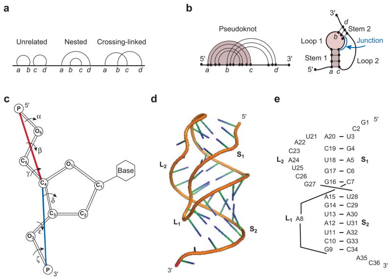
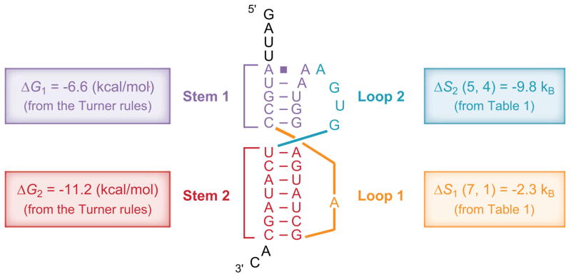
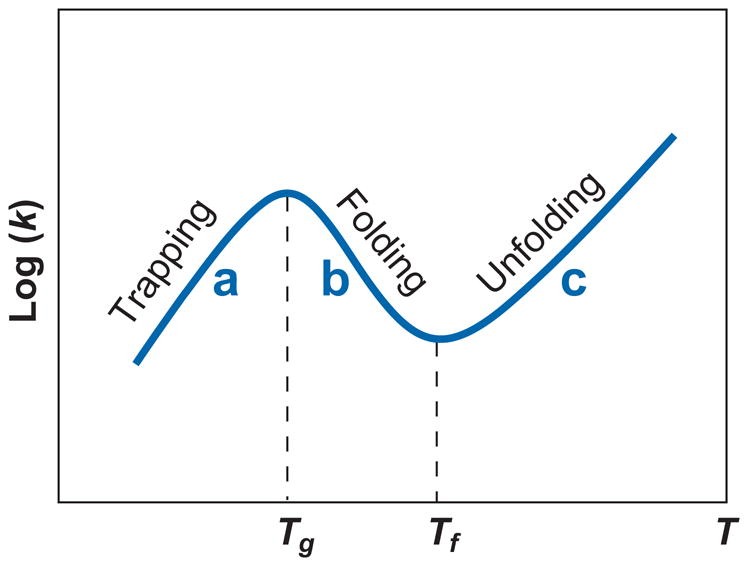
RNA folding is a remarkably complex problem that involves ion-mediated electrostatic interaction, conformational entropy, base pairing and stacking, and noncanonical interactions. During the past decade, results from a variety of experimental and theoretical studies pointed to (a) the potential ion correlation effect in Mg2+-RNA interactions, (b) the rugged energy landscapes and multistate RNA folding kinetics even for small RNA systems such as hairpins and pseudoknots, (c) the intraloop interactions and sequence-dependent loop free energy, and (d) the strong nonadditivity of chain entropy in RNA pseudoknot and other tertiary folds. Several related issues, which have not been thoroughly resolved, require combined approaches with thermodynamic and kinetic experiments, statistical mechanical modeling, and all-atom computer simulations.


Figures

Figure 1
Figure 1
(a) Graphical representation for an RNA structure, where vertices (i.e., nucleotide monomers) are connected by straight lines (i.e., backbone covalent bonds) and base pairs are denoted by curved links. (b) Graphical representation for a pseudoknot. (c) The virtual bonds (shown as the red and blue lines) for RNA backbone. (d) The three-dimensional structure and (e) base-pairing structure for a typical pseudoknot (gene 32 mRNA pseudoknot of bacteriophage T2; PDB ID: 2TPK). Loop 1 (L 1) and loop 2 (L2) span across the major (narrow, deep) groove of helix 2 (S 2) and the minor (wide, shallow) groove of helix 1 (S1), respectively. The structure in (d) is adopted from the PDB (PDB ID: 2TPK) (39).
Figure 2
Figure 2
Predicting pseudoknot stability using the loop entropy parameters in Table 1 (14). For loop 1, length of stem 2 is 7 bp, length of loop 1 is 1 nt, so the entropy is read from Table 1 as ΔS1(7, 1). Similarly, for loop 2, stem 1 is 5 bp, loop 2 is 4 nt, so the loop entropy is ΔS2(5, 4). The total free energy of the pseudoknot is ΔG = ΔG1 + ΔG2T ΔS1(7, 1) − T ΔS2(5, 4) + ΔGassemble = (−6.6) kcal/mol + (−11.2) kcal/mol + (kB T)(2.3) + (kB T)(9.8) + 1.3 kcal/mol = −9.0 kcal/mol, where ΔGassemble = 1.3 kcal/mol accounts for the multiple ways to connect the two loops (14) and kBT = 0.62 kcal/mol at T = 37°C. See Reference for more examples.
Figure 3
Figure 3
Temperature dependence of the reaction rate k (111). (a) T > Tf > Tg: The relaxation process is predominantly unfolding, with single exponential kinetics along an unzipping pathway. The apparent activation barrier [= − d ln(k)/d (1/T)] is positive, equal to the enthalpic cost of breaking the rate-limiting base stack(s). (b) Tg > T > Tf: The relaxation process is predominantly folding, with single exponential kinetics along a zipping pathway. The activation barrier is negative, equal to the stabilizing enthalpy of the rate-limiting base stack(s). (c) T < Tg: The relaxation process is predominantly folding, with multistate multiple exponential kinetics involving multiple pathways, including the zipping pathway and detrapping-refolding pathways. The activation barrier is positive, equal to the average enthalpic cost to detrap the misfolded states.
Figure 4
Figure 4
Two types of cations surrounding the P4-P6 RNA structure (17): specific binding and diffusive binding. A recent Mg2+ ion titration study suggested that two metal ions induce cooperative folding of the P4-P6 metal ion core (25).

Similar articles

Cited by

References

    1. Abrahams JP, van den Berg M, van Batenburg E, Pleij C. Prediction of RNA secondary structure, including pseudoknot, by computer simulation. Nucleic Acids Res. 1990;18:3035–44. - PMC - PubMed
    1. Andresen K, Das R, Park HY, Smith H, Kwok LW, et al. Spatial distribution of competing ions around DNA in solution. Phys Rev Lett. 2004;93:248103. - PubMed
    1. Andronescu M, Zhang ZC, Condon A. Secondary structure prediction of interacting RNA molecules. J Mol Biol. 2005;345:987–1001. - PubMed
    1. Ansari A, Kuznetsov SV, Shen Y. Configurational diffusion down a folding funnel describes the dynamics of DNA hairpins. Proc Natl Acad Sci USA. 2001;98:7771–76. - PMC - PubMed
    1. Bai Y, Das R, Millett IS, Herschlag D, Doniach S. Probing counterion modulated repulsion and attraction between nucleic acid duplexes in solution. Proc Natl Acad Sci USA. 2005;102:1035–40. Experiment demonstrates that the Mg2+-induced force is not sufficient to cause a collapsed state for two short DNA helices tethered by a neutral short loop. - PMC - PubMed

Publication types

LinkOut - more resources