Review
. 2008:37:353-73.
doi: 10.1146/annurev.biophys.37.032807.125829.

Structure-based view of epidermal growth factor receptor regulation

Affiliations
Review

Structure-based view of epidermal growth factor receptor regulation

Kathryn M Ferguson. Annu Rev Biophys. 2008.

Abstract

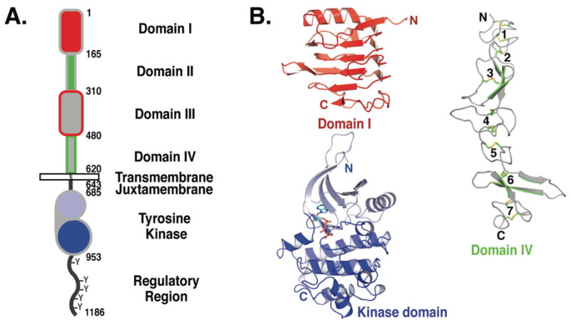
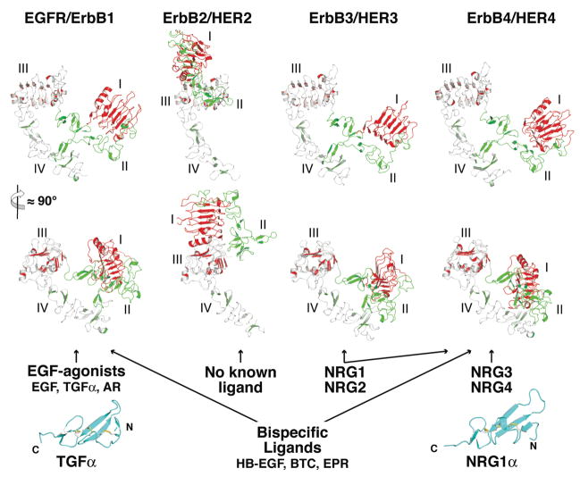
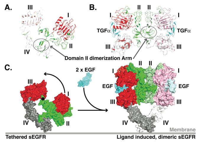
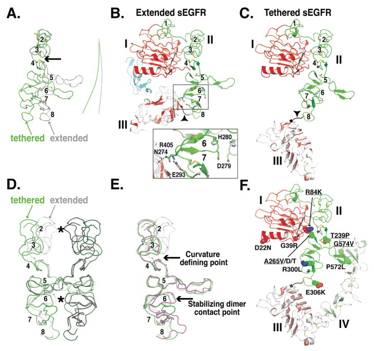
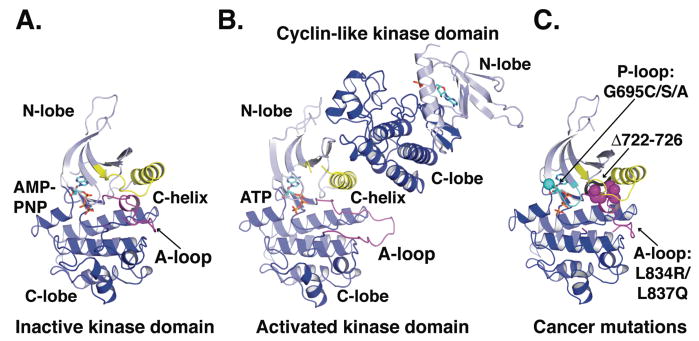
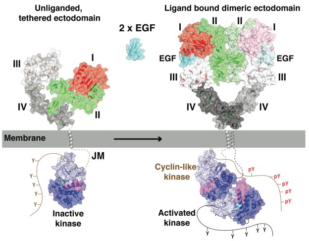
High-resolution X-ray crystal structures determined in the past six years dramatically influence our view of ligand-induced activation of the epidermal growth factor receptor (EGFR) family of receptor tyrosine kinases. Ligand binding to the extracellular region of EGFR promotes a major domain reorganization, plus local conformational changes, that are required to generate an entirely receptor-mediated dimer. In this activated complex the intracellular kinase domains associate to form an asymmetric dimer that supports the allosteric activation of one kinase. These models are discussed with emphasis on recent studies that add details or bolster the generality of this view of activation of this family of receptors. The EGFR family is implicated in several disease states, perhaps most notably in cancers. Activating tumor mutations have been identified in the intracellular and extracellular regions of EGFR. The impact of these tumor mutations on the understanding of EGFR activation and of its inhibition is discussed.


Figures

FIGURE 1
FIGURE 1. The domains of EGFR
A. The extracellular region comprises 4 domains: I–IV, sometimes referred to as L1, CR1, L2 and CR2 or L1, S1, L2 and S2. Domains I (red) and III (gray with red outline) share about 37% sequence identity, while domains II (green) and IV (gray with green outline) are cystine rich. The N-lobe of the kinase domain is in light blue and the C-lobe in darker blue. This color scheme is used in all figures unless otherwise noted. Amino acid numbers are noted for each domain boundary. The conventional numbering system is used in which amino acid one of EGFR is the assumed first amino acid of the mature protein. In some recent papers, including those defining EGFR cancer mutations, alternative numbering is used where the signal peptide of EGFR is included. To convert to this alternative scheme add 24 to numbers used here. B. Representative cartoons of the domains of EGFR. Domains I and III adopt a β-helix fold, here domain I from pdb id 1YY9 is shown. Domains II and IV adopt extended structures comprising a series of disulfide-bonded modules. Domain IV from pdb id 1YY9 is shown with the disulfides in stick representation and the disulfide-bonded modules numbered. There are two types of disulfide-bonded module. One has a single disulfide bond and the intervening loops adopt a bow-like arrangement (modules 2, 3, 5 & 6). The second type has two disulfide bonds with consecutive cysteines linked in the pattern Cys1–Cys3 and Cys2–Cys4 (modules 1, 4 & 7). The inactive kinase is shown (pdb id 2GS7) with the ATP analogue (AMP-PNP) in stick representation.
FIGURE 2
FIGURE 2. The extracellular regions of ErbB receptors and their activating ligands
Two orthogonal cartoon views of each unliganded ErbB receptor (pdb ids 1NQL, 1N8Z, 1M6B and 2AHX). The coordinates of domain III only were used to align the structures. ErbB2 is an outlier adopting an extended rather than tethered arrangement of domains (see text). Ligands are listed, grouped according to the receptors they activate. Cartoons of TGFα (left; pdb id 1MOX) and NRG1α (right; pdb id 1HRE) are shown in cyan as representative structures of the EGF-like domain of ErbB ligands. The scale for the ligands is twice that used for the receptor extracellular regions.
FIGURE 3
FIGURE 3. Ligand induced dimerization of the extracellular region of EGFR
A. Cartoon of the tethered sEGFR (pdb id 1YY9) oriented as in the upper panel of Fig. 2. B Cartoon of the TGFα induced dimer of sEGFR501 (pdb id 1MOX). The orientation of domain III is as in the lower panel of sErbB cartoons in Fig. 2. The colors of the left hand molecule have been lightened for contrast. C. A molecular surface representation of tethered sEGFR in the same orientation as in A with domains I and III in red, II in green and IV in gray. An ≈ 130° rotation about the indicated axis (black dot) plus 20 Å translation into the plane of the page is required to bring domain I from it position in the tethered structure (left) its location in the dimer (right) (22). In this model of the sEGFR dimer, domain IV is included such as to maintain the same domain III/IV relationship as in the tethered structure. Domains I, II and III in the dimer are from pdb id 1IVO and are shown in the same orientation as in B.
FIGURE 4
FIGURE 4. Conformational changes in domain II of sEGFR
A. Smoothed backbone representations of domains II from extended (gray) and from tethered (green) sEGFR. The coordinates of domain I and the first three disulfide-bonded modules of domain II (amino acids 1-225) were used to superimpose the two structures. The lines to the right indicate the curvature of the long axis of domain II. Disulfide bonds are shown (A–C & F) and disulfide-bonded modules numbered (A–E, module 1 is not shown in A, D or E). B. Cartoon of domains I, II and III from extended sEGFR (pdb id 1MOX) oriented as in A. The area of detailed (boxed) shows the domain II/III interactions that contribute to stabilizing module 6. See text for details. C. Cartoon of domains I, II and III from tethered sEGFR (pdb id 1YY9), oriented as in A. Note the very different trajectory of the end of domain II (in black and marked with an arrowhead) compared to part B. D. Domain II from the sEGFR501/TGFα dimer is shown with the left hand molecule in gray and the right hand molecule in black. Contact points across the dimer are indicated with asterisks. Using coordinates from disulfide-bonded module 5 only, domain II from tethered sEGFR has been superimposed first on the right hand extended domain II (green) and then on the left hand extended domain II (dark green) to create a model for a “dimer” of two domain II molecules in the tethered conformation. E. Domain II from extended (gray) and from tethered (green) sEGFR and from sErbB2 (magenta) shown in same orientation as D. See text for details. F. Cartoon of tethered sEGFR in the same orientation as C and with the positions of somatic mutations in glioblastoma shown in space filling representation. Those mutations that have been show to cause activation of EGFR are underlined.
FIGURE 5
FIGURE 5. Activation of the EGFR kinase domain
A. Cartoon of the EGFR kinase domain in the inactive conformation with AMP-PNP in stick representation (pdb id 2GS7). The activation loop (A-loop) is in magenta and the catalytically important C-helix is in yellow. B. Cartoon of the asymmetric EGFR kinase domain dimer (pdb id 2GS6) with the ATP moiety of the bound ATP-peptide conjugate in stick representation. The conformation of the activation loop (magenta) and position of the C-helix (yellow) are consistent with an active kinase (33). C. A cartoon view of the inactive kinase domain in the same orientation and colors as in A. The locations of somatic mutations identified in NSCLC are indicated.
FIGURE 6
FIGURE 6. Mechanism of EGFR activation
The crystal structures from EGFR are placed so as to provide a framework to consider the mechanism of activation of EGFR at the cell membrane. The same scale is used for the cartoon representations (plus transparent molecular surface) of the extracellular region and kinase domain. The TM domain is shown as an α-helix (gray), also to this same scale. Regions that have not been crystallographically defined are shown with a dashed or solid lines. The missing stretches of the inactive kinase are shown in brown, while those of the active kinase are in black. See text for details.

Similar articles

Cited by

References

    1. Adams TE, Epa VC, Garrett TP, Ward CW. Structure and function of the type 1 insulin-like growth factor receptor. Cell Mol Life Sci. 2000;57:1050–93. - PMC - PubMed
    1. Baselga J, Arteaga CL. Critical update and emerging trends in epidermal growth factor receptor targeting in cancer. J Clin Oncol. 2005;23:2445–59. - PubMed
    1. Bellot F, Moolenaar W, Kris R, Mirakhur B, Verlaan I, et al. High-affinity epidermal growth factor binding is specifically reduced by a monoclonal antibody, and appears necessary for early responses. J Cell Biol. 1990;110:491–502. - PMC - PubMed
    1. Berezov A, Chen J, Liu Q, Zhang HT, Greene MI, Murali R. Disabling receptor ensembles with rationally designed interface peptidomimetics. J Biol Chem. 2002;277:28330–39. - PubMed
    1. Boulanger MJ, Garcia KC. Shared Cytokine Signaling Receptors: Structural Insights from the Gp130 System. Adv Protein Chem. 2004;68:107–46. - PubMed

Publication types