Review
. 2008:37:465-87.
doi: 10.1146/annurev.biophys.37.032807.125842.

Bimolecular fluorescence complementation (BiFC) analysis as a probe of protein interactions in living cells

Affiliations
Review

Bimolecular fluorescence complementation (BiFC) analysis as a probe of protein interactions in living cells

Tom K Kerppola. Annu Rev Biophys. 2008.

Abstract

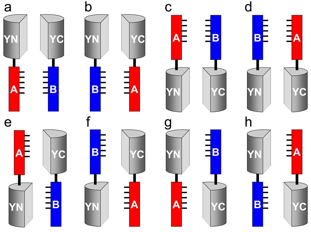
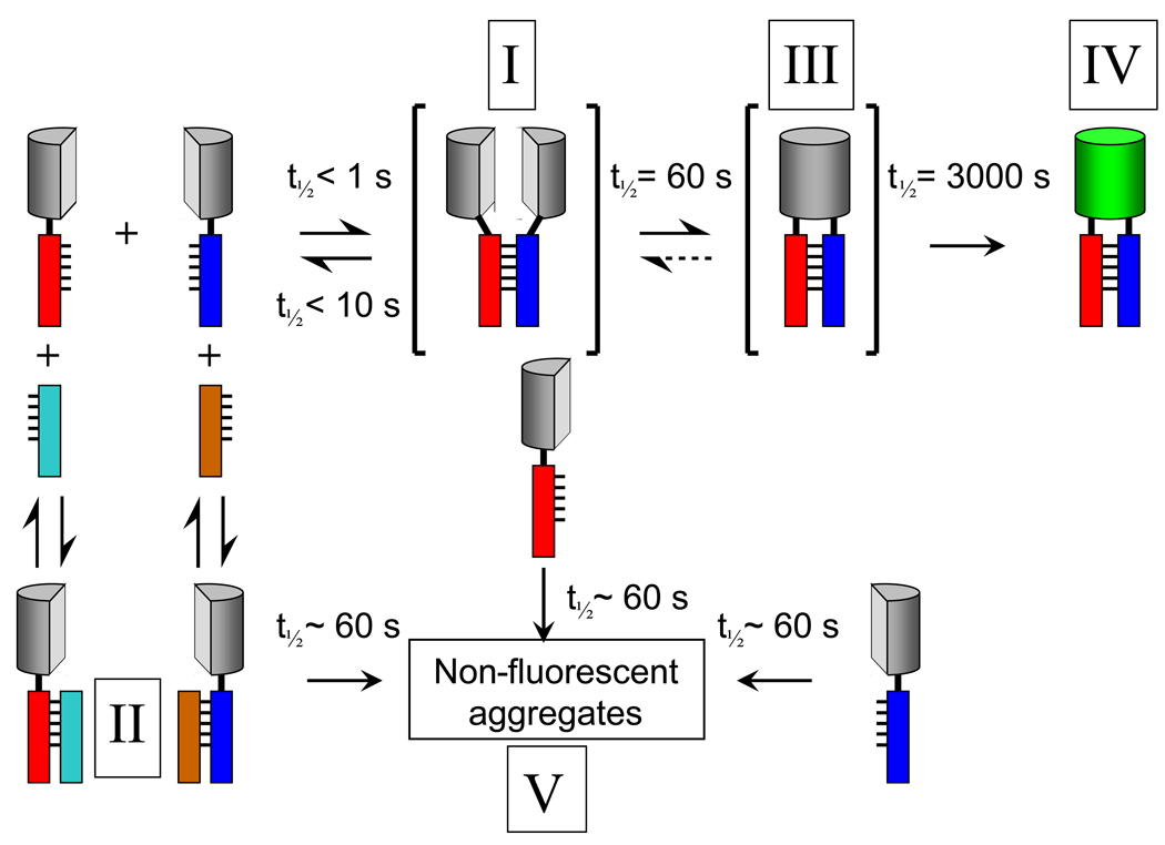
Protein interactions are a fundamental mechanism for the generation of biological regulatory specificity. The study of protein interactions in living cells is of particular significance because the interactions that occur in a particular cell depend on the full complement of proteins present in the cell and the external stimuli that influence the cell. Bimolecular fluorescence complementation (BiFC) analysis enables direct visualization of protein interactions in living cells. The BiFC assay is based on the association between two nonfluorescent fragments of a fluorescent protein when they are brought in proximity to each other by an interaction between proteins fused to the fragments. Numerous protein interactions have been visualized using the BiFC assay in many different cell types and organisms. The BiFC assay is technically straightforward and can be performed using standard molecular biology and cell culture reagents and a regular fluorescence microscope or flow cytometer.


Figures

Figure 1
Figure 1
Schematic representation of the principle of the BiFC assay. Two non-fluorescent fragments (YN and YC) of the yellow fluorescent protein (YFP) are fused to putative interaction partners (A and B).The association of the interaction partners allows formation of a bimolecular fluorescent complex.
Figure 2
Figure 2
Structures of proteins that have been used to study protein interactions using complementation approaches. The two fragments that have been used are shown in red and green based on the X-ray crystal structures of the intact proteins. In β-galactosidase, the overlap between the fragments is shown in orange. The images were generated using jmol.
Figure 3
Figure 3
Multiple combinations of fusion proteins should be tested for bimolecular fluorescence complementation. Amino- and carboxyl-terminal fusions can be used to test eight distinct combinations (a through h).
Figure 4
Figure 4
Pathway for formation of bimolecular fluorescent complexes. The pathway for fluorescent complex formation has been deduced based on in vitro studies of the dynamics of bimolecular fluorescence complementation using purified proteins (45). For a description of the steps in this pathway, please see the text.
Figure 5
Figure 5
The effects of mutations that prevent the association of the interaction partners should be tested to determine the specificity of the bimolecular fluorescence complementation (data adapted from (45)). Plasmids encoding a wild type interaction partner, B, and either the wild type (upper panel) or mutated (lower panel) forms of an interaction partner, A, fused to the fluorescent protein fragments, were transfected into cells together with an internal reference encoding CFP. The fluorescence intensities produced by bimolecular fluorescence complementation (YN-YC) and the internal reference (CFP) were measured in individual cells. The distribution of ratios between the fluorescence intensities in individual cells is plotted in each histogram.
Figure 6
Figure 6
Concurrent visualization of multiple protein complexes using multicolor fluorescence complementation analysis. (a) Two alternative interaction partners, A and B, are fused to fragments of different fluorescent proteins (YN155 and CN155 respectively). These fusions are co-expressed in cells with a shared interaction partner, Z, fused to a complementary fragment (CC155). Complexes formed by A-YN155 and Z-CC155 can be distinguished from complexes formed by B-CN155 and Z-CC155 based on their fluorescence spectra. (b) Schematic representation of the visualization of multiple protein complexes in the same cell (A-YN155-Z-CC155, cytoplasmic and perinuclear; B-CN155-Z-CC155, nuclear and perinuclear).

Similar articles

Cited by

References

    1. Abe M, Kobayashi Y, Yamamoto S, Daimon Y, Yamaguchi A, et al. FD, a bZIP protein mediating signals from the floral pathway integrator FT at the shoot apex. Science. 2005;309:1052. - PubMed
    1. Adolph D, Flach N, Mueller K, Ostareck DH, Ostareck-Lederer A. Deciphering the cross talk between hnRNP K and c-Src: the c-Src activation domain in hnRNP K is distinct from a second interaction site. Molecular and Cellular Biology. 2007;27:1758. - PMC - PubMed
    1. Ahmed KM, Dong SZ, Fan M, Li JJ. Nuclear factor-kappa B p65 inhibits mitogen-activated protein kinase signaling pathway in radioresistant breast cancer cells. Molecular Cancer Research. 2006;4:945. - PubMed
    1. Anderie I, Schulz I, Schmid A. Direct interaction between ER membrane-bound PTP1B and its plasma membrane-anchored targets. Cellular Signalling. 2007;19:582. - PubMed
    1. Aparicio F, Sanchez-Navarro JA, Pallas V. In vitro and in vivo mapping of the Prunus necrotic ringspot virus coat protein C-terminal dimerization domain by bimolecular fluorescence complementation. Journal of General Virology. 2006;87:1745. - PubMed

MeSH terms

LinkOut - more resources