. 2008 Aug;69(3):621-32.
doi: 10.1111/j.1365-2958.2008.06308.x. Epub 2008 Jun 28.

A sensor histidine kinase co-ordinates cell wall architecture with cell division in Bacillus subtilis

Affiliations

A sensor histidine kinase co-ordinates cell wall architecture with cell division in Bacillus subtilis

Tatsuya Fukushima et al. Mol Microbiol. 2008 Aug.

Abstract

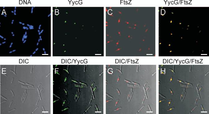
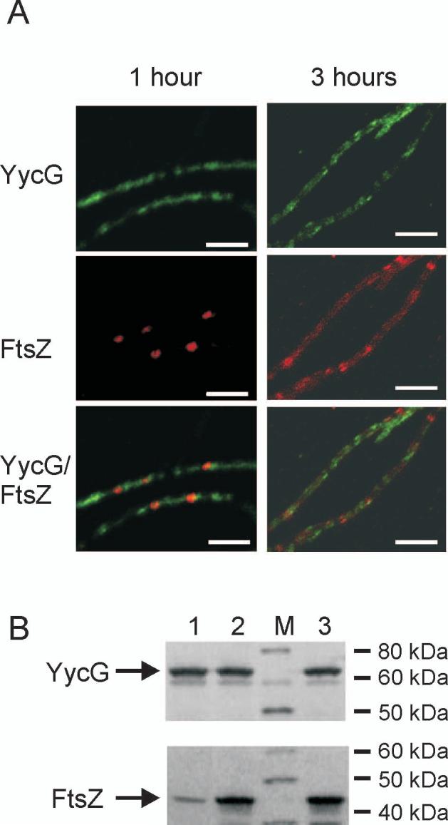
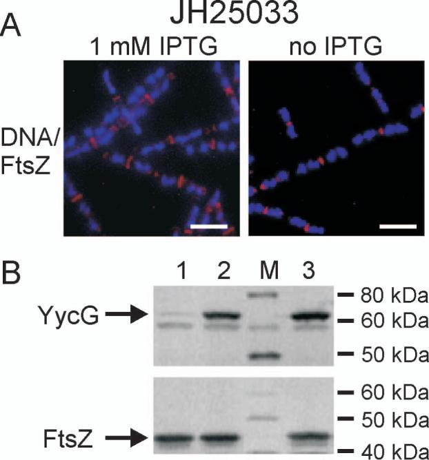
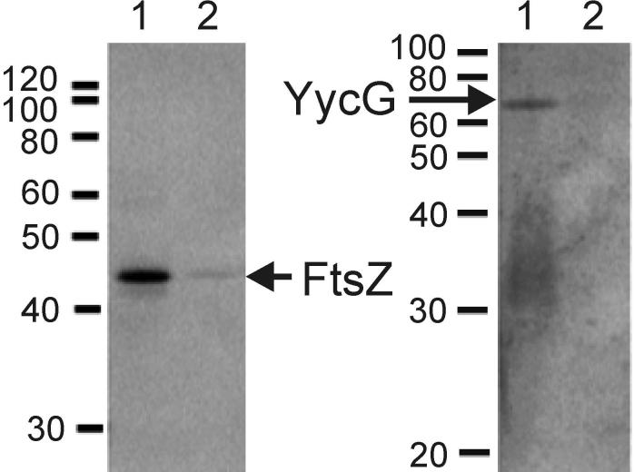
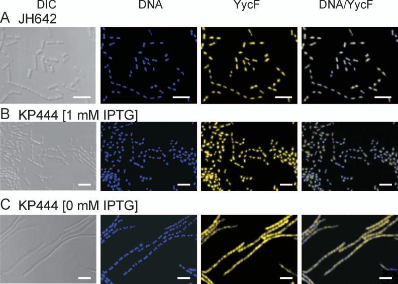
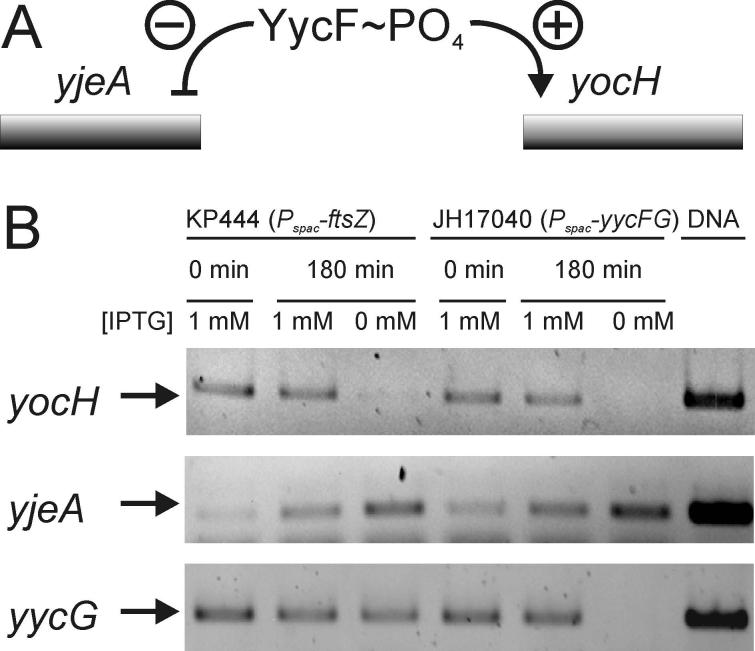
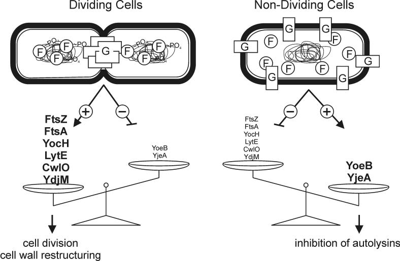
The concerted interconnection between processes driving DNA synthesis, division septum formation and cell wall synthesis and remodelling in rapidly growing bacteria requires precise co-ordination by signalling mechanisms that are, for the most part, unknown. The YycG (sensor histidine kinase)-YycF (response regulator/transcription factor) two-component system of Bacillus subtilis controls the synthesis of enzymes and their inhibitors that function in cell wall remodelling and cell separation. Here it is shown that the YycG sensor histidine kinase is a component of the division septum in growing cells. RT-PCR quantification of YycF approximately PO(4)-regulated gene transcription, in wild type and FtsZ-depleted, septum-less cells, indicated that YycG kinase activity on YycF is dependent on YycG localization to a division septum. The data support a model in which the YycG sensor kinase perceives information at the division septum and regulates the reciprocal synthesis of autolysins and autolysin inhibitors to co-ordinate growth and division with cell wall restructuring.


Figures

Figure 1
Figure 1
YycG and FtsZ co-localize to the septum in the wildtype B. subtilis strain JH642. YycG (green) and FtsZ (red) proteins were (A-D) visualized immunologically by confocal microscopy and overlain with (E-H) differential interference contrast images, DIC, in exponentially growing cells of JH642 as outlined in Materials and Methods. DNA was visualized by DAPI staining (blue). Bars indicate 5 μm.
Figure 2
Figure 2
YycG does not localize to the septum in the FtsZ-depleted strain. (A) YycG (green) and FtsZ (red) were visualized immunologically in KP444 cells following 1 and 3 hours growth at 37°C in the absence of IPTG to repress FtsZ expression. Bars indicate 5 μm. (B) Cellular protein levels of YycG and FtsZ were visualized by western blotting using whole cell protein extract derived from strain KP444 grown for one hour either in the absence (lane 1) or presence of 1 mM IPTG (lane 2) or from wild type strain JH642 (lane 3) as reference; Lane M, molecular weight standard.
Figure 3
Figure 3
FtsZ localizes to the septum independently of YycG. (A) FtsZ (red) localization was visualized immunologically in strain JH25033 grown three hours either in the presence of 1 mM IPTG (left panel) or in the absence of IPTG (right panel) to induce YycG depletion. DNA (blue) was visualized by DAPI staining. (B) Cellular protein levels of YycG and FtsZ were visualized by western blotting using whole cell protein extract derived from strain JH25033 grown three hours either in the absence (lane 1) or presence of 1 mM IPTG (lane 2) or from wild type strain JH642 (lane3) as reference; Lane M, molecular weight standard.
Figure 4
Figure 4
YycG co-precipitates with FtsZ in an immunoprecipitation assay. Exponentially growing KP444 cells in the presence of IPTG (OD525=0.3) were collected and suspended in media either supplemented with 1 mM IPTG or not. After 1 h, cultures were subjected to cross-linking and immunoprecipitation from cell extracts with purified anti-FtsZ antibodies as described in Material and Methods. FtsZ (left panel) and YycG (right panel) proteins were visualized immunologically by Western blot analysis using anti-YycG or anti-FtsZ antibody, respectively. Lanes 1 an 2 of each gel contain cross-linked sample immunoprecipitated with anti-FtsZ antibody from KP444 grown in the presence of 1 mM IPTG (lane 1) or in its absence (lane 2).
Figure 5
Figure 5
YycF localizes to the nucleoid in an FtsZ dependent manner. (A) YycF (yellow) was visualized immunologically in exponentially growing cells of wild type strain JH642. DNA (blue) was visualized by DAPI staining. The two figures were overlaid in DNA/YycF. Similarly DNA and YycF were visualized in the conditional FtsZ mutant strain KP444 grown for three hours at 37°C either in (B) the presence of 1 mM IPTG or (C) in the absence of IPTG to induce FtsZ depletion. DIC is differential interference microscopy.
Figure 6
Figure 6
FtsZ depletion mimics YycF∼PO4 depletion in the regulation of the YycFG regulon. (A) A schematic showing regulation of two genes that are under negative (yjeA) or positive (yocH) transcription control by YycF∼PO4 (B) mRNA levels of yocH, yjeA and yycG were determined in an RT-PCR approach. RNA was extracted from strain KP444 grown in the presence of 1 mM IPTG or in its absence to induce FtsZ-depletion, as described in Materials and Methods. Similarly, RNA was extracted from strain JH17040 grown with or without IPTG to observe changes in mRNA levels of the respective genes in response to yycFG depletion. RNA was transcribed into cDNA and amplified by PCR. As a control the respective genes were also amplified directly from genomic DNA (right lane).
Figure 7
Figure 7
Model for a role of YycFG in coordinating cell division with cell wall homeostasis. The present data is consistent with a model in which YycG is activated upon localization to the septum in dividing cells. This activation increases the YycF∼PO4 levels and subsequently leads to enhanced expression of genes involved in cell wall remodeling (yocH, lytE, cwlO and ydjM) and cell division (ftsA and ftsZ) and repression of genes involved in inhibiting cell wall remodeling (yoeB and yjeA). In the absence of septa in non-dividing cells YycG fails to localize, adopts a state of low activity and hence the cellular YycF pool remains unphosphorylated. Under these conditions cell wall remodeling is not required and the expression balance is tipped towards the genes involved in autolysin inhibitory processes. The precise role of the involved genes of the YycF regulon, where known, has been addressed in (Bisicchia et al., 2007).

References

    1. Baker MD, Wolanin PM, Stock JB. Signal transduction in bacterial chemotaxis. Bioessays. 2006;28:9–22. - PubMed
    1. Beall B, Lutkenhaus J. FtsZ in Bacillus subtilis is required for vegetative septation and for asymmetric septation during sporulation. Genes Dev. 1991;5:447–455. - PubMed
    1. Bi EF, Lutkenhaus J. FtsZ ring structure associated with division in Escherichia coli. Nature. 1991;354:161–164. - PubMed
    1. Biondi EG, Reisinger SJ, Skerker JM, Arif M, Perchuk BS, Ryan KR, Laub MT. Regulation of the bacterial cell cycle by an integrated genetic circuit. Nature. 2006;444:899–904. - PubMed
    1. Bisicchia P, Noone D, Lioliou E, Howell A, Quigley S, Jensen T, Jarmer H, Devine KM. The essential YycFG two-component system controls cell wall metabolism in Bacillus subtilis. Mol Microbiol. 2007;65:180–200. - PubMed

Publication types

LinkOut - more resources