. 2008 Jun 23:3:15.
doi: 10.1186/1749-8104-3-15.

Secreted APP regulates the function of full-length APP in neurite outgrowth through interaction with integrin beta1

Affiliations

Secreted APP regulates the function of full-length APP in neurite outgrowth through interaction with integrin beta1

Tracy L Young-Pearse et al. Neural Dev. .

Abstract

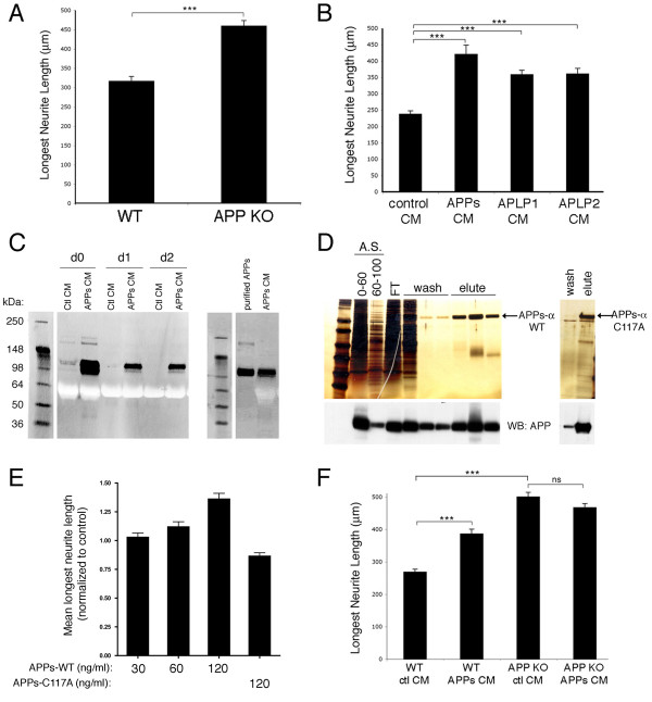
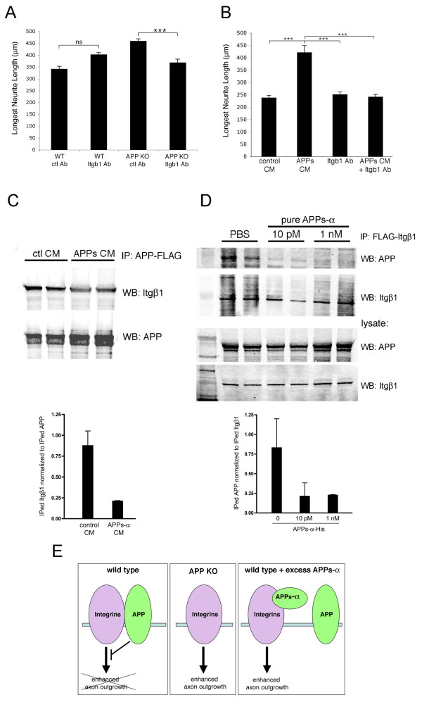
Background: Beta-amyloid precursor protein (APP) has been reported to play a role in the outgrowth of neurites from cultured neurons. Both cell-surface APP and its soluble, ectodomain cleavage product (APPs-alpha) have been implicated in regulating the length and branching of neurites in a variety of assays, but the mechanism by which APP performs this function is not understood.

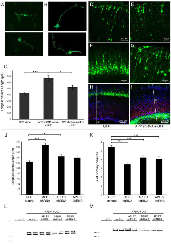
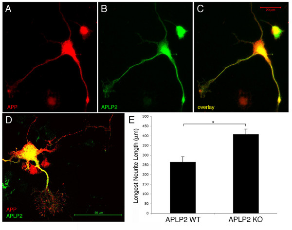
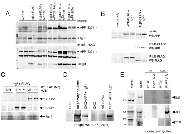
Results: Here, we report that APP is required for proper neurite outgrowth in a cell autonomous manner, both in vitro and in vivo. Neurons that lack APP undergo elongation of their longest neurite. Deletion of APLP1 or APLP2, homologues of APP, likewise stimulates neurite lengthening. Intriguingly, wild-type neurons exposed to APPs-alpha, the principal cleavage product of APP, also undergo neurite elongation. However, APPs-alpha is unable to stimulate neurite elongation in the absence of cellular APP expression. The outgrowth-enhancing effects of both APPs-alpha and the deletion of APP are inhibited by blocking antibodies to Integrin beta1 (Itgbeta1). Moreover, full length APP interacts biochemically with Itgbeta1, and APPs-alpha can interfere with this binding.

Conclusion: Our findings indicate that APPs-alpha regulates the function of APP in neurite outgrowth via the novel mechanism of competing with the binding of APP to Itgbeta1.


Figures

Figure 1
Figure 1
Loss of APP and APPs-α application each increase neurite length. E18 primary hippocampal or cortical neurons from wild-type (WT) or APP knock-out (APP KO) mice were treated as described. Three days later, the cells were fixed and immunostained for βIII-tubulin, and neurite length was measured. (a) Comparison of neurite lengths of wild type and APP knock-out hippocampal neurons. (b) Comparison of neurite lengths of wild-type hippocampal neurons treated with CM from untransfected, APPs, APLP1, or APLP2 expressing CHO cells. (c) Western blots for APPs (8E5) on CM from day 0 (d0), d1, or d2 after addition to primary neurons (left panel) or western blot of equal amounts of purified APPs-α compared to APPs-α conditioned media (right panel). (d) Silver stain of sequential purification steps of APPs-α from baculovirus-transduced insect cells. Left panel: lanes 1 and 2 are ammonium sulfate (A.S.) precipitations from 0–60% or 60–100%; lane 3 is the flow through (FT) after addition to a nickel chelating column; lanes 4–6 are washes from the column; and lanes 7–9 are elutes from column. Right panel: final purified product of APPs-α with mutation of cysteine 117 (see Materials and methods for details of purification). Bottom: western blot (WB) with an antibody that recognizes the extracellular domain of APP (8E5, Elan) (e) Quantification of neurite length after addition of purified APPs-α (wild type) at increasing concentrations or APPs-α (C117A) at 120 ng/ml. (f) E18 primary cortical neurons from APP knock-out or wild-type littermates were treated with CM from either CHO cells stably expressing APPs-α or control CHO cells. Three days later, cells were fixed and immunostained for βIII-tubulin, and neurite length was measured. Error bars represent standard error of the mean; *p < 0.05; ***p < 0.001.
Figure 2
Figure 2
APP knock-down increases neurite length in a cell-autonomous manner. (a, b) E17 primary cortical neurons were plated and transfected with plasmids encoding GFP alone (a) or GFP with APP shRNA-active (b) or with APP shRNA-inactive (not shown). Three days later, neurons were fixed and immunostained for βIII-tubulin. (c) Neurite length was quantified in GFP+, transfected cells. Error bars represent standard error of the mean; *p < 0.05; ***p < 0.001. (d-i) E17 (d, e) or E14 (f-i) cortices were electroporated with GFP (d, f, h) or GFP + APP shRNA-active (e, g, i) and harvested at postnatal day 5. Images in (d-g) are of the upper half of the cortical plate in coronal sections. (h, i) Coronal sections immunostained for Tbr1 (red), marking layer VI of the cortical plate (CP), and MAP2 (blue), marking the entire cortical plate. GFP positive, electroporated regions of E14 cortices were dissected 48 h after electroporation, dissociated, and plated. Three days later cells were fixed and immunostained for βIII-tubulin. IZ; intermediate zone; white lines delineates noted regions of the cortex (j, k) Neurite lengths (j) or the number of primary neurites (k) were quantified for GFP+ cells. Error bars represent standard error of the mean; *p < 0.05; ***p < 0.001. (l, m) FLAG-tagged murine APLP1 (l) or APLP2 (m) constructs were co-transfected into CHO cells with an empty vector or with three different shRNA constructs targeting rodent APLPs. Transfections (tf) of duplicate wells are shown. Forty-eight hours post-transfection, cells were lysed, and the western blots for FLAG on protein normalized samples are shown.
Figure 3
Figure 3
APLP2 is expressed in primary neuronal processes and loss of APLP2 increases neurite length. Primary E18 wild-type neurons were plated and fixed six days later. Neurons were immunostained for (a, c, d) APP and (b, c, d) APLP2. (d, e) E18 primary hippocampal neurons from wild-type (WT) or APLP2 knock out (KO) mice were plated. Three days later, cells were fixed and immunostained for βIII-tubulin and neurite length was measured and quantified. Error bars represent standard error of the mean; *p < 0.05.
Figure 4
Figure 4
APP and Itgβ1 biochemically interact. (a) CHO cells were transiently transfected with constructs as shown. Co-immunoprecipitations of the resultant lysates were then performed with anti-FLAG agarose. Western blots for the amino terminus of APP (anti-APP; 22C11; Chemicon) or Itgβ1 (Cell Signaling): upper panels are western blots of lysates and lower panels show western blots of FLAG-immunoprecipitations (IP). The band denoted by the single asterisk is believed to be a background band. The band denoted by the double asterisks is the heavy chain of IgG. (b) CHO cells were transiently transfected with APP and Itgβ1-FLAG or another carboxy-terminally tagged type I-transmembrane domain protein, angiotensin converting enzyme (ACE). Co-immunoprecipitations of the resultant lysates were then performed with anti-FLAG agarose and western blotted (WB) for APP or FLAG, as shown. (c) CHO cells were transiently transfected with Itgβ1-FLAG and APP, APLP1, or APLP2. Co-immunoprecipitations of the resultant lysates were then performed with anti-FLAG agarose and western blotted for APLP1, APLP2, or Itgβ1, as shown. (d) Lysates from CHO cells, E18 rat primary neurons, rat cortex, or transfected CHO cells were immunoprecipitated for Itgβ1 and western blotted for APP. (e) Lysates from total mouse brain were immunoprecipitated with antibodies directed to APP or Tbr1 (used as a control) for 30 minutes or overnight (O/N) and western blotted for Itgβ1, APP, or transferrin receptor as a negative control.
Figure 5
Figure 5
Itgβ1 blocking antibody inhibits the effects on neurite outgrowth of APPs-α or deletion of APP. (a) E18 primary cortical neurons from wild-type (WT) or APP knock-out (KO) mice were treated with Itgβ1 blocking antibody or control (ctl) antibody. (b) Wild-type E18 neurons were treated with control or APPs-rich CHO CM together with either Itgβ1 blocking or control antibody. The longest βIII-tubulin-positive neurites were quantified. Error bars represent standard error of the mean; ***p < 0.001; ns, not significant. (c, d) CHO cells were transfected with APP-FLAG and Itgβ1 (c) or Itgβ1-FLAG and APP (d). Example of western blot (WB) of co-immunoprecipitates of APP and Itgβ1 after incubation with control (ctl) CM or APPs-α CM (c) or purified His-tagged APPs-α (d) with quantification below; error bars represent standard deviation between duplicate wells. A significant decrease in co-immunoprecipitation between APP and Itgβ1 was observed between multiple experiments; p < 0.05. (e) Model for proposed functional interaction between APP and integrins in regulating neurite elongation.

Similar articles

Cited by

References

    1. Benowitz LI, Rodriguez W, Paskevich P, Muson EJ, Schenk D, Neve RL. The amyloid precursor protein is concentrated in neuronal lysosomes in normal and Alzheimer disease subjects. Exp Neurol. 1989;106:237–250. doi: 10.1016/0014-4886(89)90156-8. - DOI - PubMed
    1. McGeer PL, Akiyama H, Kawamata T, Yamada T, Walker DG, Ishii T. Immunohistochemical localization of beta-amyloid precursor protein sequences in Alzheimer and normal brain tissue by light and electron microscopy. J Neurosci Res. 1992;31:428–442. doi: 10.1002/jnr.490310305. - DOI - PubMed
    1. Jung SS, Nalbantoglu J, Cashman NR. Alzheimer's beta-amyloid precursor protein is expressed on the surface of immediately ex vivo brain cells: a flow cytometric study. J Neurosci Res. 1996;46:336–348. doi: 10.1002/(SICI)1097-4547(19961101)46:3<336::AID-JNR7>3.0.CO;2-L. - DOI - PubMed
    1. Selkoe DJ, Wolfe MS. Presenilin: running with scissors in the membrane. Cell. 2007;131:215–221. doi: 10.1016/j.cell.2007.10.012. - DOI - PubMed
    1. Esch FS, Keim PS, Beattie EC, Blacher RW, Culwell AR, Oltersdorf T, McClure D, Ward PJ. Cleavage of amyloid β-peptide during constitutive processing of its precursor. Science. 1990;248:1122–1124. doi: 10.1126/science.2111583. - DOI - PubMed

Publication types

MeSH terms