

Mannitol enhances delivery of marrow stromal cells to the brain after experimental intracerebral hemorrhage
- PMID: 18573239
- PMCID: PMC2712137
- DOI: 10.1016/j.brainres.2008.05.080
Mannitol enhances delivery of marrow stromal cells to the brain after experimental intracerebral hemorrhage
Abstract
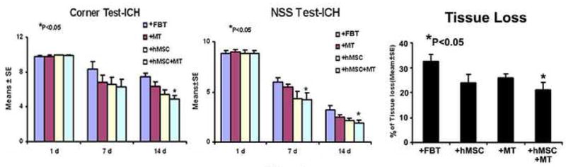
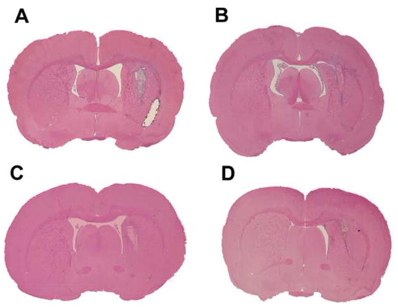
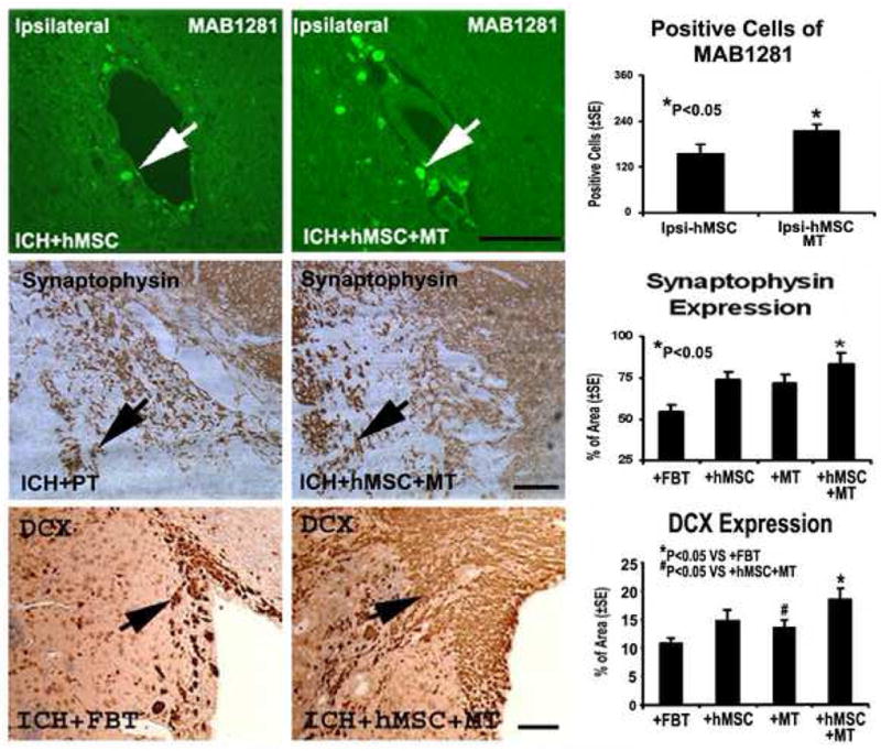
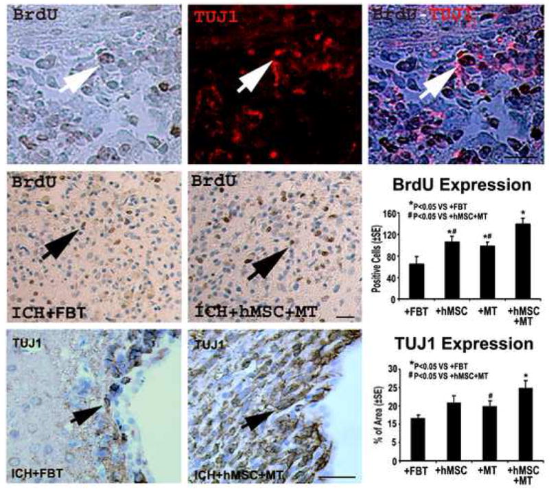
Previous studies show that intravascular injection of human bone marrow stromal cells (hBMSCs) significantly improves neurological functional recovery in a rat model of intracerebral hemorrhage (ICH). In the present study, we tested the hypothesis that mannitol improves the efficiency of intraarterial MSC delivery (i.e., fewer injected cells required for therapeutic efficacy) after ICH. There were four post-ICH groups (N=9): group 1, negative control with only intraarterial injection of 1 million human fibroblasts in phosphate-buffered saline (PBS); group 2, intravenous injection of mannitol alone in PBS (1.5 g/kg); group 3, intraarterial injection of 1 million hBMSCs alone in PBS; and group 4, intravenous injection of mannitol (1.5 g/kg) in PBS followed by intraarterial injection of 1 million hBMSCs in PBS. Group 4 exhibited significantly improved neurological functional outcome as assessed by neurological severity score (NSS) and corner test scores. Immunohistochemical staining of group 4 suggested increased synaptogenesis, proliferating immature neurons, and neuronal migration. The number of hBMSCs recruited to the injured region increased strikingly in group 4. Tissue loss was notably reduced in group 4. In summary, the beneficial effects of intraarterial infusion of MSCs are amplified with intravenous injection of mannitol. Preadministration of mannitol significantly increases the number of hBMSCs located in the ICH region, improves histochemical parameters of neural regeneration, and reduces the anatomical and pathological consequences of ICH.
Figures
Similar articles
-
Localization of bone marrow stromal cells to the injury site after intracerebral hemorrhage in rats.J Neurosurg. 2010 Feb;112(2):329-35. doi: 10.3171/2009.2.JNS08907. J Neurosurg. 2010. PMID: 19284233 Free PMC article.
-
Effects of intravenous administration of human bone marrow stromal cells after intracerebral hemorrhage in rats.J Neurosurg. 2006 Feb;104(2):313-8. doi: 10.3171/jns.2006.104.2.313. J Neurosurg. 2006. PMID: 16509507
-
Transplantation of Flk-1+ human bone marrow-derived mesenchymal stem cells promotes behavioral recovery and anti-inflammatory and angiogenesis effects in an intracerebral hemorrhage rat model.Int J Mol Med. 2013 May;31(5):1087-96. doi: 10.3892/ijmm.2013.1290. Epub 2013 Mar 5. Int J Mol Med. 2013. PMID: 23468083
-
Transplantation of bone marrow stromal cells enhances nerve regeneration of the corticospinal tract and improves recovery of neurological functions in a collagenase-induced rat model of intracerebral hemorrhage.Mol Cells. 2013 Jul;36(1):17-24. doi: 10.1007/s10059-013-2306-9. Epub 2013 Jun 25. Mol Cells. 2013. PMID: 23807046 Free PMC article.
-
New trends in hyperosmolar therapy?Curr Opin Crit Care. 2013 Apr;19(2):77-82. doi: 10.1097/MCC.0b013e32835eba30. Curr Opin Crit Care. 2013. PMID: 23385373 Free PMC article. Review.
Cited by
-
Recovery of fine motor performance after ischemic damage to motor cortex is facilitated by cell therapy in the rhesus monkey.Somatosens Mot Res. 2013 Dec;30(4):185-96. doi: 10.3109/08990220.2013.790806. Epub 2013 Jun 12. Somatosens Mot Res. 2013. PMID: 23758412 Free PMC article.
-
Present Status and Future Challenges of New Therapeutic Targets in Preclinical Models of Stroke in Aged Animals with/without Comorbidities.Int J Mol Sci. 2018 Jan 25;19(2):356. doi: 10.3390/ijms19020356. Int J Mol Sci. 2018. PMID: 29370078 Free PMC article. Review.
-
Application of stem cells and exosomes in the treatment of intracerebral hemorrhage: an update.Stem Cell Res Ther. 2022 Jun 28;13(1):281. doi: 10.1186/s13287-022-02965-2. Stem Cell Res Ther. 2022. PMID: 35765072 Free PMC article. Review.
-
Increased Endothelial Progenitor Cell Levels are Associated with Good Outcome in Intracerebral Hemorrhage.Sci Rep. 2016 Jun 27;6:28724. doi: 10.1038/srep28724. Sci Rep. 2016. PMID: 27346699 Free PMC article. Clinical Trial.
-
Matrix metalloproteinase-9 and tissue inhibitor of metalloproteinase-1 expression in early focal cerebral infarction following urokinase thrombolysis in rats.Neural Regen Res. 2012 Jun 15;7(17):1325-30. doi: 10.3969/j.issn.1673-5374.2012.17.007. Neural Regen Res. 2012. PMID: 25657663 Free PMC article.
References
-
- Boulard G, Marguinaud E, Sesay M. Osmotic cerebral oedema: the role of plasma osmolarity and blood brain barrier. Ann Fr Anesth Reanim. 2003;22:215–219. - PubMed
-
- Burke AM, Quest DO, Chien S, Cerri C. The effects of mannitol on blood viscosity. J Neurosurg. 1981;55:550–553. - PubMed
-
- Chen J, Li Y, Wang L, Lu M, Zhang X, Chopp M. Therapeutic benefit of intracerebral transplantation of bone marrow stromal cells after cerebral ischemia in rats. J Neurol Sci. 2001a;189:49–57. - PubMed
-
- Chen J, Sanberg PR, Li Y, Wang L, Lu M, Willing AE, Sanchez-Ramos J, Chopp M. Intravenous administration of human umbilical cord blood reduces behavioral deficits after stroke in rats. Stroke. 2001b;32:2682–2688. - PubMed
-
- Chen J, Zhang ZG, Li Y, Wang Y, Wang L, Jiang H, Zhang C, Lu M, Katakowski M, Feldkamp CS, Chopp M. Statins induce angiogenesis, neurogenesis, and synaptogenesis after stroke. Ann Neurol. 2003;53:743–751. - PubMed
Publication types
MeSH terms
Substances
Grants and funding
LinkOut - more resources
Full Text Sources
Other Literature Sources
Medical
