. 2008 Sep;28(17):5529-42.
doi: 10.1128/MCB.02056-07. Epub 2008 Jun 23.

Molecular identification and function of cis- and trans-acting determinants for petA transcript stability in Chlamydomonas reinhardtii chloroplasts

Affiliations

Molecular identification and function of cis- and trans-acting determinants for petA transcript stability in Chlamydomonas reinhardtii chloroplasts

Christelle Loiselay et al. Mol Cell Biol. 2008 Sep.

Abstract

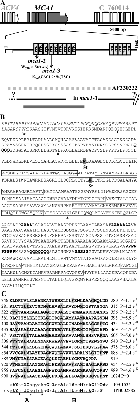
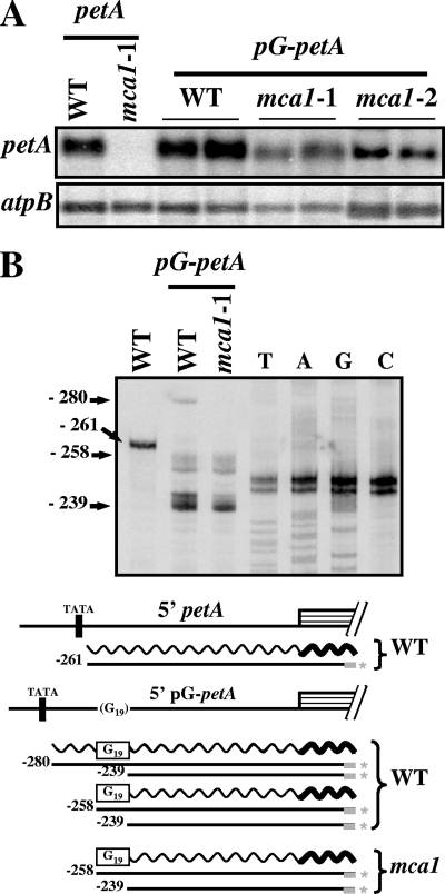
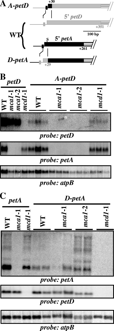
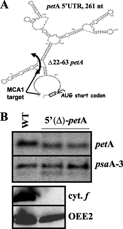
In organelles, the posttranscriptional steps of gene expression are tightly controlled by nucleus-encoded factors, most often acting in a gene-specific manner. Despite the molecular identification of a growing number of factors, their mode of action remains largely unknown. In the green alga Chlamydomonas reinhardtii, expression of the chloroplast petA gene, which codes for cytochrome f, depends on two specific nucleus-encoded factors. MCA1 controls the accumulation of the transcript, while TCA1 is required for its translation. We report here the cloning of MCA1, the first pentatricopeptide repeat protein functionally identified in this organism. By chloroplast transformation with modified petA genes, we investigated the function of MCA1 in vivo. We demonstrate that MCA1 acts on the very first 21 nucleotides of the petA 5' untranslated region to protect the whole transcript from 5'-->3' degradation but does not process the 5' end of the petA mRNA. MCA1 and TCA1 recognize adjacent targets and probably interact together for efficient expression of petA mRNA. MCA1, although not strictly required for translation, shows features of a translational enhancer, presumably by assisting the binding of TCA1 to its own target. Conversely, TCA1 participates to the full stabilization of the transcript through its interaction with MCA1.


Figures

FIG. 1.
FIG. 1.
mca1 mutant strains lack stable accumulation of petA mRNA. (A) Schematic map of the petA-petD transcription unit. At the top is shown the relevant chloroplast genomic region, with the transcripts shown below. Bent arrows indicate transcription start sites. The dashed wavy line depicts the transient petA-petD cotranscript. This cotranscript is not observed in the wild type (WT, panel B) because of its rapid processing (58, 70). Symbols (diamond, star, and circle) refer to the transcripts visible in panel B. (B and C) Accumulation of petA mRNA (B) and cytochrome f (C) in wild-type and mca1-2 strains and in an mca1-2 cw15 strain complemented by transformation with a MCA1 cDNA clone. Accumulations of atpB mRNA and of the OEE2 subunit from Photosystem II provide the respective loading controls. Sizes of the best-characterized petA transcripts are indicated on the right side of panel B.
FIG. 2.
FIG. 2.
MCA1 gene. (A) Structure of the MCA1 gene. The top line shows a schematic map of the MCA1 genomic region. MCA1 exons and 3′UTR are shown as gray boxes and a gray arrow. Adjacent gene models (ACV4 and C_760014) are also drawn as white boxes. A diagram of the protein is shown below with the position of the PPR motifs and the predicted transit peptide depicted as a white rectangle. The positions of mutations in mca1-2 and mca1-3 strains with respect to the protein sequence are indicated. From top to bottom are shown: the localization of the genomic fragment whose sequence is in the database (GenBank accession no. AF330232, dashed gray line), the approximate extend of the deletion in the mca1-1 strain (black line; note that the right border of the deletion has not been determined precisely but lies outside of the figure), and the location of the 7.7-kb DNA fragment able to restore the phototrophic growth of the mca1-2 strain upon transformation (thick gray line). (B) Sequence of the MCA1 protein. The predicted transit peptide is written in italics. PPR motifs are alternatively underlined and boxed. Stretches of four or more consecutive identical residues are in boldface. Arrowheads indicate the position of introns with respect to the coding sequence. Amino acids substituted by an amber codon in the mca1-2 and mca1-3 mutant strains are highlighted. (C) Alignment of the PPR motifs identified by program TPRpred, compared to the consensus sequences PFAM:PF01535 and InterPro:IPB002885. P values, measuring the statistical significance of the occurrence, are indicated for all repeats except repeat XI that was not detected by TPRpred (http://toolkit.tuebingen.mpg.de/tprpred) but was detected by program SMART (http://smart.embl-heidelberg.de/). Residues forming the antiparallel helices A and B are underlined. Asterisks point to the residues exposed on the external surface of helix A that could be involved in specific contacts with nucleotides (see Discussion).
FIG. 3.
FIG. 3.
The petA 5′UTR is the target of the MCA1 factor. (A) Schematic map of the chimera used for these experiments. The chloroplast genomic region encompassing the petA gene is shown in the middle line. 5′psbA-petA and 5′petA-psbB chimera, whose expression is analyzed in panels B and C, are, respectively, shown above and below the petA gene. Bent arrows indicate transcription start sites, while “K” stands for the aadA cassette, inserted in the opposite orientation with respect to petA to allow the selection of transformed strains. (B) A chimeric petA mRNA, expressed from the unrelated psbA 5′UTR, accumulates in the absence of MCA1. The accumulation of petA mRNA (either regular or the smaller chimeric 5′psbA-petA mRNA) in wild-type and mca1-2 nuclear backgrounds is shown. Two independent transformants expressing the chimeric 5′psbA-petA gene in the mca1 nuclear context are shown. The accumulation of psbB transcripts in the same strains provides a loading control. (B) Chimeric transcripts, driven by the petA 5′UTR, fail to accumulate in an mca1 mutant strain. The accumulation of psbB mRNAs (either regular or the slightly larger chimeric 5′petA-psbB mRNA) in wild-type and mca1-2 nuclear backgrounds is shown. Three independent transformants expressing the chimeric 5′petA-psbB gene are shown for each nuclear background. Accumulation of the atpB transcript in the same strains provides a loading control.
FIG. 4.
FIG. 4.
MCA1 controls the abundance, but not the localization of the 5′ end of petA transcript. The left panel shows mapping of the petA 5′ends by extension of primer RevpetA in wild-type, mca1-1, and mca1-2 strains and in the control strain ΔpetA. Mapping of the psaB 5′ end by extension of the primer RevpsaB in the same reaction mixture provides a loading control. The RevpetA primer was used to generate a sequencing ladder for comparison. Arrow points to the 5′ end of the petA transcript on the sequence shown on the left, while the promoter consensus sequence is highlighted by a gray box. Asterisks stress two 5′ termini described by Matsuda and coworkers (41). We did not find these 5′ ends in our experiments and concluded that they probably originated from nonspecific extensions. The right panel shows the 5′ end of the petA transcript analyzed by RLM-RACE after (lane +) and without (lane −) taP treatment of mRNAs. First lane, 100-bp DNA ladder.
FIG. 5.
FIG. 5.
Insertion of a poly(G) tract at the beginning of the 5′UTR stabilizes the petA mRNA. (A) Accumulation of transcripts expressed from the pG-petA chimera in two independent transformed clones derived from wild-type, mca1-1, or mca1-2 recipient strains. The accumulation of the endogenous petA transcript in wild-type and mca1-1 strains is also shown. atpB mRNA provides a loading control. (B) 5′ ends of petA transcripts in the wild type and in a transformed strain expressing the pG-petA chimera, determined by primer extension. The labeled primer RevpetA was also used to generate a sequencing ladder from template plasmid pWF-pG. The length (numbered from the initiation codon) of extension products is indicated on the left. Below are schematically shown the structure of the 5′ region of the petA gene, as well as the transcripts (wavy lines) and extension products (straight lines) found in each strain. The position of the −10 box is indicated. The thick gray bar depicts the oligonucleotide primers, labeled at the 5′ end (*).
FIG. 6.
FIG. 6.
The stability of chimeric transcripts depends on their very 5′ end. (A) Schematic representation of the chimeric genes used for these experiments. The 5′ regions of the petA and petD genes are shown in the middle. Filled boxes represent the respective 5′UTR (in black for petA and gray for petD), while dashed boxes symbolize the beginning of the coding sequences. Rectangles immediately upstream of the 5′UTRs depict the −10 boxes of the two genes. In the 5′UTR of the petA gene, “S” indicates a SwaI restriction site, lost in the D-petA chimera, used for RFLP analysis of transformants. The top and bottom lines show, respectively, the structure of the A-petD and D-petA chimeras, with the origin of the sequence elements indicated by the color code. (B) The 30 first nucleotides of the petA 5′UTR confer an MCA1-dependent stability to the A-petD transcript. The A-petD chimera was introduced in the chloroplast of wild-type, mca1-1, mca1-2, and mcd1-1 strains. The accumulation of petA, petD (and atpB as a loading control) transcripts was monitored in three independent transformed strains for each genetic background, as well as in recipient strains. The asterisk points to unspecific stain overlapping two lanes. (C) D-petA transcripts are stabilized through interactions with MCD1. The D-petA chimera was introduced in the chloroplasts of wild-type, mca1-1, mca1-2, and mcd1-1 strains. The accumulation of petA and petD (and atpB as a loading control) mRNAs was monitored in two independent transformants, as well as in recipient strains.
FIG. 7.
FIG. 7.
The D-petA chimera is still translated in the absence of TCA1. (A and B) Accumulation of cytochrome f (A) and petA transcript (B) expressed from the D-petA chimera introduced by transformation in the chloroplast genomes of tca1-8 (two independent transformants), mca1-1, and wild-type recipient strains. The accumulation of cytochrome f and of the regular petA mRNA in wild-type and tca1-8 strains is also shown. OEE2 and psbB mRNA provide the respective loading controls.
FIG. 8.
FIG. 8.
Cytochrome f can be translated from the pG-petA transcript in the absence of MCA1 but not in the absence of TCA1. (A) Accumulation of cytochrome f (and OEE2 as a loading control) detected by a specific antibody in independent transformants expressing the pG-petA chimera derived from wild-type, mca1-1, or mca1-2 recipient strains. The accumulation of cytochrome f in the recipient strains is also shown for comparison. (B) Expression of the pG-petA chimera, introduced by transformation in the chloroplast genome of the tca1-2 recipient strain. The accumulations of petA mRNA and cytochrome f are shown in the wild-type and tca1-2 recipient strains and in two independent transformants. psbB mRNA and the OEE2 protein provide the respective loading controls.
FIG. 9.
FIG. 9.
Nucleotides 22 to 63 from the petA 5′UTR are required for the translation but not for the accumulation of the mRNA. (A) In silico prediction of the possible secondary structure of the petA 5′UTR, using the program RNAfold. Note that the proposed structure lacks experimental support. (B) Transcript accumulation from the regular petA gene in the wild type and from 5′(Δ)petA in transformed strains. psaA provides a loading control. Below is shown the accumulation of cytochrome f (and OEE2 as a loading control) in the same strains.
FIG. 10.
FIG. 10.
Working model for the expression of the petA genes used in the present study. At the top is shown the expression of the endogenous petA gene in the wild type. The MCA1, TCA1, and MCD1 factors are shown with their respective targets, respectively, drawn in black, white, and gray. The interaction between the initiation codon and TCA1 is suggested. The expression of the pG-petA, on the left, and D-petA, on the right, transcripts are schematically depicted below in (from top to bottom) wild-type, mca1, and tca1 strains. For each situation, we indicated in parentheses the efficiency of the translation: +++, robust, +, reduced but significant (∼10% of the wild-type level); +/−, low (<5%); −, no synthesis of cytochrome f.

Similar articles

Cited by

References

    1. Andres, C., C. Lurin, and I. D. Small. 2007. The multifarious roles of PPR proteins in plant mitochondrial gene expression. Physiol. Plant 12914-22.
    1. Anthonisen, I. L., M. L. Salvador, and U. Klein. 2001. Specific sequence elements in the untranslated regions of rbcL and atpB gene mRNAs stabilize transcripts in the chloroplast of Chlamydomonas reinhardtii. RNA 71024-1033. - PMC - PubMed
    1. Barkan, A., and M. Goldschmidt-Clermont. 2000. Participation of nuclear gene in chloroplast gene expression. Biochimie 82559-572. - PubMed
    1. Barkan, A., M. Walker, M. Nolasco, and D. Johnson. 1994. A nuclear mutation in maize blocks the processing and translation of several chloroplast mRNAs and provides evidence for the differential translation of alternative mRNA forms. EMBO J. 133170-3181. - PMC - PubMed
    1. Bollenbach, T. J., G. Schuster, and D. B. Stern. 2004. Cooperation of endo- and exoribonucleases in chloroplast mRNA turnover. Prog. Nucleic Acids Res. Mol. Biol. 78305-337. - PubMed

Publication types

MeSH terms

LinkOut - more resources