. 2008 Sep;28(17):5147-61.
doi: 10.1128/MCB.00331-08. Epub 2008 Jun 23.

Regulation of ABCG2 expression at the 3' untranslated region of its mRNA through modulation of transcript stability and protein translation by a putative microRNA in the S1 colon cancer cell line

Affiliations

Regulation of ABCG2 expression at the 3' untranslated region of its mRNA through modulation of transcript stability and protein translation by a putative microRNA in the S1 colon cancer cell line

Kenneth K W To et al. Mol Cell Biol. 2008 Sep.

Abstract

ABCG2 is recognized as an important efflux transporter in clinical pharmacology and is potentially important in resistance to chemotherapeutic drugs. To identify epigenetic mechanisms regulating ABCG2 mRNA expression at its 3' untranslated region (3'UTR), we performed 3' rapid amplification of cDNA ends with the S1 parental colon cancer cell line and its drug-resistant ABCG2-overexpressing counterpart. We found that the 3'UTR is >1,500 bp longer in parental cells and, using the miRBase TARGETs database, identified a putative microRNA (miRNA) binding site, distinct from the recently reported hsa-miR520h site, in the portion of the 3'UTR missing from ABCG2 mRNA in the resistant cells. We hypothesized that the binding of a putative miRNA at the 3'UTR of ABCG2 suppresses the expression of ABCG2. In resistant S1MI80 cells, the miRNA cannot bind to ABCG2 mRNA because of the shorter 3'UTR, and thus, mRNA degradation and/or repression on protein translation is relieved, contributing to overexpression of ABCG2. This hypothesis was rigorously tested by reporter gene assays, mutational analysis at the miRNA binding sites, and forced expression of miRNA inhibitors or mimics. The removal of this epigenetic regulation by miRNA could be involved in the overexpression of ABCG2 in drug-resistant cancer cells.


Figures
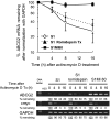
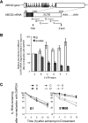
FIG. 1.
FIG. 1.
(Top) ABCG2 mRNA is more stable in resistant S1MI80 cells than in parental S1 cells. Although ABCG2 is also upregulated by romidepsin treatment (2 ng/ml for 24 h), its mRNA stability is not affected. Nascent RNA synthesis was inhibited with actinomycin D (5 μg/ml), and RNAs were harvested at 0, 4, 8, and 16 h posttreatment. RT-PCR analysis of ABCG2 mRNA was carried out to trace the remaining amount of ABCG2 mRNA with time. c-myc and GAPDH mRNA levels were also monitored as controls for fast-degrading and stable mRNAs, respectively. The value recorded was the percentage of mRNA remaining compared with the amount before the addition of actinomycin D, after normalization with GAPDH. The data shown represent the means ± SD for three independent experiments. (Bottom) Representative gel image showing RT-PCR analysis of ABCG2, c-myc, and GAPDH in cells pretreated with actinomycin D.
FIG. 2.
FIG. 2.
RT-PCR analysis investigating the relative abundances of different fragments of ABCG2 3′UTR in parental S1 and resistant S1MI80 cells. (A) Schematic showing the approximate positions of the six different overlapping fragments within the ABCG2 3′UTR analyzed by RT-PCR. Each fragment is about 500 bp long. (B) Relative abundances of different 3′UTR fragments were plotted relative to the amount in region A after normalization with GAPDH. Open bars, parental S1 cells; closed bars, resistant S1MI80 cells. The data shown represent the means ± SD for three independent experiments. Since the binding site for hsa-miR-519c is located at nt 3820 to 3841 within the ABCG2 3′UTR, only the long ABCG2 transcripts that can be regulated by hsa-miR-519c gave a PCR product for fragment F (nt 3775 to 4388). However, both the long and short ABCG2 transcripts produced PCR products for fragment A (nt 2462 to 3030). By comparing the PCR products for fragments A and F, we estimated that about 77% of ABCG2 transcripts in the parental S1 cells can be regulated by hsa-miR-519c. (C) Relative mRNA stabilities of the six different 3′UTR fragments. Total RNA was isolated at various time intervals after actinomycin D (5 μg/ml) treatment and analyzed for mRNA levels of the six 3′UTR regions by RT-PCR as described for panel A. The data from a representative experiment were plotted as the percentage of mRNA remaining for each 3′UTR fragment compared with the amount before the addition of actinomycin D, after normalization with GAPDH.
FIG. 3.
FIG. 3.
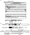
(A) Putative functional elements in the reported GenBank sequence (NM_004827) for the 3′UTR of ABCG2 mRNA. Locations of the stop codon (TAA), the canonical (AAUAAA) and noncanonical (AUUAAA) poly(A) signals, three putative AREs (UAAUUUAUUA, AUUAUUUAAUA, and GAGAAUUUAAUU), and the putative hsa-miR-519c and miR-520h binding sites within the 3′UTR of ABCG2 are marked. (B) Diagram illustrating the four clusters of putative miRNA target sites (shaded regions), as predicted by miRBase TARGETS, version 4 (19), within the 3′UTR of ABCG2. Detailed information about these putative miRNAs and their binding site sequences on the ABCG2 3′UTR are summarized in Table 2. The most upstream cluster can putatively be bound by 13 different human miRNAs (hsa-miR-518c, hsa-miR-519d, hsa-miR-302a, hsa-miR-302d, and hsa-miR-520a-h), the second site can be bound by hsa-miR-142-5p, the third site can be bound by hsa-miR-100, and the most downstream site can be bound by hsa-miR-519c. RNA hybrid free energy (ΔG) calculations and the theoretical miRNA-mRNA duplex pairing are also shown for hsa-miR-519c. The “seed” region within the hsa-miR-519c sequence, which determines the specificity of a putative miRNA, is also marked, as are locations of G-U wobbles within the putative binding site for hsa-miR-519c which may destabilize miRNA-mRNA interactions. The G-U wobble found at position 4 from the 5′ end of the hsa-miR-519c sequence may be detrimental to miRNA target recognition, but it has been demonstrated that such a G-U base pairing can usually be tolerated in the seed region (9).
FIG. 4.
FIG. 4.
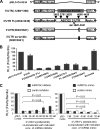
ABCG2 luciferase reporter assay. (A) Schematic diagram showing the luciferase reporter constructs used to study the 3′UTR of ABCG2 mRNA. The parental vector, pGL3-control, contains the coding region of firefly luciferase, whose expression is driven by an SV40 promoter (Promega). Nucleotide “1” refers to the first nucleotide in the ABCG2 transcript, as defined in sequence NM_004827. The following constructs were generated: 3′UTR FL (2462/4430); three truncated 3′UTR constructs (based on the FL 3′UTR construct, with progressive deletions from the 3′ end of the 3′UTR, whose 3′ ends are marked at the top of the FL construct, i.e., nt 2761, 3028, and 4060); 3′UTR(3820/3841), which contains only the 22-nt putative miRNA binding site for hsa-miR-519c; 3′UTR FL(Δ3820-3841), which contains the FL 3′UTR lacking the hsa-miR-519c binding site; 3′UTR (scramble 3820/3841), which contains the same 22 nt of the miRNA binding site, but in random order; and a 5′UTR construct (−1289/+396), prepared by inserting a fragment (−1289 to +396) from the ABCG2 5′UTR downstream of the luciferase coding sequence (serves as a control to demonstrate the specific effect of the 3′UTR on the luciferase activity of the reporter assay). The diagram is not drawn to scale. A more detailed diagram illustrating the reporter constructs can be found in Fig. S9 in the supplemental material. (B) Luciferase reporter activities of various reporter constructs, as indicated. Mean reporter activities ± SD (firefly/Renilla luciferase units) from three independent experiments are presented. pGL3-control represents the vector backbone without the ABCG2 3′UTR. (C) Luciferase reporter activity of the FL ABCG2 3′UTR FL(2462/4430) construct in cells cotransfected with either miRNA inhibitor (left) or mimic (right). Mean reporter activities ± SD were obtained as described for panel B. Statistical analysis was performed by the Student t test.
FIG. 5.
FIG. 5.
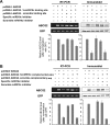
hsa-miR-519c binding site facilitates regulation of ABCG2 expression by both mRNA degradation and protein translation blockade. (A) HEK293 cells were transfected with an ABCG2 expression vector (pcDNA3-ABCG2) or one of two of its derivatives, one with a copy of the hsa-miR-519c binding sequence (pcDNA3-ABCG2-1×miRNA binding site) and the other with a scramble sequence of the hsa-miR-519c site (pcDNA3-ABCG2-scramble binding site) fused immediately after the stop codon of the ABCG2 coding sequence in the vector, and ABCG2 mRNA and protein expression was evaluated 48 h after transfection. A specific hsa-miR-519c or negative control miRNA inhibitor was also cotransfected into the cells to see if it could increase the ABCG2 expression level. ABCG2 expression levels were normalized with GFP (pEGFP-C1; DB Bioscience Clontech) and plotted in the bar graph relative to the level obtained for cells transfected with the pcDNA3-ABCG2 vector. The data shown represent the means ± SD for three independent experiments. (B) Perfect Watson-Crick pairing between hsa-miR-519c and 3′UTR sequences promotes predominantly mRNA degradation in the repression of ABCG2. HEK293 cells were transfected with similar ABCG2 expression vectors to those described for panel A. However, in the two derivative vectors, instead of the hsa-miR-519c binding site, either one copy of a 22-nt sequence complementary to the hsa-miR-519c binding site (pcDNA3-ABCG2-1×miRNA complementary sequence) or its scramble sequence (pcDNA3-ABCG2-scramble complementary sequence) was fused immediately downstream of the ABCG2 coding sequence. ABCG2 expression was found to be lower in cells transfected with the 1×miRNA complementary sequence vector than in those transfected with the plain ABCG2 expression vector without anything after the stop codon, but expression was similar at both the mRNA (−65%) and protein (−70%) levels. The scramble complementary sequence vector gave similar ABCG2 expression to that of the original ABCG2 expression vector. hsa-miR-519c was also found to be responsible for repression, because an hsa-miR-519c inhibitor, but not the scramble control miRNA inhibitor, could restore ABCG2 expression in cells transfected with the pcDNA3-ABCG2-1×miRNA complementary sequence. ABCG2 expression levels were normalized with GFP and plotted relative to the level obtained in cells transfected with the pcDNA3-ABCG2 vector.
FIG. 6.
FIG. 6.
hsa-miR-519c binding site decreases ABCG2 expression by promoting mRNA degradation. (Top) An ABCG2 expression vector (pcDNA3-ABCG2) and two of its derivatives, one with a copy of the hsa-miR-519c binding sequence (pcDNA3-ABCG2-1×miRNA binding site) and the other with a sequence complementary to that of hsa-miR-519c (pcDNA3-ABCG2-1×miRNA complementary sequence) fused immediately after the stop codon of the ABCG2 coding sequence in the vector, were transfected into HEK293 cells, and ABCG2 mRNA stability was monitored. Actinomycin D was added at 48 h posttransfection to block transcription, and total RNA was isolated at various time intervals and analyzed for ABCG2 mRNA levels by using RT-PCR. As an internal control, the level of GFP mRNA (from cotransfection of pEGFP-C1 [BD Bioscience Clontech]) was assessed and was used to normalize the ABCG2 mRNA levels. The graph shows the mean results for three independent and reproducible experiments. ABCG2 mRNA expressed from the vector with the hsa-miR-519c complementary sequence downstream of the ABCG2 coding sequence was the least stable (t1/2, ∼6 h), followed by the vector with the hsa-miR-519c binding site (t1/2, >16 h). In contrast, the ABCG2 mRNA without any 3′UTR was fairly stable for up to 24 h after actinomycin D treatment. (Bottom) Representative gel image showing the RT-PCR analysis summarized in the top panel.
FIG. 7.
FIG. 7.
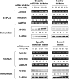
hsa-miR-519c regulates endogenous ABCG2 expression in A549 cells. A549 cells were transfected with 0 to 120 nM of either specific hsa-miR-519c inhibitor, hsa-miR-519c mimic, or the respective negative control (based on the cel-miR-67 sequence [Dharmacon]; this miRNA has been confirmed to have minimal sequence homology with miRNAs in humans, mice, and rats) for 48 h. Total RNA and whole-cell lysates were harvested for subsequent RT-PCR and Western blot analysis of ABCG2 expression. ABCG2 expression was normalized with GAPDH and reported relative to that of mock-treated cells. The transcript for hsa-miR-519c was also measured by stem-loop RT-PCR. The figure shows a representative result for three independent and reproducible experiments. The universal miRNA negative control inhibitor or mimic did not affect ABCG2, hsa-miR-519c, or GAPDH expression.
FIG. 8.
FIG. 8.
(A) Differential hsa-miR-520h expression level between parental S1 and resistant S1MI80 cells. hsa-miR-519c and hsa-miR-520h expression was measured by stem-loop RT-PCR (7, 46). U6 small nuclear RNA served as the loading control. The figure shows representative results for three independent and reproducible experiments, with means (± SD) plotted in the lower panel. (B) Location of the putative hsa-miR-520h binding site at ABCG2 3′UTR, RNA hybrid free energy (ΔG) calculations, and the theoretical miRNA-mRNA duplex pairing for hsa-miR-520h.
FIG. 9.
FIG. 9.
Proposed model for involvement of miRNA in the regulation of ABCG2 at the 3′UTR. A putative miRNA, hsa-miR-519c, binds to the 3′UTR of ABCG2 and suppresses the expression of ABCG2 in the parental cell lines. In the resistant cell lines, hsa-miR-519c cannot bind to ABCG2 mRNA because of the shorter 3′UTR, and thus mRNA degradation and/or repression of protein translation is relieved, contributing to the overexpression of ABCG2 by other mechanisms, such as permissive histone modifications at its promoter (49). hsa-miR-520h is another putative miRNA targeting ABCG2 mRNA, which was reported while the manuscript was under preparation (34). Unlike hsa-miR-519c, identified in our study, hsa-miR-520h binds to the 3′UTR of ABCG2 in both S1 and S1MI80 cells. Interestingly, S1MI80 cells express less hsa-miR-520h than do S1 cells, presumably allowing less repression of ABCG2 in the former.

Similar articles

Cited by

References

    1. Allikmets, R., L. M. Schriml, A. Hutchinson, V. Romano-Spica, and M. Dean. 1998. A human placenta-specific ATP-binding cassette gene (ABCP) on chromosome 4q22 that is involved in multidrug resistance. Cancer Res. 585337-5339. - PubMed
    1. Ambros, V. 2004. The functions of animal microRNAs. Nature 431350-355. - PubMed
    1. Bandres, E., E. Cubedo, X. Agirre, R. Malumbres, R. Zarate, N. Ramirez, A. Abajo, A. Navarro, I. Moreno, M. Monzo, and J. Garcia-Foncillas. 2006. Identification by real-time PCR of 13 mature microRNAs differentially expressed in colorectal cancer and non-tumoral tissues. Mol. Cancer 529. - PMC - PubMed
    1. Basler, C. F., and M. S. Horwitz. 1996. Subgroup B adenovirus type 35 early region 3 mRNAs differ from those of the subgroup C adenoviruses. Virology 215165-177. - PubMed
    1. Brennecke, J., A. Stark, R. B. Russell, and S. M. Cohen. 2005. Principles of microRNA-target recognition. PLoS Biol. 3e85. - PMC - PubMed

Publication types

MeSH terms