. 2008 Aug;28(16):5027-42.
doi: 10.1128/MCB.00194-08. Epub 2008 Jun 23.

STAP-2 negatively regulates both canonical and noncanonical NF-kappaB activation induced by Epstein-Barr virus-derived latent membrane protein 1

Affiliations

STAP-2 negatively regulates both canonical and noncanonical NF-kappaB activation induced by Epstein-Barr virus-derived latent membrane protein 1

Osamu Ikeda et al. Mol Cell Biol. 2008 Aug.

Abstract

The signal-transducing adaptor protein 2 (STAP-2) is a recently identified adaptor protein that contains a pleckstrin homology (PH) and Src homology 2 (SH2)-like domains, as well as a proline-rich domain in its C-terminal region. In previous studies, we demonstrated that STAP-2 binds to MyD88 and IKK-alpha or IKK-beta and modulates NF-kappaB signaling in macrophages. In the present study, we found that ectopic expression of STAP-2 inhibited Epstein-Barr virus (EBV) LMP1-mediated NF-kappaB signaling and interleukin-6 expression. Indeed, STAP-2 associated with LMP1 through its PH and SH2-like domains, and these proteins interacted with each other in EBV-positive human B cells. We found, furthermore, that STAP-2 regulated LMP1-mediated NF-kappaB signaling through direct or indirect interactions with the tumor necrosis factor receptor (TNFR)-associated factor 3 (TRAF3) and TNFR-associated death domain (TRADD) proteins. STAP-2 mRNA was induced by the expression of LMP1 in human B cells. Furthermore, transient expression of STAP-2 in EBV-positive human B cells decreased cell growth. Finally, STAP-2 knockout mouse embryonic fibroblasts showed enhanced LMP1-induced cell growth. These results suggest that STAP-2 acts as an endogenous negative regulator of EBV LMP1-mediated signaling through TRAF3 and TRADD.


Figures

FIG. 1.
FIG. 1.
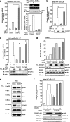
STAP-2 negatively regulates LMP1-induced NF-κB activation and IL-6 gene expression. (a) 293T cells in 12-well plates were transfected with HA-tagged LMP1 (10 ng) and NF-κB-LUC (100 ng) and/or increasing amounts of Myc-tagged STAP-2 (30, 150, or 300 ng). At 48 h after transfection, the cells were harvested, and the luciferase activities were measured. HeLa cells in 24-well plates were transfected with HA-tagged LMP1 (3 ng) and NF-κB-LUC (200 ng) and/or increasing amounts of Myc-tagged STAP-2, using jetPEI. At 48 h after transfection, the cells were harvested and assayed for their luciferase activities using a dual-luciferase reporter assay system. An aliquot of each total cell lysate (TCL) was analyzed by immunoblotting (IB) with an anti-HA or anti-Myc antibody. (b) HeLa/pcDNA3 and HeLa/STAP-2 in 12-well plates were transfected with or without increasing amounts of HA-tagged LMP1 (0, 0.1, 0.3, 0.5, or 1.5 μg), using jetPEI. At 36 or 48 h after transfection, the cells were lysed, and total extracts were immunoblotted with anti-IκBα, anti-p52, anti-HA, anti-Myc, or anti-actin antibodies. (c) 293T cells in 12-well plates were transfected with HA-tagged LMP1 (3 ng) and pIL-6-LUC (400 ng) and/or increasing amounts of Myc-tagged STAP-2 (30, 150, or 300 ng). At 48 h after transfection, the cells were harvested, and the luciferase activities were measured. HeLa cells in 24-well plates were transfected with HA-tagged LMP1 (50 ng) and pIL-6-LUC (100 ng) and/or increasing amounts of Myc-tagged STAP-2, using jetPEI. At 48 h after transfection, the cells were harvested and assayed for their luciferase activities using a dual-luciferase reporter assay system. An aliquot of each TCL was analyzed by immunoblotting with an anti-HA or anti-Myc antibody. (d) HeLa/pcDNA3 and HeLa/STAP-2 in 12-well plates were transfected with or without increasing amounts of HA-tagged LMP1 (0, 0.1, 0.5, or 1.5 μg), using jetPEI. At 48 h after transfection, total RNA samples were extracted and analyzed for their IL-6 expression levels by RT and quantitative real-time PCR analyses. Data represent the levels of IL-6 mRNA normalized by that of G3PDH mRNA as an internal control and are expressed relative to the value at time zero. Data represent the means of duplicate PCR determinations, which generally varied by less than 10%. An aliquot of each TCL was analyzed by immunoblotting with an anti-HA or anti-Myc antibody.
FIG. 2.
FIG. 2.
STAP-2 physiologically associates with LMP1. (a) 293T cells (1 × 10⁷ cells/well) were transfected with HA-tagged LMP1 (10 μg) with or without Myc-tagged STAP-2 (5 μg). At 48 h after transfection, the cells were lysed, immunoprecipitated with an anti-HA antibody, and immunoblotted (IB) with an anti-Myc or an anti-HA antibody. An aliquot of each total cell lysate (TCL) was immunoblotted with the anti-Myc antibody (bottom panel). (b) Human B lymphoma IB4 and Raji cells (2 × 10⁸ cells/well) were lysed, immunoprecipitated (IP) with control mouse IgG or anti-LMP1 MAb (S12), and immunoblotted with an anti-STAP-2 antibody or anti-LMP1 MAb. (c) HeLa cells in 12-well plates were transfected with HA-tagged LMP1 (1 μg) and Myc-tagged STAP-2 (1 μg), using jetPEI. At 48 h after transfection, the cells were fixed, incubated with anti-HA and anti-Myc antibodies and visualized with FITC- and rhodamine-conjugated secondary antibodies. The same slides were also stained with DAPI to detect the nuclei.
FIG. 3.
FIG. 3.
The PH and SH2-like domains of STAP-2 interact with the CTAR1 and CTAR2 domains of LMP1. (a) Schematic diagram of the domain structures of STAP-2 and its GST-fused mutant fragments. (b) 293T cells (1 × 10⁷ cells/well) were transfected with HA-tagged LMP1 (10 μg) with or without GST or GST-fused STAP-2 deletion mutants (8 μg). At 48 h after transfection, the cells were lysed, immunoprecipitated (IP) with an anti-HA antibody and immunoblotted (IB) with an anti-GST or anti-HA antibody. An aliquot of each total cell lysate (TCL) was immunoblotted with the anti-GST antibody. (c) Schematic diagram of the domain structures of LMP1 and its mutant fragments. (d) 293T cells (1 × 10⁷ cells/well) were transfected with Myc-tagged STAP-2 (5 μg) with or without LMP1 deletion mutants (10 μg). At 48 h after transfection, the cells were lysed, immunoprecipitated with an anti-Myc antibody, and immunoblotted with an anti-FLAG or anti-Myc antibody. An aliquot of each TCL was immunoblotted with the anti-FLAG antibody. (e) 293T cells (1 × 10⁷ cells/well) were transfected with Myc-tagged STAP-2 (5 μg) with or without GST or GST-fused LMP1 deletion mutants (10 μg). At 48 h after transfection, the cells were lysed, immunoprecipitated with an anti-Myc antibody, and immunoblotted with an anti-GST or anti-Myc antibody. An aliquot of each TCL was immunoblotted with the anti-GST antibody. (f and g) 293T cells (1 × 10⁷ cells/well) were transfected with GST-fused CTAR1 (f) or CTAR2 (g) (5 μg) with or without FLAG-tagged STAP-2 PH or STAP-2 SH2 (10 μg). At 48 h after transfection, the cells were lysed, pulled down with glutathione-Sepharose, and immunoblotted with an anti-FLAG or anti-GST antibody. An aliquot of each TCL was immunoblotted with the anti-FLAG antibody. Asterisks indicate the migration positions of the STAP-2 (b, F, and g), LMP1 (d), and GST-fused LMP1 (e) deletion mutants.
FIG. 4.
FIG. 4.
Functional role of the PH domain of STAP-2 in LMP1-induced signals. (a) Schematic diagram of the domain structures of the STAP-2 deletion mutant fragments. (b) 293T cells in 12-well plates were transfected with HA-tagged LMP1 (10 ng) and NF-κB-LUC (100 ng) and/or increasing amounts of Myc-tagged STAP-2 mutants (30, 150, or 300 ng). At 48 h after transfection, the cells were harvested, and the luciferase activities were measured. An aliquot of each total cell lysate (TCL) was analyzed by immunoblotting (IB) with an anti-HA or anti-Myc antibody. (c) Schematic diagram of the domain structures of LMP1 with a point mutation. (d) 293T cells in 12-well plates were transfected with FLAG-tagged LMP1 mutants (50 ng) and NF-κB-LUC (100 ng) and/or increasing amounts of Myc-tagged STAP-2 (30, 150 or 300 ng). At 48 h after transfection, the cells were harvested, and the luciferase activities were measured. An aliquot of each TCL was analyzed by immunoblotting with an anti-FLAG or anti-Myc antibody.
FIG. 5.
FIG. 5.
Molecular interactions among STAP-2, LMP1, and TRAF3. (a) 293T cells (1 × 10⁷ cells/well) were transfected with GST-fused STAP-2 (10 μg) with or without FLAG-tagged TRAF1, TRAF2, TRAF3, TRAF5, or TRAF6 (15 μg). At 48 h after transfection, the cells were lysed, pulled down with glutathione-Sepharose, and immunoblotted (IB) with an anti-FLAG or anti-GST antibody. An aliquot of each total cell lysate (TCL) was immunoblotted with the anti-FLAG antibody. (b) 293T cells (1 × 10⁷ cells) were transfected with FLAG-tagged TRAF3 (8 μg) with or without GST or GST-fused STAP-2 deletion mutants (10 μg). At 48 h after transfection, the cells were lysed, immunoprecipitated (IP) with an anti-FLAG antibody and immunoblotted with an anti-GST or anti-FLAG antibody. An aliquot of each TCL was immunoblotted with the anti-GST antibody. (c) Schematic diagrams of the domain structures of the TRAF3 deletion mutant fragments. (d) 293T cells (1 × 10⁷ cells/well) were transfected with Myc-tagged STAP-2 (5 μg) with or without FLAG-tagged TRAF3 deletion mutants (10 μg). At 48 h after transfection, the cells were lysed, immunoprecipitated with an anti-Myc antibody, and immunoblotted with an anti-FLAG or anti-Myc antibody. An aliquot of each TCL was immunoblotted with the anti-FLAG antibody. (e) 293T cells (1 × 10⁷) were transfected with HA-tagged LMP1 (10 μg) or FLAG-tagged TRAF3 (10 μg) and/or GST-fused STAP-2 (8 μg). At 48 h after transfection, the cells were lysed, immunoprecipitated with anti-LMP1 MAb, and immunoblotted with an anti-TRAF3 or anti-GST antibody or an anti-HA antibody. An aliquot of each TCL was immunoblotted with the anti-TRAF3 or the anti-GST antibody. (f) 293T cells (1 × 10⁷ cells/well) were transfected with HA-tagged LMP1 (10 μg) or FLAG-tagged TRAF3 (10 μg) and/or GST-fused STAP-2 (8 μg). At 48 h after transfection, the cells were lysed, immunoprecipitated with anti-HA antibody, and immunoblotted with an anti-GST or anti-FLAG or anti-HA antibody. An aliquot of each TCL was immunoblotted with the anti-GST or anti-FLAG antibody. (g) MEFs in 10-cm dishes were retrovirally transfected with a control vector or the LMP1 expression vector. At 48 h after transfection, the cells were lysed, immunoprecipitated with control mouse IgG or an anti-TRAF3 antibody and immunoblotted with an anti-LMP1 or anti-TRAF3 antibody. An aliquot of each TCL was analyzed by immunoblotting with the anti-LMP1 antibody. (h) 293T cells in 12-well plates were transfected with FLAG-tagged LMP1 mutants (50 ng) and NF-κB-LUC (100 ng) and/or increasing amounts of FLAG-tagged TRAF3 (10 or 100 ng). At 48 h after transfection, the cells were harvested, and the luciferase activities were measured. An aliquot of each TCL was immunoblotted with an anti-FLAG antibody. Asterisks indicate the migration positions of the TRAFs (a), GST-fused STAP-2 deletion mutants (b), and TRAF3 deletion mutants (d).
FIG. 6.
FIG. 6.
Reduction of endogenous STAP-2 or TRAF3 enhances LMP1-induced NF-κB activation. (a) HeLa cells in 24-well plates were transfected with a control siRNA or siRNA targeting human STAP-2. The cells were then transfected with HA-tagged LMP1 (3 ng) and NF-κB-LUC (100 ng), using jetPEI. At 48 h after transfection, the cells were harvested, and the luciferase activities were measured. At least three independent experiments were carried out for each assay. *, P was <0.01. Total RNA samples isolated from these cells were subjected to RT-PCR analysis using STAP-2 or G3PDH primers. An aliquot of each total cell lysate (TCL) was analyzed by immunoblotting (IB) with an anti-LMP1 antibody. NS, nonspecific band. The STAP-2 expression levels were also quantified by RT and quantitative real-time PCR analysis. Data represent the levels of STAP-2 mRNA normalized by that of G3PDH mRNA as an internal control and are expressed relative to the value at time zero. Data represent the means of duplicate PCR determinations, which generally varied by less than 10%. (b) MEFs in 24-well plates were retrovirally transfected with a control vector or the LMP1 expression vector. The cells were then transfected with NF-κB-LUC (100 ng) using Lipofectamine 2000. At 48 h after transfection, the cells were harvested, and the luciferase activities were measured. At least three independent experiments were carried out for each assay. *, P was <0.01. (c) HeLa cells in 24-well plates were transfected with a control siRNA or siRNAs targeting human TRAF3 (no. 1 and no. 2). The cells were then transfected with HA-tagged LMP1 (3 ng) and NF-κB-LUC (100 ng), using jetPEI. At 48 h after transfection, the cells were harvested, and the luciferase activities were measured. At least three independent experiments were carried out for each assay. *, P was <0.01. An aliquot of each total cell lysate (TCL) was analyzed by immunoblotting with an anti-LMP1, an anti-TRAF3, or an anti-actin antibody. (d) HeLa cells in 12-well plates were transfected with a control siRNA or siRNAs targeting human TRAF3 (no. 1 and no. 2). The cells were then transfected with HA-tagged LMP1 (1.5 μg), using jetPEI. At 48 h after transfection, total RNA samples were extracted and analyzed for their IL-6 expression levels by RT and quantitative real-time PCR analysis. Data represent the levels of IL-6 mRNA normalized by that of G3PDH mRNA as an internal control and are expressed relative to data from control siRNA-treated samples without LMP1. Data represent the means of duplicate PCR determinations, which generally varied by less than 10%. An aliquot of each TCL was also analyzed by immunoblotting with the indicated antibodies. (e) Human Raji B cells (2 × 10⁶ cells/well) were transfected with a control siRNA or TRAF3 siRNA (no. 2) using a MicroPorator according to the manufacturer's instructions. The siRNA-transfected Raji B cells were cultured for an additional 36 h and lysed. An aliquot of each TCL was analyzed by immunoblotting with the indicated antibodies. (f) HeLa cells in 24-well plates were transfected with a control siRNA or TRAF3 siRNA (no. 2). The cells were then transfected with NF-κB-LUC (100 ng) with or without FLAG-tagged LMP1 (aa 1 to 231) (100 ng) and Myc-tagged STAP-2 (100 ng), using jetPEI. At 48 h after transfection, the cells were harvested, and the luciferase activities were measured. At least three independent experiments were carried out for each assay. *, P was <0.01. An aliquot of each TCL was analyzed by immunoblotting with an anti-FLAG, an anti-Myc, an anti-TRAF3, or an anti-actin antibody.
FIG. 7.
FIG. 7.
TRAF6, TRADD, and RIP1, but not IKK-α, IKK-β, TRAF1, TRAF2, and BS69, overcame STAP-2 mediated suppression of LMP-induced NF-κB activation. (a) 293T cells in 24-well plates were transfected with HA-tagged LMP1 (10 ng) and NF-κB-LUC (100 ng) with or without Myc-tagged STAP-2 (100 ng) and/or increasing amounts of Myc-tagged IKK-α or IKK-β (1.0, 10, or 100 ng). At 48 h after transfection, the cells were harvested, and the luciferase activities were measured. An aliquot of each total cell lysate (TCL) was immunoblotted with an anti-HA or an anti-Myc antibody. (b) 293T cells in 24-well plates were transfected with HA-tagged LMP1 (10 ng) and NF-κB-LUC (100 ng) with or without Myc-tagged STAP-2 (100 ng) and/or increasing amounts of FLAG-tagged TRAF1 (1.0, 10, or 100 ng). At 48 h after transfection, the cells were harvested, and the luciferase activities were measured. An aliquot of each TCL was immunoblotted with an anti-HA, an anti-Myc, or an anti-FLAG antibody. (c) 293T cells in 24-well plates were transfected with HA-tagged LMP1 (10 ng) and NF-κB-LUC (100 ng) with or without Myc-tagged STAP-2 (100 ng) and/or increasing amounts of FLAG-tagged TRAF2 (1.0, 10, or 100 ng). At 48 h after transfection, the cells were harvested, and the luciferase activities were measured. An aliquot of each TCL was immunoblotted (IB) with an anti-HA, an anti-Myc, or an anti-FLAG antibody. (d) 293T cells in 24-well plates were transfected with HA-tagged LMP1 (10 ng) and NF-κB-LUC (100 ng) with or without Myc-tagged STAP-2 (100 ng) and/or increasing amounts of HA-tagged BS69 (1.0, 10, or 100 ng). At 48 h after transfection, the cells were harvested, and the luciferase activities were measured. An aliquot of each TCL was immunoblotted with an anti-HA or an anti-Myc antibody. (e) 293T cells in 24-well plates were transfected with HA-tagged LMP1 (10 ng) and NF-κB-LUC (100 ng) with or without Myc-tagged STAP-2 (100 ng) and/or increasing amounts of Myc-tagged TRAF6 (1.0, 10, or 100 ng). At 48 h after transfection, the cells were harvested, and the luciferase activities were measured. An aliquot of each TCL was immunoblotted with an anti-HA, an anti-Myc, or an anti-TRAF6 antibody. (f) 293T cells in 24-well plates were transfected with HA-tagged LMP1 (10 ng) and NF-κB-LUC (100 ng) with or without Myc-tagged STAP-2 (100 ng) and/or increasing amounts of HA-tagged TRADD (1.0, 10, or 100 ng). At 48 h after transfection, the cells were harvested, and the luciferase activities were measured. An aliquot of each TCL was immunoblotted with an anti-Myc or an anti-HA antibody. (g) 293T cells in 24-well plates were transfected with HA-tagged LMP1 (10 ng) and NF-κB-LUC (100 ng) with or without Myc-tagged STAP-2 (100 ng) and/or increasing amounts of FLAG-tagged RIP1 (1.0, 10, or 100 ng). At 48 h after transfection, the cells were harvested, and the luciferase activities were measured. An aliquot of each TCL was immunoblotted with an anti-HA, an anti-Myc, or an anti-FLAG antibody.
FIG. 8.
FIG. 8.
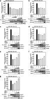
Reduction of endogenous TRAF6 or TRADD decreases LMP1-induced NF-κB activation. (a to d) HeLa cells in 24-well plates were transfected with a control siRNA or siRNA targeting human TRAF6, TRADD, RIP1, or RIP2. The cells were then transfected with HA-tagged LMP1 (5 ng) and NF-κB-LUC (100 ng), using jetPEI. At 48 h after transfection, the cells were harvested, and the luciferase activities were measured. At least three independent experiments were carried out for each assay. *, P was <0.01. An aliquot of each total cell lysate (TCL) from TRAF6 or TRADD siRNA-treated cells was analyzed by immunoblotting (IB) with the respective antibodies. Total RNA samples isolated from RIP1or RIP2 siRNA-treated cells were subjected to RT-PCR analysis using RIP1, RIP2, or G3PDH primers.
FIG. 9.
FIG. 9.
STAP-2 displaces TRADD from LMP1. (a and b) 293T cells (1 × 10⁷ cells/well) were transfected with Myc-tagged STAP-2 (5 μg) with or without HA-tagged TRADD or FLAG-tagged RIP1 (10 μg). At 48 h after transfection, the cells were lysed, immunoprecipitated (IP) with an anti-HA (a) or anti-FLAG (b) antibody and immunoblotted (IB) with an anti-Myc or an anti-FLAG antibody. An aliquot of each total cell lysate (TCL) was immunoblotted with the anti-Myc antibody. (c) 293T cells (1 × 10⁷) were transfected with FLAG-tagged LMP1 (10 μg) or HA-tagged TRADD (10 μg) and/or Myc-tagged STAP-2 (5 μg). At 48 h after transfection, the cells were lysed, immunoprecipitated with anti-FLAG antibody, and immunoblotted with an anti-HA, an anti-Myc, or an anti-FLAG antibody. An aliquot of each TCL was immunoblotted with the anti-HA or anti-Myc antibody. (d) 293T cells (1 × 10⁷ cells/well) were transfected with HA-tagged LMP1 (10 μg) or FLAG-tagged RIP1 (10 μg) and/or Myc-tagged STAP-2 (5 μg). At 48 h after transfection, the cells were lysed, immunoprecipitated with anti-HA antibody and immunoblotted with an anti-FLAG, an anti-Myc, or an anti-HA antibody. An aliquot of each TCL was immunoblotted with the anti-FLAG or anti-Myc antibody.
FIG. 10.
FIG. 10.
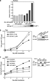
LMP1 induces STAP-2 mRNA expression in human B cells, and STAP-2 acts as an endogenous negative regulator of LMP1-mediated signaling. (a) Human Ramos B cells expressing tTA-control or tTA-LMP1 (1 × 10⁷ cells/well) were incubated with or without Dox (1 μg/ml) for the indicated periods. The STAP-2 expression levels were quantified by RT and quantitative real-time PCR analysis. Data represent the levels of STAP-2 mRNA normalized by that of G3PDH mRNA as an internal control and are expressed relative to the values at time zero. Data represent the means of duplicate PCR determinations, which generally varied by less than 10%. An aliquot of each total cell lysate (TCL) was analyzed by immunoblotting (IB) with an anti-LMP1 antibody. (b) Human Raji B cells (2 × 10⁶ cells/well) were nucleofected with empty vector, the Myc-tagged STAP-2 WT, or the STAP-2 ΔPH construct (2 μg), using 100 μl of V solution (Amaxa Biosystems) according to the manufacturer's instructions. Nucleofected Raji cells (3 × 10³ cells/well) were cultured in 96-well plates for the indicated periods, and the final numbers of cells were measured using a Cell Counting Kit-8. Data are the means of triplicate experiments, which generally varied by less than 10%. Similar results were obtained in three independent experiments. An aliquot of each TCL was analyzed by immunoblotting with an anti-Myc or an anti-LMP1 antibody. (c) MEFs (2 × 10³ cells/well) in 96-well plates were retrovirally transfected with a control GFP vector or an LMP1 expression vector and cultured for the indicated periods. The cell numbers were measured using a Cell Counting Kit-8. Data are the means of triplicate experiments, which generally varied by less than 10%. Similar results were obtained in three independent experiments. An aliquot of each TCL was analyzed by immunoblotting with an anti-LMP1 or an anti-GFP antibody.
FIG. 11.
FIG. 11.
Schematic showing the proposed function of STAP-2 in the suppression of LMP1-induced NF-κB activation. LMP1 consists of an N-terminal tail, six transmembrane domains, and a long cytoplasmic C-terminal domain which contains two NF-κB activating domains, CTAR1 and CTAR2. STAP-2 directly interacts with LMP1 and suppresses LMP1-induced NF-κB activation through both CTAR1 and CTAR2. STAP-2 suppresses CTAR1-mediated noncanonical NF-κB activation through direct interactions with TRAF3. Alternatively, STAP-2 regulates CTAR2-mediated canonical NF-κB activation by displacing TRADD from LMP1. LMP1-induced JNK activation is mediated by BS69 and TRAF6.

Similar articles

Cited by

References

    1. Brinkmann, M. M., and T. F. Schulz. 2006. Regulation of intracellular signalling by the terminal membrane proteins of members of the Gammaherpesvirinae. J. Gen. Virol. 871047-1074. - PubMed
    1. Brodeur, S. R., G. Cheng, D. Baltimore, and D. A. Thorley-Lawson. 1997. Localization of the major NF-kappaB-activating site and the sole TRAF3 binding site of LMP-1 defines two distinct signaling motifs. J. Biol. Chem. 27219777-19784. - PubMed
    1. Busch, L. K., and G. A. Bishop. 2001. Multiple carboxyl-terminal regions of the EBV oncoprotein, latent membrane protein 1, cooperatively regulate signaling to B lymphocytes via TNF receptor-associated factor (TRAF)-dependent and TRAF-independent mechanisms. J. Immunol. 1675805-5813. - PubMed
    1. Chaudhary, P. M., A. Jasmin, M. T. Eby, and L. Hood. 1999. Modulation of the NF-kappa B pathway by virally encoded death effector domains-containing proteins. Oncogene 185738-5746. - PubMed
    1. Cheng, G., and D. Baltimore. 1996. TANK, a co-inducer with TRAF2 of TNF and CD40L-mediated NF-kappaB activation. Genes Dev. 10963-973. - PubMed

Publication types

MeSH terms

LinkOut - more resources