

Characterization of human cellular immune responses to novel Mycobacterium tuberculosis antigens encoded by genomic regions absent in Mycobacterium bovis BCG
- PMID: 18573897
- PMCID: PMC2519442
- DOI: 10.1128/IAI.00199-08
Characterization of human cellular immune responses to novel Mycobacterium tuberculosis antigens encoded by genomic regions absent in Mycobacterium bovis BCG
Abstract
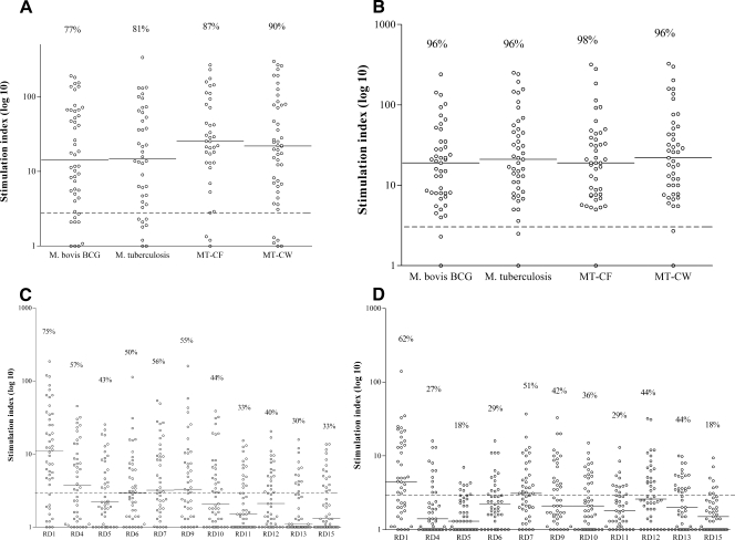
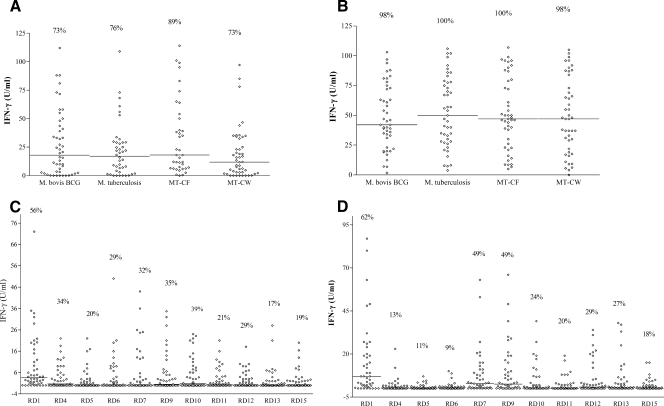
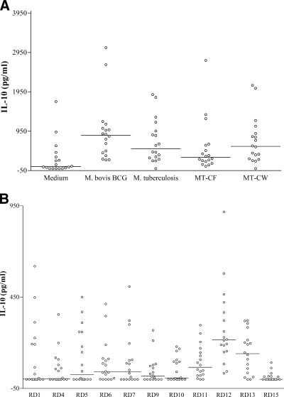
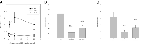
Comparative genomics has identified several regions of differences (RDs) between the infectious Mycobacterium tuberculosis and the vaccine strains of Mycobacterium bovis BCG. We aimed to evaluate the cellular immune responses induced by antigens encoded by genes predicted in 11 RDs. Synthetic peptides covering the sequences of RD1, RD4 to RD7, RD9 to RD13, and RD15 were tested for antigen-induced proliferation and secretion of Th1 cytokine, gamma interferon (IFN-gamma), by peripheral blood mononuclear cells (PBMC) obtained from culture-proven pulmonary tuberculosis (TB) patients and M. bovis BCG-vaccinated healthy subjects. Among the peptide pools, RD1 induced the best responses in both donor groups and in both assays. In addition, testing of TB patients' PBMC for secretion of proinflammatory cytokines (tumor necrosis factor alpha [TNF-alpha], interleukin 6 [IL-6], IL-8, and IL-1beta), Th1 cytokines (IFN-gamma, IL-2, and TNF-beta), and Th2 cytokines (IL-4, IL-5, and IL-10) showed differential effects of RD peptides in the secretion of IFN-gamma and IL-10, with high IFN-gamma/IL-10 ratios (32 to 5.0) in response to RD1, RD5, RD7, RD9, and RD10 and low IFN-gamma/IL-10 ratios (<1.0) in response to RD12, RD13, and RD15. Peptide-mixing experiments with PBMC from healthy subjects showed that secretion of large quantities of IL-10 in response to RD12 and RD13 correlated with inhibition of Th1 responses induced by RD1 peptides. In conclusion, our results suggest that M. tuberculosis RDs can be divided into two major groups--one group that activates PBMC to preferentially secrete IFN-gamma and another group that activates preferential secretion of IL-10--and that these two groups of RDs may have roles in protection against and pathogenesis of TB, respectively.
Figures
Similar articles
-
In silico analysis and experimental validation of Mycobacterium tuberculosis -specific proteins and peptides of Mycobacterium tuberculosis for immunological diagnosis and vaccine development.Med Princ Pract. 2013;22 Suppl 1(Suppl 1):43-51. doi: 10.1159/000354206. Epub 2013 Aug 31. Med Princ Pract. 2013. PMID: 24008694 Free PMC article. Review.
-
Cytokines in response to proteins predicted in genomic regions of difference of Mycobacterium tuberculosis.Microbiol Immunol. 2011 Apr;55(4):267-78. doi: 10.1111/j.1348-0421.2011.00307.x. Microbiol Immunol. 2011. PMID: 21244466
-
Characterization of human cellular immune responses to Mycobacterium tuberculosis proteins encoded by genes predicted in RD15 genomic region that is absent in Mycobacterium bovis BCG.FEMS Immunol Med Microbiol. 2010 Jul 1;59(2):177-87. doi: 10.1111/j.1574-695X.2010.00677.x. Epub 2010 Apr 7. FEMS Immunol Med Microbiol. 2010. PMID: 20482628
-
Identification of Mycobacterium tuberculosis-specific genomic regions encoding antigens inducing protective cellular immune responses.Indian J Exp Biol. 2009 Jun;47(6):498-504. Indian J Exp Biol. 2009. PMID: 19634716
-
Immunogenic potential of latency associated antigens against Mycobacterium tuberculosis.Vaccine. 2014 Feb 3;32(6):712-6. doi: 10.1016/j.vaccine.2013.11.065. Epub 2013 Dec 2. Vaccine. 2014. PMID: 24300592 Review.
Cited by
-
Cloning of the Recombinant Cytochrome P450 Cyp141 Protein of Mycobacterium tuberculosis as a Diagnostic Target and Vaccine Candidate.Iran Red Crescent Med J. 2014 Nov 5;16(11):e18001. doi: 10.5812/ircmj.18001. eCollection 2014 Nov. Iran Red Crescent Med J. 2014. PMID: 25763215 Free PMC article.
-
Immunological Characterization of Proteins Expressed by Genes Located in Mycobacterium tuberculosis-Specific Genomic Regions Encoding the ESAT6-like Proteins.Vaccines (Basel). 2021 Jan 7;9(1):27. doi: 10.3390/vaccines9010027. Vaccines (Basel). 2021. PMID: 33430286 Free PMC article. Review.
-
Comparative evaluation of MPT83 (Rv2873) for T helper-1 cell reactivity and identification of HLA-promiscuous peptides in Mycobacterium bovis BCG-vaccinated healthy subjects.Clin Vaccine Immunol. 2011 Oct;18(10):1752-9. doi: 10.1128/CVI.05260-11. Epub 2011 Aug 18. Clin Vaccine Immunol. 2011. PMID: 21852544 Free PMC article.
-
A novel tuberculosis DNA vaccine in an HIV-1 p24 protein backbone confers protection against Mycobacterium tuberculosis and simultaneously elicits robust humoral and cellular responses to HIV-1.Clin Vaccine Immunol. 2012 May;19(5):723-30. doi: 10.1128/CVI.05700-11. Epub 2012 Mar 29. Clin Vaccine Immunol. 2012. PMID: 22461526 Free PMC article.
-
In silico analysis and experimental validation of Mycobacterium tuberculosis -specific proteins and peptides of Mycobacterium tuberculosis for immunological diagnosis and vaccine development.Med Princ Pract. 2013;22 Suppl 1(Suppl 1):43-51. doi: 10.1159/000354206. Epub 2013 Aug 31. Med Princ Pract. 2013. PMID: 24008694 Free PMC article. Review.
References
-
- Al-Attiyah, R., A. S. Mustafa, A. T. Abal, N. M. Madi, and P. Andersen. 2003. Restoration of mycobacterial antigen-induced proliferation and interferon-γ responses in peripheral blood mononuclear cells of tuberculosis patients upon effective chemotherapy. FEMS Immunol. Med. Microbiol. 38249-256. - PubMed
-
- Al-Attiyah, R., and A. S. Mustafa. 2004. Computer-assisted prediction of HLA-DR binding and experimental analysis for human promiscuous Th1-cell peptides in the 24 kDa secreted lipoprotein (LppX) of Mycobacterium tuberculosis. Scand. J. Immunol. 5916-24. - PubMed
-
- Al-Attiyah, R., N. Madi, A. S. El-Shamy, H. Wiker, P. Andersen, and A. S. Mustafa. 2006. Cytokine profiles in tuberculosis patients and healthy subjects in response to complex and single antigens of Mycobacterium tuberculosis. FEMS Immunol. Med. Microbiol. 47254-261. - PubMed
-
- Amoudy, H. A., M. B. Al-Turab, and A. S. Mustafa. 2006. Identification of transcriptionally active open reading frames within the RD1 genomic segment of Mycobacterium tuberculosis. Med. Princ. Pract. 15137-144. - PubMed
Publication types
MeSH terms
Substances
LinkOut - more resources
Full Text Sources
Other Literature Sources
Miscellaneous
