

CD46 Contributes to the severity of group A streptococcal infection
- PMID: 18573902
- PMCID: PMC2519399
- DOI: 10.1128/IAI.00109-08
CD46 Contributes to the severity of group A streptococcal infection
Abstract
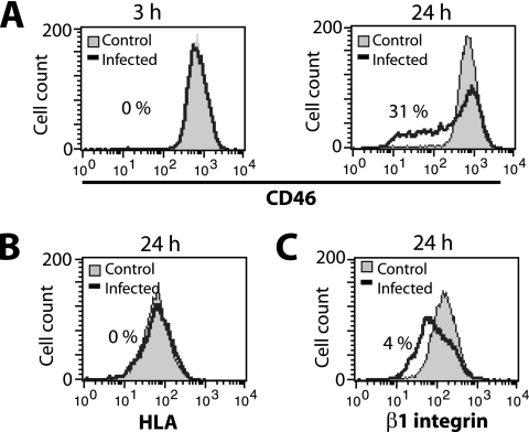
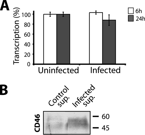
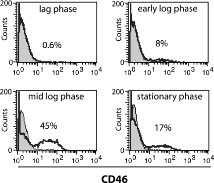
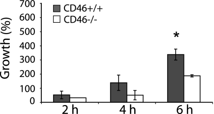
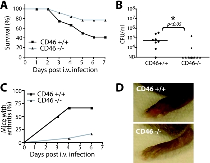
Streptococcus pyogenes (group A Streptococcus) is a human pathogen that causes a wide variety of diseases ranging from uncomplicated superficial infections to severe infections such as streptococcal toxic shock syndrome and necrotizing fasciitis. These bacteria interact with several host cell receptors, one of which is the cell surface complement regulator CD46. In this study, we demonstrate that infection of epithelial cells with S. pyogenes leads to the shedding of CD46 at the same time as the bacteria induce apoptosis and cell death. Soluble CD46 attached to the streptococcal surface, suggesting that bacteria might bind available extracellular CD46 as a strategy to survive and avoid host defenses. The protective role of human CD46 was demonstrated in ex vivo whole-blood assays showing that the growth of S. pyogenes was enhanced in blood from mice expressing human CD46. Finally, in vivo experimental infection showed that bacteremia levels, arthritis frequency, and mortality were higher in CD46 transgenic mice than in nontransgenic mice. Taken together, these results argue that bacterial exploitation of human CD46 enhances bacterial survival and represents a novel pathogenic mechanism that contributes to the severity of group A streptococcal disease.
Figures

Similar articles
-
CD46 transgenic mouse model of necrotizing fasciitis caused by Streptococcus pyogenes infection.Infect Immun. 2009 Nov;77(11):4806-14. doi: 10.1128/IAI.00577-09. Epub 2009 Sep 8. Infect Immun. 2009. PMID: 19737905 Free PMC article.
-
Dermal mast cells reduce progressive tissue necrosis caused by subcutaneous infection with Streptococcus pyogenes in mice.J Med Microbiol. 2011 Jan;60(Pt 1):128-134. doi: 10.1099/jmm.0.020495-0. Epub 2010 Sep 30. J Med Microbiol. 2011. PMID: 20884771
-
A highly susceptible CD46 transgenic mouse model of subcutaneous infection with Streptococcus dysgalactiae subspecies equisimilis.J Infect Chemother. 2016 Apr;22(4):229-34. doi: 10.1016/j.jiac.2016.01.001. Epub 2016 Feb 19. J Infect Chemother. 2016. PMID: 26908231
-
Binding of complement regulatory proteins to group A Streptococcus.Vaccine. 2008 Dec 30;26 Suppl 8:I75-8. doi: 10.1016/j.vaccine.2008.11.054. Vaccine. 2008. PMID: 19388169 Review.
-
Immunopathogenesis of streptococcal deep tissue infections.Curr Top Microbiol Immunol. 2013;368:173-88. doi: 10.1007/82_2012_282. Curr Top Microbiol Immunol. 2013. PMID: 23224738 Review.
Cited by
-
Preclinical safety, pharmacokinetics, pharmacodynamics, and biodistribution studies with Ad35K++ protein: a novel rituximab cotherapeutic.Mol Ther Methods Clin Dev. 2016 Mar 30;5:16013. doi: 10.1038/mtm.2016.13. eCollection 2016. Mol Ther Methods Clin Dev. 2016. PMID: 27069950 Free PMC article.
-
CD46 plasticity and its inflammatory bias in multiple sclerosis.Arch Immunol Ther Exp (Warsz). 2011 Feb;59(1):49-59. doi: 10.1007/s00005-010-0109-7. Epub 2011 Jan 26. Arch Immunol Ther Exp (Warsz). 2011. PMID: 21267793 Free PMC article. Review.
-
Presenilin/gamma-secretase cleaves CD46 in response to Neisseria infection.J Immunol. 2010 Jan 15;184(2):694-701. doi: 10.4049/jimmunol.0900522. Epub 2009 Dec 16. J Immunol. 2010. PMID: 20018629 Free PMC article.
-
Genetic Dissection of the Host Tropism of Human-Tropic Pathogens.Annu Rev Genet. 2015;49:21-45. doi: 10.1146/annurev-genet-112414-054823. Epub 2015 Sep 25. Annu Rev Genet. 2015. PMID: 26407032 Free PMC article. Review.
-
Inflammatory Joint Disease Is a Risk Factor for Streptococcal Sepsis and Septic Arthritis in Mice.Front Immunol. 2020 Oct 7;11:579475. doi: 10.3389/fimmu.2020.579475. eCollection 2020. Front Immunol. 2020. PMID: 33117382 Free PMC article.
References
-
- Berggard, K., E. Johnsson, E. Morfeldt, J. Persson, M. Stalhammar-Carlemalm, and G. Lindahl. 2001. Binding of human C4BP to the hypervariable region of M protein: a molecular mechanism of phagocytosis resistance in Streptococcus pyogenes. Mol. Microbiol. 42539-551. - PubMed
-
- Chaussee, M. A., A. V. Dmitriev, E. A. Callegari, and M. S. Chaussee. 2008. Growth phase-associated changes in the transcriptome and proteome of Streptococcus pyogenes. Arch. Microbiol. 18927-41. - PubMed
Publication types
MeSH terms
Substances
LinkOut - more resources
Full Text Sources
Medical
