. 2008 Jul 7;205(7):1535-41.
doi: 10.1084/jem.20080159. Epub 2008 Jun 23.

IL-12- and IL-23-modulated T cells induce distinct types of EAE based on histology, CNS chemokine profile, and response to cytokine inhibition

Affiliations

IL-12- and IL-23-modulated T cells induce distinct types of EAE based on histology, CNS chemokine profile, and response to cytokine inhibition

Mark A Kroenke et al. J Exp Med. .

Abstract

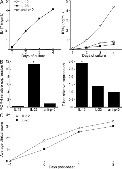
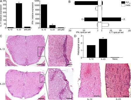
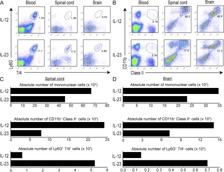
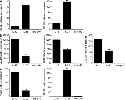
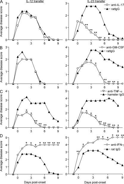
The interleukin (IL)-12p40 family of cytokines plays a critical role in the development of experimental autoimmune encephalomyelitis (EAE). However, the relative contributions of IL-12 and IL-23 to the pathogenic process remain to be elucidated. Here, we show that activation of uncommitted myelin-reactive T cells in the presence of either IL-12p70 or IL-23 confers encephalogenicity. Adoptive transfer of either IL-12p70- or IL-23-polarized T cells into naive syngeneic hosts resulted in an ascending paralysis that was clinically indistinguishable between the two groups. However, histological and reverse transcription-polymerase chain reaction analysis of central nervous system (CNS) tissues revealed distinct histopathological features and immune profiles. IL-12p70-driven disease was characterized by macrophage-rich infiltrates and prominent NOS2 up-regulation, whereas neutrophils and granulocyte-colony-stimulating factor (CSF) were prominent in IL-23-driven lesions. The monocyte-attracting chemokines CXCL9, 10, and 11 were preferentially expressed in the CNS of mice injected with IL-12p70-modulated T cells, whereas the neutrophil-attracting chemokines CXCL1 and CXCL2 were up-regulated in the CNS of mice given IL-23-modulated T cells. Treatment with anti-IL-17 or anti-granulocyte/macrophage-CSF inhibited EAE induced by transfer of IL-23-polarized, but not IL-12p70-polarized, cells. These findings indicate that autoimmunity can be mediated by distinct effector populations that use disparate immunological pathways to achieve a similar clinical outcome.


Figures

Figure 1.
Figure 1.
IL-12– and IL-23–modulated myelin-specific T cells induce EAE in naive hosts. (A) PLP139–151/IFA-primed T cells were cultured with antigen under conditions favorable to the generation of Th1 cells (IL-12, IFN-γ, αIL-4, and αIL-23p19), Th17 cells (IL-23, IL-1, αIFN-γ, and αIL-4), or lineage-uncommitted cells (αIL-12p40). Supernatants were harvested every 24 h and analyzed by ELISA. (B) RNA was extracted at 96 h from LNC cultures as described in A for measurement of transcription factors by real-time RT-PCR. Levels were normalized to GAPDH. The data are shown as fold induction over the level in cells cultured under neutral conditions. (C) Purified T cells from IL-12– and IL-23–modulated cultures were transferred to naive syngeneic recipients (25 × 106 cells/mouse). Mice were killed at peak disease for histological analysis. The data shown are representative of four independent experiments. Asterisks indicate a significant difference when compared with the control group. *, P < 0.05 compared with recipients of lineage uncommitted T cells.
Figure 2.
Figure 2.
IL-12– and IL-23–modulated T cells induce forms of EAE with distinct cytokine profiles and histological characteristics. (A) RNA was isolated from spinal cords of the recipients of IL-12– and IL-23–modulated T cells at the time of peak disease. Cytokine mRNA levels were measured by real-time RT-PCR and normalized to GAPDH. The data are shown as fold increase over levels in naive cords. (B) Mononuclear cells were isolated from pooled spinal cords at peak EAE for measurement of cytokines by ELISPOT assay. (C) Representative spinal cord sections are from adoptive transfer recipients that were killed upon reaching a clinical score of 3. Bars: left, 200 μm; right, 50 μm. (D) Optic nerves from representative mice with a clinical score of 3 were fixed and stained with hematoxylin and eosin. Histological scores were calculated from a total of 25 sections (obtained from 10 optic nerves) per group using ImageJ software. Bar, 100 μm. **, P < 0.001; *, P < 0.05 compared with recipients of IL-12–modulated cells.
Figure 3.
Figure 3.
CNS inflammatory infiltrates induced by IL-12– and IL-23–modulated T cells differ in cellular composition. (A and B) Mice were killed at peak EAE, and flow cytometry was performed on mononuclear cells isolated from pooled brains and spinal cords (five to six mice/group). Peripheral blood monocytes were analyzed in parallel. Plots in A are gated on class II cells. (C and D) Absolute cell numbers were calculated as the product of the average number of CNS mononuclear cells harvested per mouse (pooled from brain and spinal cord) and the percentage of CD11b+ class II+ or Ly6G+7/4+ cells (determined by flow cytometry). The data shown are representative of three independent experiments.
Figure 4.
Figure 4.
IL-12– and IL-23–modulated T cells induce distinct panels of chemokines and effector molecules in the CNS. Real-time RT-PCR was performed using RNA isolated from individual spinal cords at peak disease. Levels of ELR+ CXC chemokines (A), ELR CXC chemokines (B), and G-CSF and NOS2 (C) were normalized to GAPDH and averaged over four to six mice per group. The data shown are representative of three independent experiments. **, P < 0.001; *, P < 0.05 by comparison to recipients of IL-12–modulated T cells.
Figure 5.
Figure 5.
EAE mediated by IL-12– or IL-23–polarized T cells responds differently to specific immunomodulatory interventions. Adoptive transfer recipients were injected with neutralizing antibodies specific for IL-17 (A), GM-CSF (B), TNF-α (C), IFN-γ (D), or isotype control. Antibody injections were given either every 4 d (A, C, and D) or every 2 d (B) from day 4 after transfer (0.4 mg/dose). Each treatment group consisted of five to seven mice. Data shown are representative of two or more experiments with similar results. **, P < 0.005; *, P < 0.05 compared with the group treated with isotype control.

Comment in

  • A rush to judgment on Th17.
    Steinman L. Steinman L. J Exp Med. 2008 Jul 7;205(7):1517-22. doi: 10.1084/jem.20072066. J Exp Med. 2008. PMID: 18591407 Free PMC article.

References

    1. Fassbender, K., A. Ragoschke, S. Rossol, A. Schwartz, O. Mielke, A. Paulig, and M. Hennerici. 1998. Increased release of interleukin-12p40 in MS: association with intracerebral inflammation. Neurology. 51:753–758. - PubMed
    1. Moldovan, I.R., R.A. Rudick, A.C. Cotleur, S.E. Born, J.C. Lee, M.T. Karafa, and C.M. Pelfrey. 2003. Interferon gamma responses to myelin peptides in multiple sclerosis correlate with a new clinical measure of disease progression. J. Neuroimmunol. 141:132–140. - PubMed
    1. van Boxel-Dezaire, A.H., S.C. Hoff, B.W. van Oosten, C.L. Verweij, A.M. Drager, H.J. Ader, J.C. van Houwelingen, F. Barkhof, C.H. Polman, and L. Nagelkerken. 1999. Decreased interleukin-10 and increased interleukin-12p40 mRNA are associated with disease activity and characterize different disease stages in multiple sclerosis. Ann. Neurol. 45:695–703. - PubMed
    1. Merrill, J.E., D.H. Kono, J. Clayton, D.G. Ando, D.R. Hinton, and F.M. Hofman. 1992. Inflammatory leukocytes and cytokines in the peptide-induced disease of experimental allergic encephalomyelitis in SJL and B10.PL mice. Proc. Natl. Acad. Sci. USA. 89:574–578. - PMC - PubMed
    1. Platt, J.L., B.W. Grant, A.A. Eddy, and A.F. Michael. 1983. Immune cell populations in cutaneous delayed-type hypersensitivity. J. Exp. Med. 158:1227–1242. - PMC - PubMed

Publication types

MeSH terms