. 2008 Jul 7;205(7):1611-9.
doi: 10.1084/jem.20080297. Epub 2008 Jun 23.

TAK1 is required for the survival of hematopoietic cells and hepatocytes in mice

Affiliations

TAK1 is required for the survival of hematopoietic cells and hepatocytes in mice

Minghui Tang et al. J Exp Med. .

Abstract

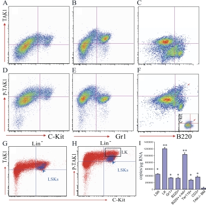
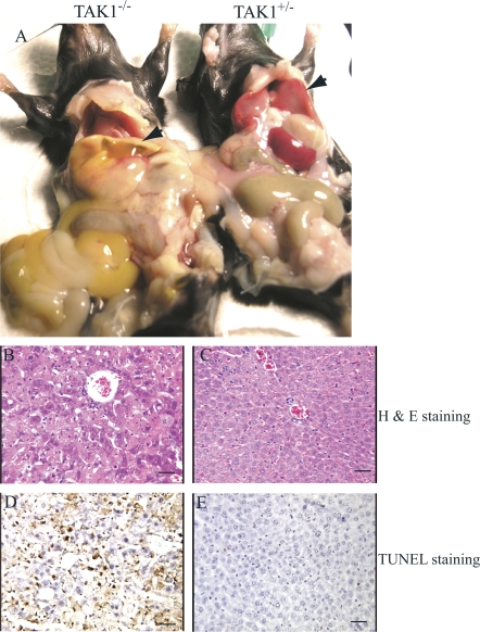
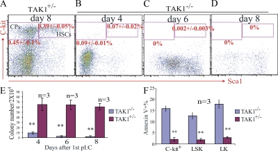
Transforming growth factor beta-activated kinase 1 (TAK1), a member of the MAPKKK family, is a key mediator of proinflammatory and stress signals. Activation of TAK1 by proinflammatory cytokines and T and B cell receptors induces the nuclear localization of nuclear factor kappaB (NF-kappaB) and the activation of c-Jun N-terminal kinase (JNK)/AP1 and P38, which play important roles in mediating inflammation, immune responses, T and B cell activation, and epithelial cell survival. Here, we report that TAK1 is critical for the survival of both hematopoietic cells and hepatocytes. Deletion of TAK1 results in bone marrow (BM) and liver failure in mice due to the massive apoptotic death of hematopoietic cells and hepatocytes. Hematopoietic stem cells and progenitors were among those hematopoietic cells affected by TAK1 deletion-induced cell death. This apoptotic cell death is autonomous, as demonstrated by reciprocal BM transplantation. Deletion of TAK1 resulted in the inactivation of both JNK and NF-kappaB signaling, as well as the down-regulation of expression of prosurvival genes.


Figures

Figure 1.
Figure 1.
TAK1 is expressed and activated in hematopoietic cells, including HSC/Ps. BM cells were collected into lyse/fix buffer immediately after the mice were killed to fix the BM cells and lyse the RBCs simultaneously. BM-nucleated cells were washed three times with PBS containing 5% FBS to completely remove all traces of the lyse/fix buffer. The cells were stained with cell surface markers as indicated and then permeabilized using permeabilization buffer for TAK1 and p-TAK1 staining. (A–C) High level of TAK1 protein expression in c-kit+ hematopoietic cells and Gr1+ myeloid cells, but low expression in B220+ B cells. (D–F) TAK activity (shown here by p-TAK1 levels) is higher in c-kit+ hematopoietic cells (including B220+c-kit+ B cell progenitors, red arrow in F). (G and H) No obvious difference in TAK1 expression in LSK-HSC/Ps and LK-CPs, but increased TAK1 activity in LK-CPs compared with LSK-HSC/Ps. (I) TAK1 is expressed in all lineages of hematopoietic cells, including HSC/Ps, as shown by quantitative RT-PCR. BM cells from TAK1 knockout mice (4 d after mutation induction) were used as negative controls. Compared with differentiated cells, including Gr1+ myeloid cells, B220+ B cells, Ter119+-nucleated erythoid cells, and CD3+ T cells, TAK1 RNA expression was significantly higher in LK-CPs and B cell progenitors (B220+c-kit+). Both * and ** are P < 0.01. *, compared with TAK1−/− BM cells; **, compared with mature hematopoietic cells, including Gr1+, B220+, and CD3+ cells.
Figure 2.
Figure 2.
Hepatocyte apoptosis and liver failure in TAK1−/− mice.TAK1−/− mice were killed on day 8 after the first polyI:C injection. (A) Both jaundice and ascites were observed in TAK1−/− mice. Livers of the TAK1−/− mice were pale and jaundiced (left specimen, arrowhead) compared with TAK1+/− control (right specimen, arrow). Nuclear condensation due to apoptosis was observed in >30–50% of hepatocytes of TAK1−/− mice as shown by hematoxylin and eosin staining of liver sections (B), which is confirmed by TUNEL staining (D). Liver sections from TAK1+/− mice were used as controls. (C and E) Bars, 100 μm.
Figure 3.
Figure 3.
Pancytopenia and BM failure in TAK1−/− mice. Peripheral blood, BM, spleens, and thymuses were collected on day 8 after the first polyI:C injection. WBC count (A), plt in peripheral blood (B), and nucleated cell number in the BM (C), spleen (D), and thymus (E) were measured in TAK1−/−, TAK1+/−, and WT mice, as well as in noninjected control mice. BM sections from TAK1+/− (F) and TAK1−/− (G) mice at day 8 after the first polyI:C injection were stained with hematoxylin and eosin. BM sections from TAK1+/− (H) and TAK1−/− (I) mice at day 4 after the first polyI:C injection were stained with TUNEL to display the apoptotic cells (indicated by red color staining). Bars, 100 μm. *, P < 0.05; **, P < 0.01.
Figure 4.
Figure 4.
HSC/Ps are affected by TAK1 deletion–induced apoptotic cell death in TAK1−/− mice. BM was collected from TAK1+/− and TAK1−/− mice on days 4, 6, and 8 after the first polyI:C injection. After lysis of RBCs, BM-nucleated cells were stained with cell surface markers and analyzed by flow cytometry for HSCs and CPs. Progressive loss of HSCs and CPs in TAK1−/− mouse BM is shown by lineage marker and Sca1 c-kit staining (B–D). Day 8 TAK1+/− BM cells were used as a control (A). (E) Progressive reduction of colony-forming ability of TAK1−/− BM cells after TAK1 deletion. (F) BM cells were collected from TAK1−/− and TAK1+/− mice 16 h after a single polyI:C injection, and apoptosis was checked by annexin V staining. *, P < 0.05; **, P < 0.01.
Figure 5.
Figure 5.
Cell-autonomous defects of TAK1−/− HSC/Ps. Reciprocal transplantation assay was performed by transplantation of TAK1−/− BM (before mutation was induced) into lethally irradiated WT mice (A) and, reciprocally, WT BM into lethally irradiated TAK1−/− mice (before mutation was induced; C). TAK1 deletion was induced 1 mo after transplantation by injecting the recipient mice with polyI:C every other day for a total of three injections. TAK1+/− mice were used as respective controls in this experiment (B and D). On day 8 after the first polyI:C injection, recipient mice were killed for analysis. BM sections from the recipients showed that TAK1−/− HSC/Ps failed to grow in the WT BM environment (A), whereas the TAK1−/− BM microenvironment was able to support the growth of WT HSC/Ps (C). (E–H) Significant reduction of WBC counts and plt in peripheral blood, and nucleated cells in the BM and spleen in WT recipients that received TAK1−/− BM. (E) Obvious increase in WBC counts in TAK1−/− recipients that received WT BM. (I and J) Equal numbers of TAK1−/− BM cells (CD45.2+, before mutation was induced) were mixed with WT (CD45.1+) BM cells and transplanted into lethally irradiated WT (CD45.1+) recipients. 6 wk after transplantation, TAK1 mutation was induced by injecting the recipient mice with polyI:C every other day for a total of three injections. TAK1+/− BM cells were transplanted in parallel as a control. The contribution of the TAK1−/− BM HSC/Ps to hematopoiesis in the recipient mice (shown as CD45.2+ percentage in peripheral blood) was significantly lower than that of control BM HSC/Ps. Data shown in I and J were analyzed on day 15 after the first polyI:C injection. *, P < 0.05; **, P < 0.01.

References

    1. Orelio, C., and E. Dzierzak. 2007. Bcl-2 expression and apoptosis in the regulation of hematopoietic stem cells. Leuk. Lymphoma. 48:16–24. - PubMed
    1. Motoyama, N., F. Wang, K.A. Roth, H. Sawa, K. Nakayama, K. Nakayama, I. Negishi, S. Senju, Q. Zhang, S. Fujii, et al. 1995. Massive cell death of immature hematopoietic cells and neurons in Bcl-x-deficient mice. Science. 267:1506–1510. - PubMed
    1. Opferman, J.T., H. Iwasaki, C.C. Ong, H. Suh, S. Mizuno, K. Akashi, and S.J. Korsmeyer. 2005. Obligate role of anti-apoptotic MCL-1 in the survival of hematopoietic stem cells. Science. 307:1101–1104. - PubMed
    1. Brandt, J.E., K. Bhalla, and R. Hoffman. 1994. Effects of interleukin-3 and c-kit ligand on the survival of various classes of human hematopoietic progenitor cells. Blood. 83:1507–1514. - PubMed
    1. Kimura, S., A.W. Roberts, D. Metcalf, and W.S. Alexander. 1998. Hematopoietic stem cell deficiencies in mice lacking c-Mpl, the receptor for thrombopoietin. Proc. Natl. Acad. Sci. USA. 95:1195–1200. - PMC - PubMed

Publication types

MeSH terms