. 2008 Jun 30;181(7):1141-54.
doi: 10.1083/jcb.200709091. Epub 2008 Jun 23.

Chibby cooperates with 14-3-3 to regulate beta-catenin subcellular distribution and signaling activity

Affiliations

Chibby cooperates with 14-3-3 to regulate beta-catenin subcellular distribution and signaling activity

Feng-Qian Li et al. J Cell Biol. .

Abstract

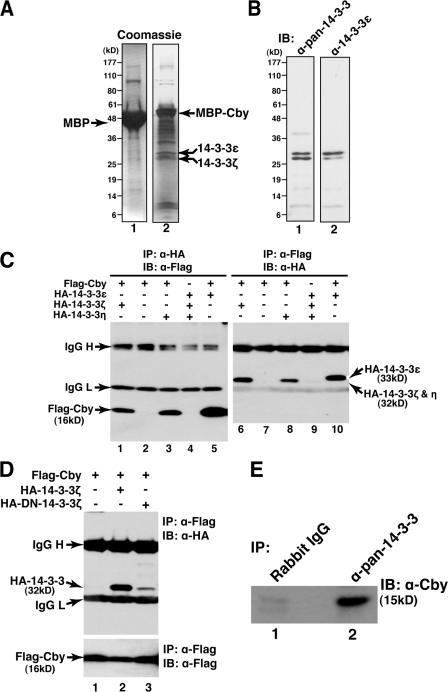
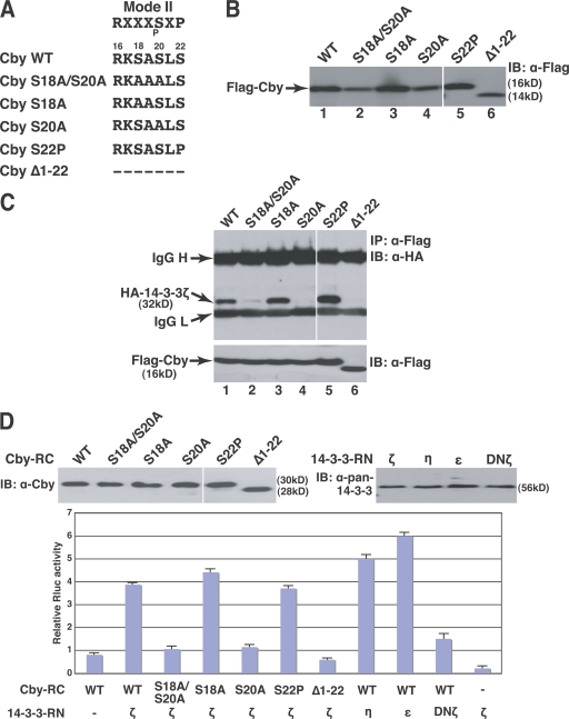
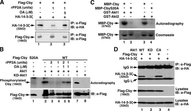
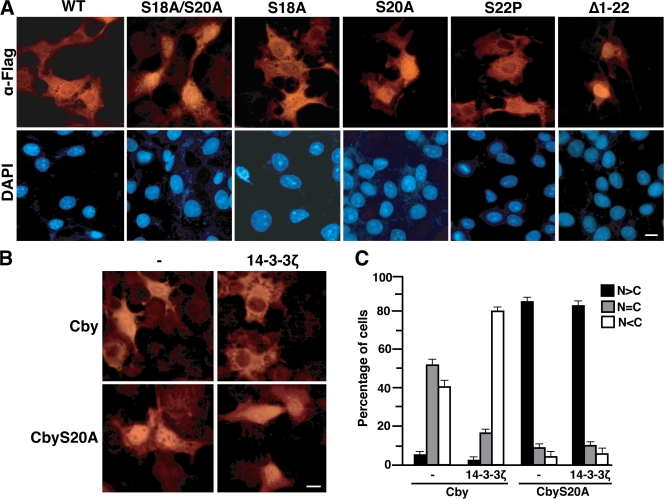
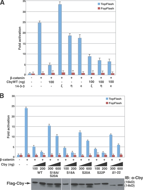
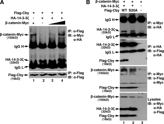
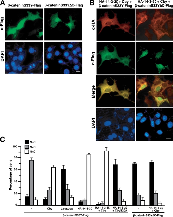
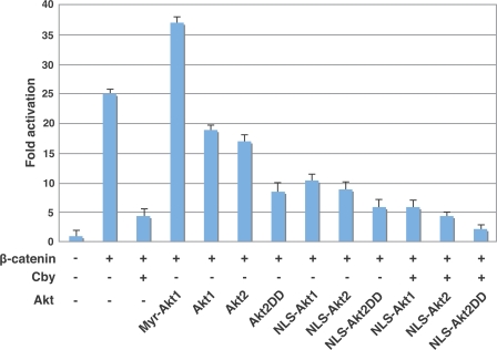
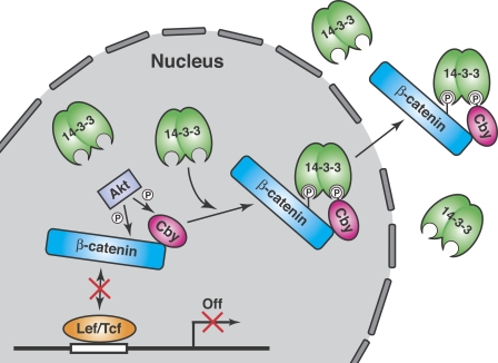
beta-Catenin functions in both cell-cell adhesion and as a transcriptional coactivator in the canonical Wnt pathway. Nuclear accumulation of beta-catenin is the hallmark of active Wnt signaling and is frequently observed in human cancers. Although beta-catenin shuttles in and out of the nucleus, the molecular mechanisms underlying its translocation remain poorly understood. Chibby (Cby) is an evolutionarily conserved molecule that inhibits beta-catenin-mediated transcriptional activation. Here, we identified 14-3-3epsilon and 14-3-3zeta as Cby-binding partners using affinity purification/mass spectrometry. 14-3-3 proteins specifically recognize serine 20 within the 14-3-3-binding motif of Cby when phosphorylated by Akt kinase. Notably, 14-3-3 binding results in sequestration of Cby into the cytoplasm. Moreover, Cby and 14-3-3 form a stable tripartite complex with beta-catenin, causing beta-catenin to partition into the cytoplasm. Our results therefore suggest a novel paradigm through which Cby acts in concert with 14-3-3 proteins to facilitate nuclear export of beta-catenin, thereby antagonizing beta-catenin signaling.


Figures

Figure 1.
Figure 1.
Physical interaction between Cby and 14-3-3 proteins. (A) Purification of 14-3-3 proteins as Cby-binding partners by an MBP pull-down approach. Lysates from HEK293T cells were incubated with bacterially expressed and purified MBP (lane 1) or MBP-Cby fusion protein (lane 2). Associated proteins were precipitated, separated by a 4–20% gradient SDS-PAGE, and visualized by Coomassie staining. (B) Immunoblotting with an anti–pan–14-3-3 antibody confirmed the copurification of 14-3-3 proteins with MBP-Cby (lane 1). An anti–14-3-3ε–specific antibody detected the upper band more intensely (lane 2). (C) HEK293T cells were transiently transfected with the indicated combinations of expression plasmids for Flag-Cby and HA–14-3-3 (ε, ζ, and η). Total cell lysates were immunoprecipitated with anti-HA antibodies and detected with anti-Flag antibodies (left) or vice versa (right). All three isoforms of 14-3-3 were expressed at comparable levels (not depicted). IgG H, IgG heavy chain; IgG L, light chain. (D) DN-14-3-3 exhibits significantly reduced affinity for Cby. Lysates from HEK293T cells transfected with Flag-Cby alone or together with HA–14-3-3ζ or HA–DN-14-3-3ζ were immunoprecipitated with anti-Flag antibodies and analyzed by Western blotting with anti-HA antibodies. Precipitated Flag-Cby levels were similar in all lanes, as shown in the bottom panel. (E) Coimmunoprecipitation of endogenous proteins. Cell lysates from nontransfected HEK293T cells were immunoprecipitated with control IgG (lane 1) or anti–pan–14-3-3 antibodies (lane 2) and immunoblotted for Cby.
Figure 2.
Figure 2.
Binding of 14-3-3 to Cby requires serine 20 within the N-terminal 14-3-3–binding motif of Cby. (A) Cby contains a mode II 14-3-3–binding consensus sequence RXXXpSXP, where pS represents phosphoserine. Shown are the Cby point and deletion mutants used in this study. CbyΔ1-22 lacks the N-terminal 22 amino acids. (B) Expression levels of Cby mutants. Lysates from HEK293T cells transfected with an equal amount of an expression plasmid for Flag-tagged WT or mutant Cby were subjected to Western blotting with an anti-Flag antibody. (C) Cby serine 20 is crucial for binding to 14-3-3. HEK293T cells were transfected with Flag-tagged WT or mutant Cby and HA–14-3-3ζ, followed by coimmunoprecipitation with anti-Flag antibodies and immunoblotting with anti-HA antibodies. Note that to compensate for protein levels, the amounts of DNA for CbyS18A/S20A, CbyS20A, and CbyΔ1-22 were appropriately increased for transfection, and hence, roughly similar expression levels were observed for all the Cby mutants (bottom). IgG H, IgG heavy chain; IgG L, light chain. (D) Confirmation of Cby–14-3-3 interactions by split hRluc protein fragment–assisted complementation. WT or mutant Cby was fused in-frame to the C-terminal portion of hRluc (Cby-RC), whereas 14-3-3 was fused to its N-terminal part (14-3-3-RN). These expression constructs (200 ng each) were transfected into HEK293T cells as indicated, and Renilla luciferase activities were measured 24 h after transfection. A firefly luciferase plasmid was cotransfected to normalize transfection efficiency. Immunoblotting with anti-Cby antibodies (top left) or anti–pan–14-3-3 antibodies (top right) showed that these fusion proteins were stably expressed. Transfections were carried out in triplicate and the means ± SD are shown. White lines indicate that intervening lanes have been spliced out.
Figure 3.
Figure 3.
Cby seine 20 is phosphorylated by Akt, facilitating its interaction with 14-3-3. (A) Interaction between Cby and 14-3-3 depends on phosphorylation. HEK293T cells were transfected with a Flag-Cby expression vector, and cell lysates were immunoprecipitated using anti-Flag affinity beads. The precipitated materials were preincubated in the presence or absence of rPP2A and its inhibitor OA. These samples were then mixed with an equal amount of total cell lysates from HEK293T cells expressing HA–14-3-3ζ, and the beads were precipitated, washed, and analyzed by SDS-PAGE and Western blotting with anti-HA antibodies. (B) Akt phosphorylates Cby at serine 20. HEK293T cells were transfected with an expression plasmid for Flag-tagged CbyWT or S20A, and Flag-Cby was immunoprecipitated using anti-Flag affinity beads. The bead-bound materials were incubated with [γ-32P]ATP and affinity-purified HA-tagged Akt1 or KD-Akt1 from transfected HEK293T cells in the presence or absence of rPP2A and OA as indicated. The reaction mixtures were then resolved by SDS-PAGE and visualized by autoradiography. (C) In vitro Akt kinase assay. Recombinant MBP-Cby or MBP-CbyS20A was incubated with active GST-Akt1 or Akt2 in the presence of [γ-32P]ATP. Proteins were separated by SDS-PAGE and detected by autoradiography. Roughly equal amounts of MBP-Cby fusion proteins were observed in all lanes upon Coomassie blue staining (bottom). (D) Coexpression of KD-Akt1 abolishes the interaction between Cby and 14-3-3. HEK293T cells were cotransfected with Flag-Cby, HA–14-3-3ζ, and either WT, KD, or myristoylated, constitutively active (CA) Akt1; and cell lysates were immunoprecipitated with anti-Flag antibodies followed by Western blot analysis with anti-HA antibodies. IgG H, IgG heavy chain; IgG L, light chain.
Figure 4.
Figure 4.
14-3-3 proteins sequester Cby in the cytoplasm. (A) The 14-3-3–binding motif in Cby is critical for the regulation of its subcellular localization. COS7 cells were transiently transfected with an expression vector encoding Flag-tagged WT or mutant Cby. Cells were fixed 24 h after transfection and immunostained with an anti-Flag antibody. Nuclei were stained with DAPI. (B) 14-3-3 facilitates the cytoplasmic relocation of Cby but not CbyS20A. COS7 cells were cotransfected with an expression construct for Flag-tagged Cby or CbyS20A and an empty control or HA–14-3-3ζ plasmid, followed by immunostaining with an anti-Flag antibody. Bars, 10 μm. (C) Quantitative analysis of the results in B. The subcellular localization of Flag-Cby was scored as follows: N > C, predominantly nuclear; N = C, evenly distributed between the nucleus and cytoplasm; N < C, predominantly cytoplasmic. Error bars represent the means ± SD of three independent experiments.
Figure 5.
Figure 5.
Association of Cby with 14-3-3 influences β-catenin signaling. (A) Effects of different 14-3-3 isoforms on β-catenin–mediated transcriptional activation were evaluated by TopFlash assays. (B) The ability of Cby mutants to repress β-catenin signaling was tested by TopFlash assays. HEK293T cells were transfected with 10 ng of TopFlash or mutant FopFlash luciferase reporter with or without 10 ng of an expression vector for stabilized β-catenin (β-catenin–Myc), 200 ng of a HA-tagged 14-3-3 plasmid, and the indicated amounts of a Flag-tagged Cby expression vector. Luciferase activity was measured 24 h after transfection and normalized to Renila luciferase activity used as an internal control. Transfections were performed in triplicate and the means ± SD are shown. Western blot analysis with anti-Cby antibodies showed that Cby proteins were expressed at similar levels. Note that, to compensate protein levels, higher amounts of DNA for CbyS18A/20A, CbyS20A, and CbyΔ1-22 were used for transfection. White lines indicate that intervening lanes have been spliced out.
Figure 6.
Figure 6.
Cby, 14-3-3, and β-catenin form a complex. (A) Coimmunoprecipitation of β-catenin and 14-3-3 with Cby. Total cell lysates from HEK293T cells expressing Flag-Cby, HA-14-3-3ζ, and stabilized β-catenin–Myc in the indicated combinations were immunoprecipitated with anti-Flag antibodies and subjected to Western blotting with a mixture of anti-Flag and anti-HA antibodies. (B) Cby–14-3-3 interaction is crucial for stable tripartite complex formation. HEK293T cells were cotransfected with expression constructs for β-catenin–Myc, HA–14-3-3ζ, and Flag-tagged CbyWT or S20A as shown. Cell lysates were immunoprecipitated with either anti-Myc or anti-HA antibodies and immunoblotted with the indicated antibodies. IgG H, IgG heavy chain; IgG L, light chain.
Figure 7.
Figure 7.
Cby and 14-3-3 relocate β-catenin to the cytoplasm. (A) A β-catenin mutant incapable of binding Cby preferentially localizes to the nucleus. COS7 cells were transfected with either stabilized β-cateninS33Y–Flag or a C-terminal truncation mutant (β-cateninS33YΔC–Flag) defective in Cby binding and then immunostained with anti-Flag antibodies. (B) β-CateninS33Y–Flag or β-cateninΔC–Flag were coexpressed with HA–14-3-3ζ and untagged Cby in COS7 cells. After 24 h, cells were double stained with anti-Flag (green) and anti-HA (red) antibodies. (C) Quantification of β-catenin localization. The subcellular distribution of Flag-tagged β-catenin was scored as in Fig. 4 C. Error bars represent the means ± SD of three independent experiments. N > C, predominantly nuclear; N = C, evenly distributed between the nucleus and cytoplasm; N < C, predominantly cytoplasmic. Bars, 10 μm.
Figure 8.
Figure 8.
Nuclear Akt enhances inhibition of β-catenin signaling by Cby. HEK293T cells were transiently transfected with 10 ng of TopFlash luciferase reporter together with the indicated combinations of expression constructs for stabilized β-catenin (β-catenin–Myc; 10 ng), Cby (100 ng), and different forms of Akt (200 ng). Luciferase activity was measured as described in the legend for Fig. 5. Myr-Akt1 predominantly localized to the plasma membrane, whereas NLS-Akt was exclusively nuclear. The other Akt forms were primarily distributed throughout the cytoplasm (not depicted). Transfections were carried out in triplicate and the means ± SD are shown.
Figure 9.
Figure 9.
Dual mechanism model for inhibition of β-catenin signaling activity by Cby. In the nucleus, Cby binds to β-catenin and competes with Tcf/Lef transcription factors, leading to repression of target gene expression. In addition, phosphorylation of Cby and β-catenin by Akt facilitates 14-3-3 binding, resulting in nuclear export of β-catenin to the cytoplasm. These two distinct mechanisms might be required to achieve full repression of β-catenin transcriptional activity. For details, see Discussion.

Similar articles

Cited by

References

    1. Aitken, A. 2006. 14-3-3 proteins: a historic overview. Semin. Cancer Biol. 16:162–172. - PubMed
    1. Andjelkovic, M., D.R. Alessi, R. Meier, A. Fernandez, N.J.C. Lamb, M. Frech, P. Cron, P. Cohen, J.M. Lucocq, and B.A. Hemmings. 1997. J. Biol. Chem. 272:31515–31524. - PubMed
    1. Borgatti, P., A.M. Martelli, G. Tabellini, A. Bellacosa, S. Capitani, and L.M. Neri. 2003. Threonine 308 phosphorylated form of Akt translocates to the nucleus of PC12 cells under nerve growth factor stimulation and associate with the nuclear matrix protein nucleolin. J. Cell. Physiol. 196:79–88. - PubMed
    1. Brazil, D.P., J. Park, and B.A. Hemmings. 2002. PKB binding proteins. Getting in on the Akt. Cell. 111:293–303. - PubMed
    1. Brunet, A., A. Bonni, M.J. Zigmond, M.Z. Lin, P. Juo, L.S. Hu, M.J. Anderson, K.C. Arden, J. Blenis, and M.E. Greenberg. 1999. Akt promotes cell survival by phosphorylating and inhibiting a Forkhead transcription factor. Cell. 96:857–868. - PubMed

Publication types

MeSH terms