. 2008 Sep 1;112(5):1662-72.
doi: 10.1182/blood-2008-01-128413. Epub 2008 Jun 23.

Transient blockade of the inducible costimulator pathway generates long-term tolerance to factor VIII after nonviral gene transfer into hemophilia A mice

Affiliations

Transient blockade of the inducible costimulator pathway generates long-term tolerance to factor VIII after nonviral gene transfer into hemophilia A mice

Baowei Peng et al. Blood. .

Abstract

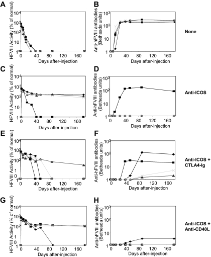
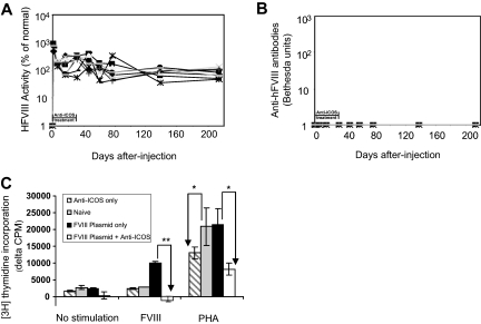
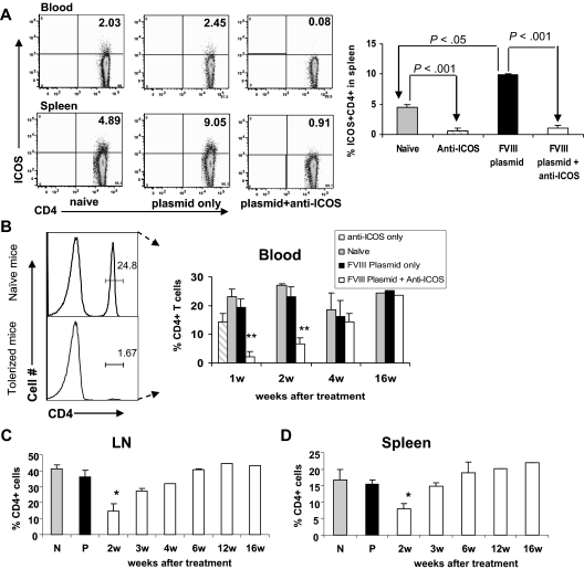
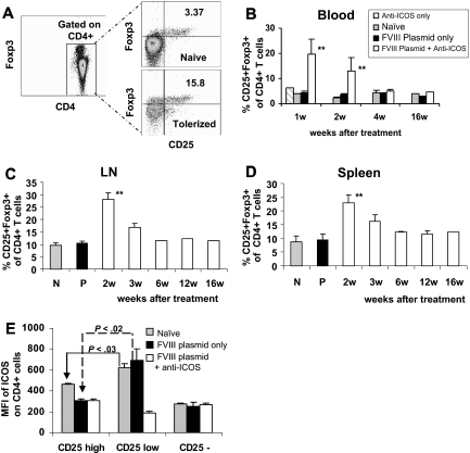
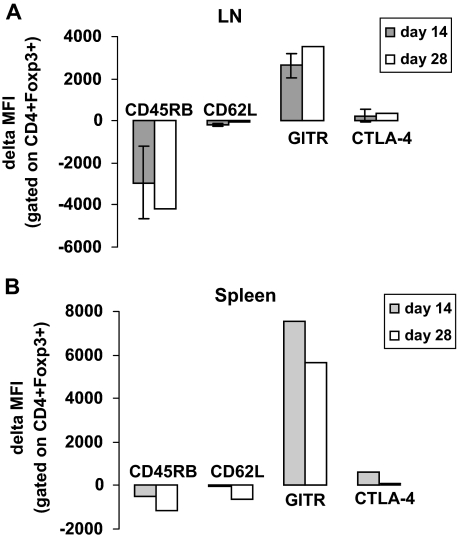
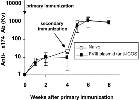
Formation of inhibitory antibodies is a common problem encountered in clinical treatment for hemophilia. Human factor VIII (hFVIII) plasmid gene therapy in hemophilia A mice also leads to strong humoral responses. We demonstrate that short-term therapy with an anti-ICOS monoclonal antibody to transiently block the inducible costimulator/inducible costimulator ligand (ICOS/ICOSL) signaling pathway led to sustained tolerance to hFVIII in hFVIII plasmid-treated hemophilia A mice and allowed persistent, high-level FVIII functional activity (100%-300% of normal). Anti-ICOS treatment resulted in depletion of ICOS(+)CD4(+) T cells and activation of CD25(+)Foxp3(+) Tregs in the peripheral blood, spleen, and lymph nodes. CD4(+) T cells from anti-ICOS-treated mice did not proliferate in response to hFVIII stimulation and produced high levels of regulatory cytokines, including interleukin-10 and transforming growth factor-beta. Moreover, CD4(+)CD25(+) Tregs from tolerized mice adoptively transferred dominant tolerance in syngeneic hFVIII plasmid-treated hemophilia A mice and reduced the production of antibodies against FVIII. Anti-ICOS-treated mice tolerized to hFVIII generated normal primary and secondary antibody responses after immunization with the T-dependent antigen, bacteriophage Phix 174, indicating maintenance of immune competency. Our data indicate that transient anti-ICOS monoclonal antibody treatment represents a novel single-agent immunomodulatory strategy to overcome the immune responses against transgene product after gene therapy.


Figures

Figure 1
Figure 1
Naked plasmid transfer of hFVIII plasmids into hemophilia A mice with short-term anti-ICOS treatment either alone or in combination with CTLA4-Ig or antimurine CD40L antibody (MR1). A total of 50 μg of the plasmid in 2 mL of saline solution was injected into the tail vein of hemophilia A mice (n = 4) in 5 to 8 seconds. Immunosuppressive agents were administered intraperitoneally. Anti-ICOS mAb was administrated 8 times in a 2-week period, CTLA4-Ig was injected 2 times at day 0 and 2, and anti-CD40L mAb was administered 5 times over a 2-week period. Circulating hFVIII activity was evaluated in plasma at regular intervals by a modified clotting assay (A,C,E,G) and confirmed by a chromogenic COATEST assay. Inhibitory antibody titers were evaluated by Bethesda assay and expressed as Bethesda units/mL (B,D,F,H). (A,B) No immunosuppression. (C,D) Anti-ICOS mAb alone. (E,F) Anti-ICOS + CTLA4-Ig. (G,H) Anti-ICOS + anti-CD40L mAb. The respective set of figures (A,B, C,D, E,F, and G,H) show data from the same experiment, and an individual mouse was represented by the same symbol in a set.
Figure 2
Figure 2
Effects of extended anti-ICOS treatment in mice after hFVIII plasmid transfer. hFVIII plasmid-treated mice (n = 9) were injected intraperitoneally with anti-ICOS 16 times during a 4-week period. (A) Persistent high levels of FVIII were expressed in mice as shown by hFVIII activity. (B) No inhibitors developed in mice as evaluated by Bethesda assay. (C) Absence of in vitro proliferation of CD4+ T cells isolated from tolerized mice in response to stimulation with hFVIII. Eight weeks after hFVIII plasmid transfer and anti-ICOS treatment, spleen CD4+ T cells were isolated and stimulated with hFVIII or PHA or cultured without stimulation for 72 hours in the presence of irradiated APCs. A total of 1 μCi of 3H-thymidine per well was added for the last 18 hours of culture. Spleen CD4+ T cells from mice treated with anti-ICOS alone, hFVIII plasmid alone, and untreated naive mice were used as controls. Data are presented as the mean counts per minute plus or minus SD by subtracting data obtained from T cells only in the absence of APCs and are representative of 2 separate experiments. *P < .05, **P < .01, compared with groups indicated by arrows.
Figure 3
Figure 3
Depletion of CD4+ICOS+ T cells in mice with extended 4-week anti-ICOS treatment. Lymphocytes isolated from secondary lymphoid organs of 4 groups of mice, namely, untreated control, anti-ICOS alone, hFVIII plasmid only, and hFVIII plasmid plus anti-ICOS–treated mice, were stained for ICOS and CD4 markers. (A) Left panel, representative dot plots of flow cytometry of blood and spleen samples at day 2 after indicated treatment. Right panel, statistical analysis of percentage of ICOS+CD4+ cells in 4 groups of mice (n = 3). (B-D) Time course of percentage of CD4+ T cells in peripheral blood (B), LNs (C), and spleen (D) against isolated total mononuclear cells. *P < .05, **P < .01, compared with groups of naive, anti-ICOS only, and plasmid-only treated mice. N and P indicate untreated naive mice and mice treated with hFVIII plasmid only, respectively.
Figure 4
Figure 4
Immunomodulation of gene therapy with extended anti-ICOS treatment results in increased percentage of CD4+ CD25+ Foxp3+ cells. Lymphocytes of secondary lymphoid organs from 3 groups of mice treated with different regimens were stained for CD4, CD25, and intracellularly for Foxp3. (A) Representative dot plots of flow cytometry of blood samples at day 14. Time course of percentage of CD25+Foxp3+ was evaluated in CD4+ T cells isolated from (B) peripheral blood, (C) LNs, and (D) spleen. **P < .01, compared with groups of naive, anti-ICOS alone, and plasmid only treated mice. (E) ICOS expression on CD4+CD25high, CD4+CD25low, and CD4+CD25 cell populations isolated from spleen of 3 groups of mice at day 2 to 4 after plasmid transfer. Results were shown as MFI of ICOS.
Figure 5
Figure 5
Immunomodulation of gene therapy with extended anti-ICOS treatment-induced activation of CD4+Foxp3+ Tregs. Lymphocytes isolated from secondary lymphoid organs of hFVIII plasmid plus anti-ICOS–treated mice were stained for various regulatory surface and intracellular markers, including CD45RB, CD62L, GITR, and CTLA4 at day 14 and 28 after plasmid transfer. For analysis, cells were gated on CD4+Foxp3+ population. (A) Expression of regulatory markers in LNs. (B) Expression of regulatory markers in spleen. The data represent delta MFI obtained by subtracting the background MFI observed in untreated naive mice.
Figure 6
Figure 6
CD4+ and CD4+CD25+ T lymphocytes from hFVIII plasmid plus extended anti-ICOS–treated mice exert dominant tolerance after adoptive transfer. (A,B) A total of 5 × 10⁶ CD4+ cells, 1 × 10⁶ CD4+CD25 cells, or 1 × 10⁶ CD4+CD25+ cells from tolerized mice 4 weeks after gene transfer were adoptively transferred into naive hemophilia A mice. The recipient mice were subsequently challenged with hFVIII plasmid 1 day after adoptive transfer. (A) hFVIII activity and (B) anti-hFVIII antibodies were examined in recipient mice at day 14 after adoptive transfer of indicated cell populations isolated from tolerized mice. (C,D) 5 × 10⁶ CD4+ cells were obtained from donor mice 8 weeks after gene transfer and adoptively transferred into naive hemophilia A mice. The recipient mice were then challenged with hFVIII plasmid 1 day after adoptive transfer. (C) hFVIII activity and (D) anti-hFVIII antibodies were measured in recipient mice at day 7 and day 14 after adoptive transfer of total CD4+ T cells from mice treated as indicated.
Figure 7
Figure 7
Tolerance induced by anti-ICOS immunomodulation is specific to FVIII. Six months after anti-ICOS mAb treatment, tolerized hemophilia A mice (n = 4) were challenged twice 4 weeks apart with the neoantigen bacteriophage Φx 174 (2 × 10⁸ PFU/each challenge). Untreated hemophilia A mice (n = 4) were used as control. Phage-neutralizing antibody activity was expressed as the rate of phage inactivation (Kv) using a standard formula. Mice receiving no bacteriophage did not produce neutralizing antibody (data not shown). All mice showed adequate antibody titers, amplification, and isotype switching (100% IgG) after secondary immunization.

Similar articles

Cited by

References

    1. Fields PA, Kowalczyk DW, Arruda VR, et al. Role of vector in activation of T cell subsets in immune responses against the secreted transgene product factor IX. Mol Ther. 2000;1:225–235. - PubMed
    1. Chao H, Mao L, Bruce AT, Walsh CE. Sustained expression of human factor VIII in mice using a parvovirus-based vector. Blood. 2000;95:1594–1599. - PubMed
    1. Greengard JS, Jolly PJ. Animal testing of retroviral-mediated gene therapy for factor VIII deficiency. Thromb Haemost. 1999;82:555–561. - PubMed
    1. VandenDriessche T, Vanslembrouck V, Goovaerts I, et al. Long-term expression of human coagulation factor VIII and correction of hemophilia A after in vivo retroviral gene transfer in factor VIII-deficient mice. Proc Natl Acad Sci U S A. 1999;96:10379–10384. - PMC - PubMed
    1. Ye P, Thompson AR, Sarkar R, et al. Naked DNA transfer of Factor VIII induced transgene-specific, species-independent immune response in hemophilia A mice. Mol Ther. 2004;10:117–126. - PubMed

Publication types

MeSH terms