. 2008 Aug 29;283(35):24089-102.
doi: 10.1074/jbc.M803422200. Epub 2008 Jun 23.

Functional characterization of fingers subdomain-specific monoclonal antibodies inhibiting the hepatitis C virus RNA-dependent RNA polymerase

Affiliations

Functional characterization of fingers subdomain-specific monoclonal antibodies inhibiting the hepatitis C virus RNA-dependent RNA polymerase

Andrei Nikonov et al. J Biol Chem. .

Abstract

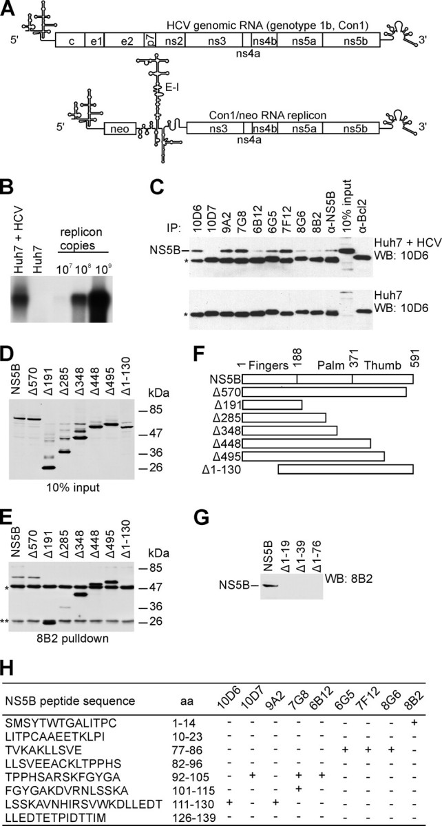
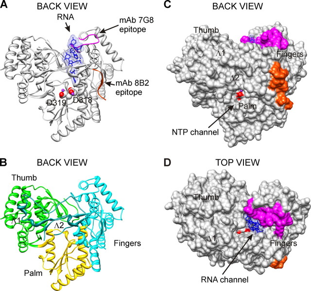
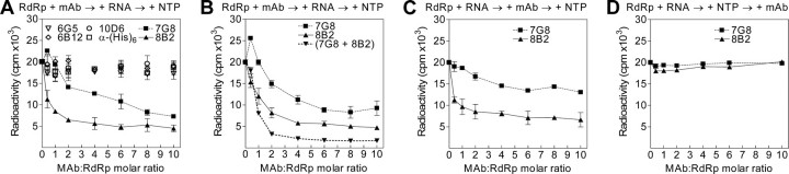
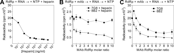
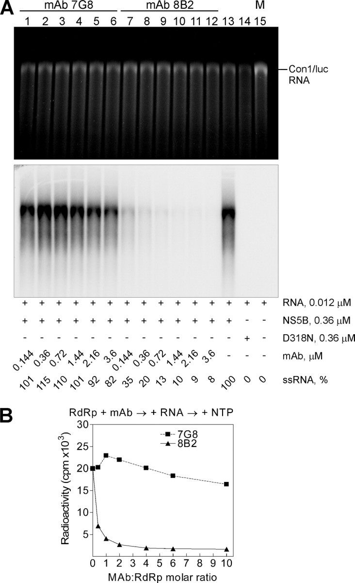
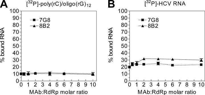
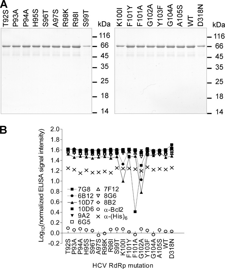
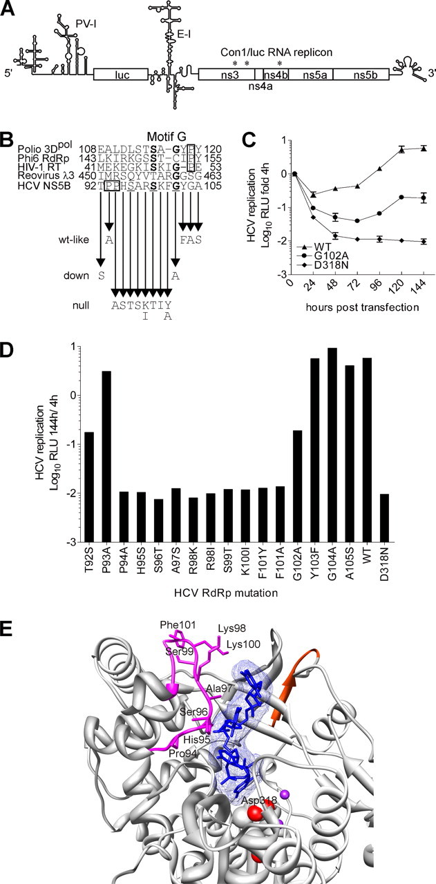
The hepatitis C virus (HCV) RNA-dependent RNA polymerase (RdRp), encoded by nonstructural protein 5B (NS5B), is absolutely essential for the viral replication. Here we describe the development, characterization, and functional properties of the panel of monoclonal antibodies (mAbs) and specifically describe the mechanism of action of two mAbs inhibiting the NS5B RdRp activity. These mAbs recognize and bind to distinct linear epitopes in the fingers subdomain of NS5B. The mAb 8B2 binds the N-terminal epitope of the NS5B and inhibits both primer-dependent and de novo RNA synthesis. mAb 8B2 selectively inhibits elongation of RNA chains and enhances the RNA template binding by NS5B. In contrast, mAb 7G8 binds the epitope that contains motif G conserved in viral RdRps and inhibits only primer-dependent RNA synthesis by specifically targeting the initiation of RNA synthesis, while not interfering with the binding of template RNA by NS5B. To reveal the importance of the residues of mAb 7G8 epitope for the initiation of RNA synthesis, we performed site-directed mutagenesis and extensively characterized the functionality of the HCV RdRp motif G. Comparison of the mutation effects in both in vitro primer-dependent RdRp assay and cellular transient replication assay suggested that mAb 7G8 epitope amino acid residues are involved in the interaction of template-primer or template with HCV RdRp. The data presented here allowed us to describe the functionality of the epitopes of mAbs 8B2 and 7G8 in the HCV RdRp activity and suggest that the epitopes recognized by these mAbs may be useful targets for antiviral drugs.


Figures

FIGURE 1.
FIGURE 1.
Features of mAbs directed against HCV NS5B RdRp. A, schematic representation of HCV genomic RNA and subgenomic replicon Con1/neo construct (25, 29) used for stable cell line selection (5′, HCV 5′-UTR; neo, neomycin phosphotransferase gene; E-I, encephalomyocarditis virus IRES; 3′, HCV 3′-UTR). Reported secondary structures for 5′-UTR (60), 3′-UTR (61), and encephalomyocarditis virus IRES (62) were used in the illustration. B, detection of Con1/neo subgenomic replicon in G418 selected Huh7 cells. Northern blot analysis was performed using total cellular RNA and antisense ns5b-specific probe. 32P-Labeled Con1/neo RNA was used as a marker. C, immunoprecipitation (IP)/Western blot (WB) analysis. Various mAbs were used to immunoprecipitate the NS5B either from the lysate of the Huh7 cell line containing selectable subgenomic HCV RNA replicon depicted in A (Huh7 + HCV) or from the Huh7 cell line lysate (Huh7). NS5B polyclonal antibodies (α-NS5B) and α-Bcl2 mAb, directed against cellular protein, were used as positive and negative controls, respectively. *, immunoglobulin heavy chain; protein blots were probed with NS5B-specific mAb 10D6, and reactivity was detected with secondary horseradish peroxidase-conjugated antibody of a murine origin. D, Western blot analysis of bovine papilloma virus E2 protein 3F12 epitope-tagged (63) NS5B truncated construct expression in COS7 cells. Molecular mass standards are indicated on the right. E, immunoprecipitation of the proteins shown in D using mAb 8B2. **, immunoglobulin light chain; protein blots were probed with bovine papilloma virus E2-specific mAb 3F12, and reactivity was detected as in C. F, schematic diagram of wild-type HCV NS5B (shown on top) and deletion constructs used for immunoprecipitation. Polymerase subdomains are indicated. Numbers refer to the first residue in a given subdomain. G, Western blot analysis of mAb 8B2 binding to N-terminal truncations of the NS5B protein. H, results of the epitope mapping for NS5B mAbs. Wells of MaxiSorp™ plate (Nunc) were coated with 5 μg of peptide and blocked with bovine serum albumin. HCV NS5B-specific mAbs were incubated with the peptides, and ELISA reactivity was detected by horseradish peroxidase-conjugated antibody; aa, amino acid residues.
FIGURE 2.
FIGURE 2.
mAbs 8B2 and 7G8 epitopes. The localization of mAbs 8B2 and 7G8 epitopes is shown on the three-dimensional x-ray structure of HCV RdRp complex with oligonucleotide RNA (Protein Data Bank accession code 1nb7, HCV J4 strain (45)). A and B, ribbon representations of the back view are shown. Solid molecular surface representations of back and top views are shown in C and D. The spatial orientation of HCV RdRp is identical for A-C. The epitopes of mAbs 8B2 (amino acids Ser1-Ala9) and 7G8 (amino acids Thr92-Ala105) are colored red-orange and magenta, respectively. Catalytic aspartate residues are represented as spheres, and their oxygen atoms are displayed as red. Manganese atoms are shown as spheres and depicted in purple. B, Fingers, Palm, and Thumb are depicted in cyan, gold, and green, respectively. Two loops emanating from Fingers subdomain and making extensive contacts with Thumb are designated Λ1 (amino acids Ile11-Ala45) and Λ2 (Met139-Ile160) and depicted in dark cyan. The RNA, colored blue, is represented as sticks embedded in transparent mesh molecular surface. Figures were prepared with UCSF, Chimera package (64, 65).
FIGURE 3.
FIGURE 3.
mAbs 8B2 and 7G8 inhibit the primer-dependent NS5B RdRp activity. RdRp assays were performed as specified under “Experimental Procedures” using poly(rC)/(rG)12 template-primer RNA. The order of reagent addition is indicated at the top of the graphs. The x axis displays mAb molar excess over NS5B, and the y axis shows the total amount of synthesized 32P-labeled RNA product in counts/min. The means of replicate (at least three) independent experiments with standard deviation values are indicated. A, selection of the mAbs inhibiting RdRp activity of HCV NS5B. B, NS5B RdRp activity inhibition by mAbs 8B2 and 7G8 separately and in combination. When applied in combination, mAbs 8B2 and 7G8 were used at the same concentration as for the individual mAb experiments. C, effect of NS5B and template-primer RNA preincubation on the inhibition of NS5B RdRp activity by mAbs 7G8 and 8B2. D, RdRp assay was initiated by the addition of NTP and incubated for 15 min prior to the addition of mAbs.
FIGURE 4.
FIGURE 4.
In primer-dependent RdRp assay, RNA elongation and RNA synthesis initiation are inhibited by mAbs 8B2 and 7G8, respectively. A, determination of heparin concentration required for a single cycle primer-dependent RNA synthesis by HCV RdRp in vitro (representative dose-effect curve). After NS5B preincubation with template-primer RNA, RdRp assay was initiated by the simultaneous addition of NTP and various heparin concentrations (x axis). After NS5B preincubation with mAbs, the RNA template-primer was added, and RdRp assays were initiated by addition of NTP either in the presence (B) or in the absence (C) of a constant amount of heparin. The NTP and heparin were added simultaneously. The molar ratio of mAb to NS5B RdRp is plotted versus the total amount of synthesized 32P-labeled RNA product (counts/min). Note the scale of the y axis in B and C differs 10-fold.
FIGURE 5.
FIGURE 5.
mAb 8B2 blocks primer-independent (de novo) RdRp activity of HCV NS5B in vitro. A, standard de novo RdRp assay was performed using an in vitro transcribed Con1/luc RNA template and increasing concentrations of mAbs 7G8 (lanes 1-6) and 8B2 (lanes 7-12). After a 2-h incubation at 25 °C, RNA was precipitated and analyzed on denaturing formaldehyde-agarose gel. Shown are ethidium bromide staining and the autoradiogram of the same gel. The critical additives and ssRNA yield normalized to the RdRp reaction without mAbs (lane 13) are indicated below the panels. The position of the Con1/luc RNA template is indicated on the right. M is the marker lane with transcribed and purified Con1/luc ssRNA (9510 nucleotides) used for the RdRp assay. D318N (lane 14) is an inactive form of the NS5B. The mAb:RdRp molar ratio value of 1 corresponds to 0.36 μm mAb concentration. B, quantitative PhosphorImager analysis of the reactions shown in A. The analysis of a single gel is presented. The curves show the efficiency of the ssRNA synthesis on Con1/luc RNA template by NS5B in the presence of mAbs 7G8 and 8B2.
FIGURE 6.
FIGURE 6.
The binding of HCV subgenomic RNA by NS5B is enhanced in the presence of mAb 8B2. RNA binding assays were performed with a 32P-labeled poly(rC)/(rG)12 template-primer RNA (A) or HCV Con1/luc replicon RNA (B) in the presence of increasing concentrations of mAbs. The NS5B-RNA complexes were immobilized onto nitrocellulose filters (BA85, Schleicher & Schuell), and bound radioactivity was measured by liquid scintillation counting. The means of replicate (three) independent experiments with standard deviation values are indicated.
FIGURE 7.
FIGURE 7.
Purification of recombinant HCV NS5B proteins with single amino acid changes produced in E. coli and their conformational integrity. A, SDS-polyacrylamide gels (12% polyacrylamide) stained with Coomassie Brilliant Blue G-250. Consecutive lanes correspond to different mutant enzymes. 1/100 of each mutant enzyme produced from 300 ml of corresponding bacterial culture (the expression was induced with isopropyl thiogalactoside under constant conditions) was analyzed. Marker proteins sizes (in kDa) are indicated to the right from each gel. B, structural integrity of recombinant HCV NS5B mutant proteins depicted in A. Wells of MaxiSorp™ plate (Nunc) were coated with 200 ng of corresponding purified NS5B mutant protein in phosphate-buffered saline and blocked with bovine serum albumin. Equal amounts of HCV NS5B-specific and control (α-His and α-Bcl2) mAbs were incubated with the mutant proteins, and ELISA reactivity was detected by horseradish peroxidase-conjugated antibody. The normalized ELISA signal intensity was calculated as the logarithm of ELISA signals ratio obtained for the reaction with and without corresponding primary mAb. The experiment was performed in duplicate. Because of low variation, error bars are not visible on the graph.
FIGURE 8.
FIGURE 8.
mAb 7G8 epitope site-directed mutations and their effect on HCV Con1/luc replicon transient replication. A, scheme of Con1/luc replicon (PV-I, poliovirus IRES; luc, firefly luciferase gene; E-I, encephalomyocarditis virus IRES; *, adaptive mutations (26, 27)). Reported secondary structure of poliovirus IRES (66) was used in the illustration. B, HCV NS5B peptide sequence, corresponding to mAb 7G8 epitope, was aligned based on protein secondary structure with the corresponding peptide sequences of HIV-1 RT and RdRps from poliovirus (3Dpol), phage ϕ6 (P2), and reovirus (λ3). Highly conserved residues of HCV NS5B and motif G are underlined and shown in boldface, respectively. The Pro residues flanking motif G are boxed. The arrows indicate NS5B amino acid residue changes that were made in Con1/luc replicon. The wt-like, down, and null refer to the resulting mutations features; wt-like, wild-type-like. C, representative Con1/luc replication kinetic curves in Huh7-Lunet cells characterizing wild-type (WT, WT-like), down (G102A), and null (D318N) mutations. Relative light units (RLU) were measured 4, 24, 48, 72, 96, 120, and 144 h after transfection. Relative light units were normalized to the input replication signal measured at 4 h post-transfection (logarithmic scale) and total protein content in the lysate. Note that, because of low variation, error bars are in most cases invisible. D, transient replication assay of mutant Con1/luc replicons in Huh7-Lunet cells. The HCV replication efficiency is given as luciferase activity (logarithmic scale) 144 h after transfection normalized to the input measured at 4 h and to the total protein content in the lysate. E, shown is the ribbon representation of a close-up view for mAb 7G8 epitope on the three-dimensional structure of HCV RdRp complex with RNA oligonucleotide (Protein Data Bank code 1nb7). The residues, which upon change produce the null mutations in transient replication assay, are depicted as sticks. The color codes are the same as in Fig. 2. The NS5B amino acid residue 98 of genotype 1b is Arg and Lys in the Con1 (this study) and J4 isolates, respectively. E was prepared with UCSF Chimera package (64, 65).

Similar articles

Cited by

References

    1. Choo, Q. L., Kuo, G., Weiner, A. J., Overby, L. R., Bradley, D. W., and Houghton, M. (1989) Science 244 pa359-362 - PubMed
    1. Hoofnagle, J. H. (2002) Hepatology 36 Suppl. 1, 21-29 - PubMed
    1. Perz, J. P., Farrington, L. A., Pecoraro, C., Hutin, Y. J., and Armstrong, G. L. (2004) 42nd Annual Meeting of the Infectious Diseases Society of America, September 30-October 3, p. 214, Boston, MA
    1. Grakoui, A., Wychowski, C., Lin, C., Feinstone, S. M., and Rice, C. M. (1993) J. Virol. 67 1385-1395 - PMC - PubMed
    1. Bartenschlager, R., Ahlborn-Laake, L., Mous, J., and Jacobsen, H. (1993) J. Virol. 67 3835-3844 - PMC - PubMed

Publication types

MeSH terms