. 2008 Oct;56(10):901-10.
doi: 10.1369/jhc.2008.951665. Epub 2008 Jun 23.

Pericellular hyaluronan coat visualized in live cells with a fluorescent probe is scaffolded by plasma membrane protrusions

Affiliations

Pericellular hyaluronan coat visualized in live cells with a fluorescent probe is scaffolded by plasma membrane protrusions

Kirsi Rilla et al. J Histochem Cytochem. 2008 Oct.

Abstract

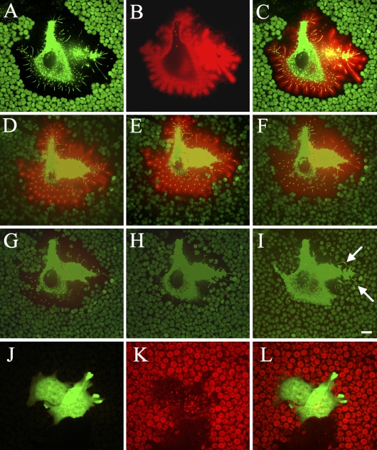
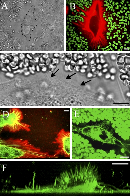
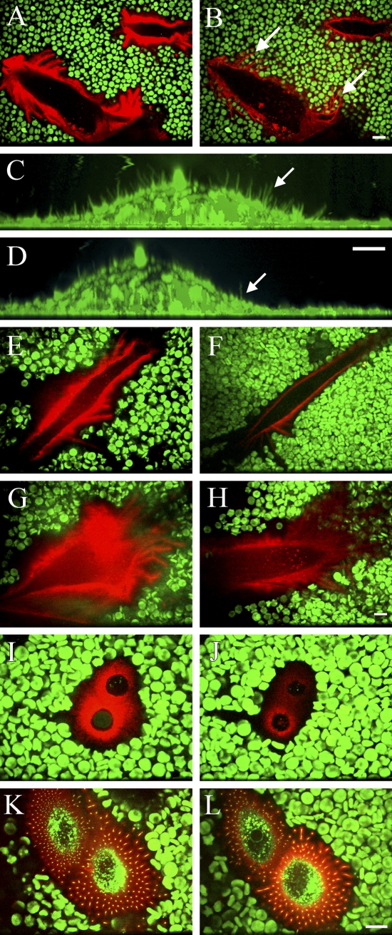
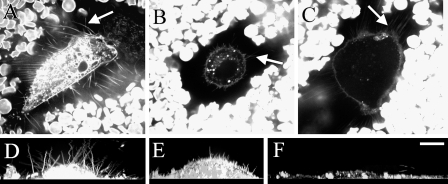
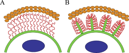
Many cell types wear up to 20-mum-wide hyaluronidase-sensitive surface coats, detected by exclusion of sedimenting particles like fixed erythrocytes. The structure of the coat is enigmatic, being apparently too thick to be accounted by random coils or even extended chains of just hyaluronan attached to cell surface. We have shown that hyaluronan synthesis enforced by green fluorescent protein-hyaluronan synthase transfection creates microvillous protrusions. The idea that the plasma membrane protrusions rather than hyaluronan alone is responsible for the exclusion space was studied with a fluorescent probe for hyaluronan and a dye with membrane affinity, applied to live cell cultures. Mesothelial and smooth muscle cells, fibroblasts, and chondrocytes, all known for their endogenously active hyaluronan synthesis, showed hyaluronan-coated plasma membrane protrusions, barely visible in phase contrast microscopy. Treatment with hyaluronidase and inhibition of hyaluronan synthesis caused retraction of the protrusions unless they were attached to substratum. Hyaluronan and the exclusion space were reduced, but did not disappear, by purified hyaluronan hexasaccharides that compete with hyaluronan attached to CD44. The results suggest that slender plasma membrane protrusions are an inherent feature of hyaluronan coats, form their scaffold, and largely result from ongoing hyaluronan synthesis in their plasma membrane. This manuscript contains online supplemental material at http://www.jhc.org. Please visit this article online to view these materials.


Figures

Figure 1
Figure 1
Confocal images of hyaluronan-coated microvilli on live MCF-7 cells transfected with green fluorescent protein (GFP)-hyaluronan synthase (HAS)3. In single optical sections, the exclusion space is indicated by erythrocytes, and GFP-HAS3 in cell protrusions is shown (A), hyaluronan around the same cell visualized with a hyaluronan binding probe (B), and a composite image (C). (D–I) Three-dimensional images composed of horizontal stacks of the same cell before (D) and 1 (E), 2 (F), 3 (G), 4 (H), and 5 min (I) after the addition of Streptomyces hyaluronidase (10 TRU/ml). Arrows in I point out hyaluronidase-resistant extensions adhered to the substratum. Red, hyaluronan; green particles, red blood cell autofluorescence; green color in the cell, GFP-HAS3. Control cells transfected with a GFP-expressing vector are presented in J–L; green color, GFP; red particles, red blood cells; red color in the cell, hyaluronan. Bar = 10 μm.
Figure 2
Figure 2
Hyaluronan-coated plasma membrane extensions in live LP-9 cells. In regular phase contrast microscopy, a live human mesothelial cell is surrounded by a wide space that excludes erythrocytes (A). Hyaluronan-rich streaks (red) around a mesothelial cell correspond to areas free of erythrocytes (green), suggesting underlying cell protrusions (B). High-resolution phase contrast microscopy shows thin lines probably representing the protrusions (C, arrows), confirmed by double staining for hyaluronan and the FM1-43 membrane marker (D). Protrusions illustrated by the membrane marker FM1-43 and the exclusion space by erythrocytes (both green) are shown together in E. A vertical view of a living LP-9 cell labeled with FM1-43 also shows extensions protruding upward (F). Bar = 10 μm.
Figure 3
Figure 3
Role of hyaluronan in the maintenance of the exclusion space and membrane protrusions. LP-9 mesothelial cells (A,B) and fibroblasts (C,D) before (A,C) and after (B,D) treatment with the Streptomyces hyaluronidase (10 turbidity reducing units/ml, 20 min). Note that the extensions adhered to the substratum do not retract concomitantly with the removal of hyaluronan (B, arrows), whereas most of the upward oriented, shorter extensions do retract (C,D, arrows). The exclusion space of a mesothelial cell and hyaluronan before (E) and after (F) treatment with the hyaluronan synthesis inhibitor, 4-methylumbelliferone (4-MU; 1 mM, 4 hr). A mesothelial cell (G,H), chondrocyte (I,J), and GFP-Has overexpressing MCF-7 cell (K,L) before (G,I,K) and after (H,J,L) treatment with hyaluronan hexasaccharides (0.2 mg/ml, 2 hr). Single optical sections are shown in A, B, and E–L, and vertical sections from compressed image stacks are shown in C and D. The same cell is represented in each pair of figures. Hyaluronan, red; FM1-43 membrane marker, green (C,D); GFP-HAS3, green (K–L); erythrocytes, green. Bar = 10 μm.
Figure 4
Figure 4
Plasma membrane protrusions (arrows) and the exclusion space of different cells (A–C). Membranes were labeled with FM1-43, and sedimenting erythrocytes show the exclusion space in single optical sections (A–C). Plasma membrane morphology as a side view obtained from compressed series of optical sections (D–F). Human tumor stromal fibroblast (A,D), bovine primary chondrocyte (B,E), and rabbit aortic smooth muscle cell (C,F) secreting ∼305, 44, and 21 ng hyaluronan/10,000 cells/24 hr, respectively (Table 1). Bar = 10 μm.
Figure 5
Figure 5
Schematic models for the hyaluronan coat. (A) A model in which extended hyaluronan chains (red), attached to plasma membrane (green) by hyaluronan receptors or hyaluronan synthases, account for the whole thickness of the exclusion space. (B) These data suggest a model where plasma membrane protrusions give a scaffold to the hyaluronan coat that excludes erythrocytes (orange ovals). The protrusions are supported by hyaluronan on their surface. Blue circles in the cell represent nucleus.

References

    1. Bard JB, McBride WH, Ross AR (1983) Morphology of hyaluronidase-sensitive cell coats as seen in the SEM after freeze-drying. J Cell Sci 62:371–383 - PubMed
    1. Brinck J, Heldin P (1999) Expression of recombinant hyaluronan synthase (HAS) isoforms in CHO cells reduces cell migration and cell surface CD44. Exp Cell Res 252:342–351 - PubMed
    1. Clarris BJ, Fraser JR (1968) On the pericellular zone of some mammalian cells in vitro. Exp Cell Res 49:181–193 - PubMed
    1. Cohen M, Klein E, Geiger B, Addadi L (2003) Organization and adhesive properties of the hyaluronan pericellular coat of chondrocytes and epithelial cells. Biophys J 85:1996–2005 - PMC - PubMed
    1. de la Motte CA, Hascall VC, Drazba J, Bandyopadhyay SK, Strong SA (2003) Mononuclear leukocytes bind to specific hyaluronan structures on colon mucosal smooth muscle cells treated with polyinosinic acid:Polycytidylic acid: Inter-alpha-trypsin inhibitor is crucial to structure and function. Am J Pathol 163:121–133 - PMC - PubMed

Publication types