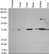
Tissue distribution of human AKR1C3 and rat homolog in the adult genitourinary system
- PMID: 18574251
- PMCID: PMC2516960
- DOI: 10.1369/jhc.2008.951384
Tissue distribution of human AKR1C3 and rat homolog in the adult genitourinary system
Abstract
Human aldo-keto reductase (AKR) 1C3 (type 2 3alpha-hydroxysteroid dehydrogenase/type 5 17beta-hydroxysteroid dehydrogenase) catalyzes androgen, estrogen, and prostaglandin metabolism. AKR1C3 is therefore implicated in regulating ligand access to the androgen receptor, estrogen receptor, and peroxisome proliferator activating receptor gamma in hormone target tissues. Recent reports on close relationships between ARK1C3 and various cancers including breast and prostate cancers implicate the involvement of AKR1C3 in cancer development or progression. We previously described the characterization of an isoform-specific monoclonal antibody against AKR1C3 that does not cross-react with related, >86% sequence identity, human AKR1C1, AKR1C2, or AKR1C4, human aldehyde reductase AKR1A1, or rat 3alpha-hydroxysteroid dehydrogenase (AKR1C9). In this study, a clone of murine monoclonal antibody raised against AKR1C3 was identified and characterized for its recognition of rat homolog. Tissue distribution of human AKR1C3 and its rat homolog in adult genitourinary systems including kidney, bladder, prostate, and testis was studied by IHC. A strong immunoreactivity was detected not only in classically hormone-associated tissues such as prostate and testis but also in non-hormone-associated tissues such as kidney and bladder in humans and rats. The distribution of these two enzymes was comparable but not identical between the two species. These features warrant future studies of AKR1C3 in both hormone- and non-hormone-associated tissues and identification of the rodent homolog for establishing animal models.
Figures


Similar articles
-
Characterization of a monoclonal antibody for human aldo-keto reductase AKR1C3 (type 2 3alpha-hydroxysteroid dehydrogenase/type 5 17beta-hydroxysteroid dehydrogenase); immunohistochemical detection in breast and prostate.Steroids. 2004 Dec;69(13-14):795-801. doi: 10.1016/j.steroids.2004.09.014. Steroids. 2004. PMID: 15582534
-
Increased expression of type 2 3alpha-hydroxysteroid dehydrogenase/type 5 17beta-hydroxysteroid dehydrogenase (AKR1C3) and its relationship with androgen receptor in prostate carcinoma.Endocr Relat Cancer. 2006 Mar;13(1):169-80. doi: 10.1677/erc.1.01048. Endocr Relat Cancer. 2006. PMID: 16601286
-
Differential expression of type 2 3α/type 5 17β-hydroxysteroid dehydrogenase (AKR1C3) in tumors of the central nervous system.Int J Clin Exp Pathol. 2010 Mar 25;3(8):743-54. Int J Clin Exp Pathol. 2010. PMID: 21151387 Free PMC article.
-
Inhibitors of type 5 17β-hydroxysteroid dehydrogenase (AKR1C3): overview and structural insights.J Steroid Biochem Mol Biol. 2011 May;125(1-2):95-104. doi: 10.1016/j.jsbmb.2010.11.004. Epub 2010 Nov 16. J Steroid Biochem Mol Biol. 2011. PMID: 21087665 Free PMC article. Review.
-
AKR1C3 as a target in castrate resistant prostate cancer.J Steroid Biochem Mol Biol. 2013 Sep;137:136-49. doi: 10.1016/j.jsbmb.2013.05.012. Epub 2013 Jun 6. J Steroid Biochem Mol Biol. 2013. PMID: 23748150 Free PMC article. Review.
Cited by
-
Expression of AKR1C3 in renal cell carcinoma, papillary urothelial carcinoma, and Wilms' tumor.Int J Clin Exp Pathol. 2009 Nov 15;3(2):147-55. Int J Clin Exp Pathol. 2009. PMID: 20126582 Free PMC article.
-
Aldo-keto reductase family 1 member C3 (AKR1C3) is a biomarker and therapeutic target for castration-resistant prostate cancer.Mol Med. 2013 Jan 22;18(1):1449-55. doi: 10.2119/molmed.2012.00296. Mol Med. 2013. PMID: 23196782 Free PMC article.
-
Suppressed expression of type 2 3alpha/type 5 17beta-hydroxysteroid dehydrogenase (AKR1C3) in endometrial hyperplasia and carcinoma.Int J Clin Exp Pathol. 2010 Jul 5;3(6):608-17. Int J Clin Exp Pathol. 2010. PMID: 20661409 Free PMC article.
-
Transcriptomic signatures of cellular and humoral immune responses in older adults after seasonal influenza vaccination identified by data-driven clustering.Sci Rep. 2018 Jan 15;8(1):739. doi: 10.1038/s41598-017-17735-x. Sci Rep. 2018. PMID: 29335477 Free PMC article.
-
Expression of AKR1C3 and CNN3 as markers for detection of lymph node metastases in colorectal cancer.Clin Exp Med. 2015 Aug;15(3):333-41. doi: 10.1007/s10238-014-0298-1. Epub 2014 Jun 17. Clin Exp Med. 2015. PMID: 24934327 Free PMC article.
References
-
- Azadzoi KM, Heim VK, Tarcan T, Siroky MB (2004) Alteration of urothelial-mediated tone in the ischemic bladder: role of eicosanoids. Neurourol Urodyn 23:258–264 - PubMed
-
- Barbier O, Lapointe H, El Alfy M, Hum DW, Belanger A (2000) Cellular localization of uridine diphosphoglucuronosyltransferase 2B enzymes in the human prostate by in situ hybridization and immunohistochemistry. J Clin Endocrinol Metab 85:4819–4826 - PubMed
-
- Campean V, Theilig F, Paliege A, Breyer M, Bachmann S (2003) Key enzymes for renal prostaglandin synthesis: site-specific expression in rodent kidney (rat, mouse). Am J Physiol Renal Physiol 285:F19–32 - PubMed
-
- de Jongh R, van Koeveringe GA, van Kerrebroeck PE, Markerink-van Ittersum M, de Vente J, Gillespie JI (2007) The effects of exogenous prostaglandins and the identification of constitutive cyclooxygenase I and II immunoreactivity in the normal guinea pig bladder. BJU Int 100:419–429 - PubMed
-
- Desmond JC, Mountford JC, Drayson MT, Walker EA, Hewison M, Ride JP, Luong QT, et al. (2003) The aldo-keto reductase AKR1C3 is a novel suppressor of cell differentiation that provides a plausible target for the non-cyclooxygenase-dependent antineoplastic actions of nonsteroidal anti-inflammatory drugs. Cancer Res 63:505–512 - PubMed
Publication types
MeSH terms
Substances
LinkOut - more resources
Full Text Sources
Other Literature Sources
Molecular Biology Databases
Research Materials
Miscellaneous

