. 2008 Aug;88(8):865-72.
doi: 10.1038/labinvest.2008.56. Epub 2008 Jun 23.

Expression of specific hepatocyte and cholangiocyte transcription factors in human liver disease and embryonic development

Affiliations

Expression of specific hepatocyte and cholangiocyte transcription factors in human liver disease and embryonic development

Pallavi B Limaye et al. Lab Invest. 2008 Aug.

Abstract

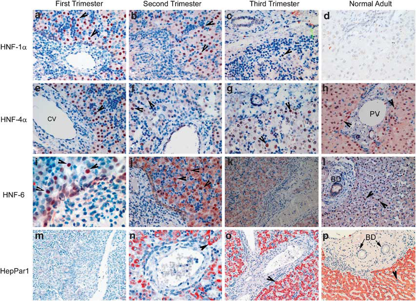
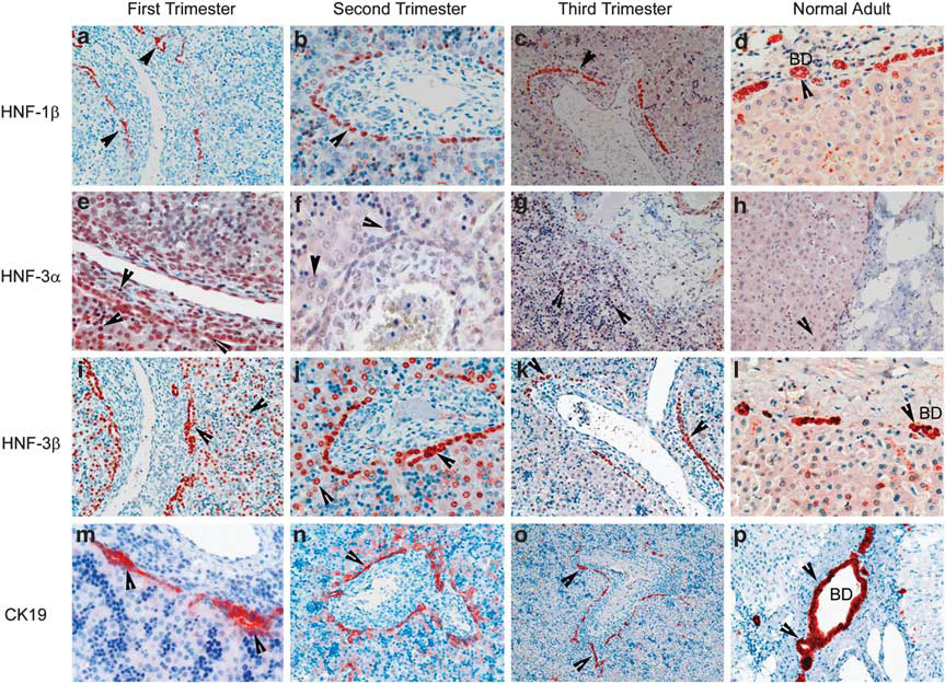
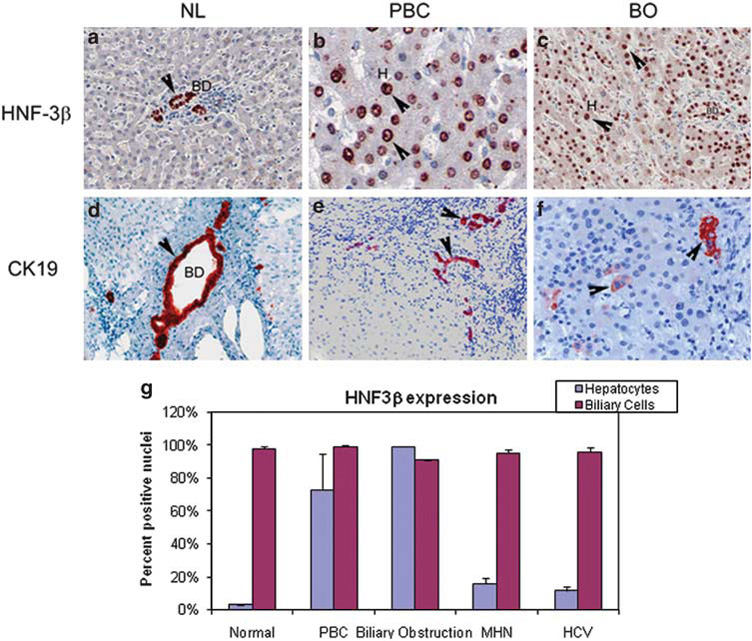
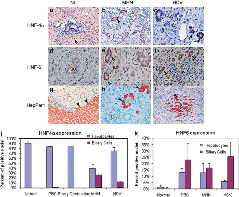
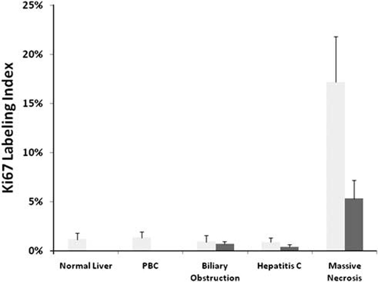
Transcription factors are major determinants of cell-specific gene expression in all cell types. Studies in rodent liver have shown that alterations in transcription factor expression determine lineage specification during fetal liver development and signify transdifferentiation of cells of the biliary compartment into 'oval' cells and eventually hepatocytes in adult liver. We examined the cellular localization of hepatocyte- or BEC-associated transcription factors in human fetal and adult liver and in diseases in which transdifferentiation between hepatocytes and biliary cells may play a role. In the normal adult human liver, hepatocyte nuclear factor (HNF)4 alpha and HNF6 appeared exclusively in hepatocytes; HNF1beta, HNF3alpha, and HNF3beta were observed only in BEC. During fetal development both BEC and hepatocytes expressed HNF3alpha, HNF3beta, and HNF6. HNF1alpha was expressed only in fetal hepatocytes. We further examined expression of transcription factors in massive hepatic necrosis and in specific types of chronic liver disease. Hepatocyte-associated transcription factors HNF4 alpha and HNF6 also appeared in BEC in massive hepatic necrosis and chronic hepatitis C virus infection. Similarly, HNF3beta that is expressed only in BEC in normal adult liver was also observed in hepatocytes in primary biliary cirrhosis and chronic biliary obstruction. These data mimic previous findings in rodents in which hepatocyte-associated transcription factors appear in biliary cells prior to emergence of oval cells, which function as progenitor cells for hepatocytes when the regenerative capacity of the latter is compromised.


Conflict of interest statement

DISCLOSURE/DUALITY OF INTEREST

The authors state no conflict of interest.

Figures

Figure 1
Figure 1. Immunohistochemical staining of hepatocyte-specific transcription factors in the developing human fetal liver and in normal human adult liver
Nuclear staining of HNF1α (a–d) appears only in fetal hepatocytes in the first and second trimester. HNF4α (e–h) is seen only in hepatocyte nuclei at all stages of fetal and adult liver. HNF6 (i–l) is expressed in both hepatocytes and biliary cells in the first two trimesters, following which its expression is limited to hepatocytes. Cytoplasmic staining of HepPar1 was seen only in hepatocytes. It was weakly positive in the first trimester and strongly positive in the second and third trimester and adult liver (m–p). Arrows indicate positive staining. CV, central vein; PV, portal vein; BD, bile duct.
Figure 2
Figure 2. Immunohistochemical staining of biliary-specific transcription factors in the developing fetal liver and in normal adult liver
Nuclear staining of HNF1β (a–d) and HNF3β (i–l) is seen in biliary cells of all stages. HNF3α (e–h) is expressed in biliary cells only in the first trimester. Cytoplasmic staining of CK19 in BEC in fetal and adult liver (m–p). The photomicrographs also illustrate the formation of ductal plate during the different trimesters. Arrows indicate positive staining. BD, bile duct; CV, central vein; PV, portal vein.
Figure 3
Figure 3. Expression of biliary-associated transcription factor HNF3β in hepatocytes, in cases of late-stage primary biliary cirrhosis (PBC) and chronic biliary obstruction (BO)
(a–f) Immunohistochemical staining of HNF3β and CK19 in normal vs PBC and BO. Hepatocytes are negative whereas bile ducts are positive for HNF3β in normal adult liver (a) however, hepatocytes stain positive for HNF3β in PBC (b) and BO (c). In normal adult liver, hepatocytes are negative (d) whereas bile ducts are positive for CK19 in PBC (e) and BO (f). Arrows indicate positive staining. BD, bile duct; CV, central vein; PV, portal vein. (g) Quantitative analysis of HNF3β immunostaining in the liver diseases in hepatocytes vs BEC. The bars indicate the mean and standard error from cell counts of separate cases, each disease from a number of separate specimens as described in ‘Materials and Methods’. Blue, hepatocytes; red, biliary cells.
Figure 4
Figure 4. Expression of hepatocyte-associated transcription factors in biliary epithelial cell (BEC) in different hepatic diseases
(a–i) Immunohistochemical staining of HNF4α, HNF6, and HepPar1 in normal vs massive hepatic necrosis and end-stage HCV cirrhotic liver. BEC in normal adult liver are negative for HNF4α (a) and HNF6 (d). BEC in massive hepatic necrosis (MHN) express HNF4α (b) and HNF6 (e). BEC also expresses HNF6 in MHN (c) and HCV (f). The appearance of the single-gene hepatocyte marker HEPAR is also prominent is many ductular cells. Arrows indicate positive staining. BD, bile duct; CV, central vein; PV, portal vein. (j) Quantitative analysis of HNF4α immunostaining in liver disease in hepatocytes vs BEC. (k) Quantitative analysis of HNF6 immunostaining in liver disease in hepatocytes vs BEC. Both HNF4α and HNF6 emerge in BECs in diseases associated with chronic hepatocyte injury. In both (j and k), the bars indicate the mean and standard error from cell counts of separate cases, each disease from a number of separate specimens as described in ‘Materials and Methods’. Blue, hepatocytes; red, biliary cells.
Figure 5
Figure 5
Proliferation rates of hepatocytes and BEC in different hepatic diseases, as measured by the nuclear expression of Ki67, a marker of nuclear DNA synthesis. The bars indicate the mean and standard error from cell counts of separate cases, each disease from a number of separate specimens as described in ‘Materials and methods’. Light gray, hepatocytes; dark gray, biliary cells.

References

    1. Duncan SA. Transcriptional regulation of liver development. Dev Dyn. 2000;219:131–142. - PubMed
    1. Lee CS, Friedman JR, Fulmer JT, et al. The initiation of liver development is dependent on Foxa transcription factors. Nature. 2005;435:944–947. - PubMed
    1. Nijjar SS, Wallace L, Crosby HA, et al. Altered Notch ligand expression in human liver disease: further evidence for a role of the Notch signaling pathway in hepatic neovascularization and biliary ductular defects. Am J Pathol. 2002;160:1695–1703. - PMC - PubMed
    1. Nijjar SS, Crosby HA, Wallace L, et al. Notch receptor expression in adult human liver: a possible role in bile duct formation and hepatic neovascularization. Hepatology. 2001;34:1184–1192. - PubMed
    1. Crosby HA, Hubscher SG, Joplin RE, et al. Immunolocalization of OV-6, a putative progenitor cell marker in human fetal and diseased pediatric liver. Hepatology. 1998;28:980–985. - PubMed

Publication types

Substances