Review
. 2008 Jul;8(7):559-68.
doi: 10.1038/nri2314.

Interferon-inducible antiviral effectors

Affiliations
Review

Interferon-inducible antiviral effectors

Anthony J Sadler et al. Nat Rev Immunol. 2008 Jul.

Abstract

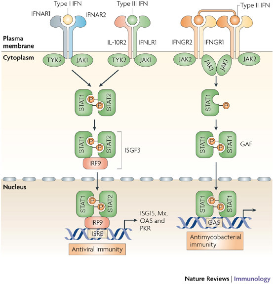
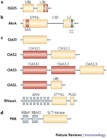
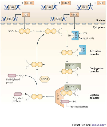
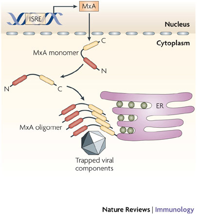
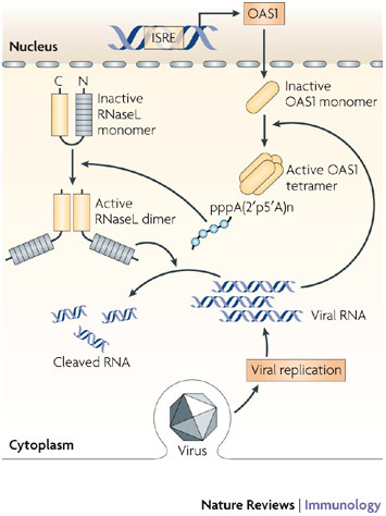
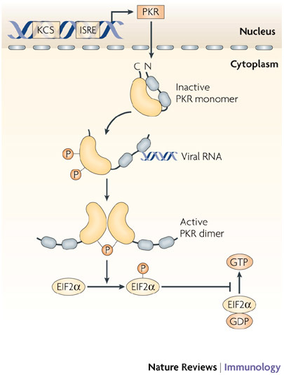
Since the discovery of interferons (IFNs), considerable progress has been made in describing the nature of the cytokines themselves, the signalling components that direct the cell response and their antiviral activities. Gene targeting studies have distinguished four main effector pathways of the IFN-mediated antiviral response: the Mx GTPase pathway, the 2',5'-oligoadenylate-synthetase-directed ribonuclease L pathway, the protein kinase R pathway and the ISG15 ubiquitin-like pathway. As discussed in this Review, these effector pathways individually block viral transcription, degrade viral RNA, inhibit translation and modify protein function to control all steps of viral replication. Ongoing research continues to expose additional activities for these effector proteins and has revealed unanticipated functions of the antiviral response.


Figures

Figure 1
Figure 1. Interferon receptor signalling.
The action of the interferons (IFNs) is mediated through three receptor complexes: a heterodimer of IFNα receptor 1 (IFNAR1) and IFNAR2 binds type I IFNs; the interleukin-10 receptor 2 (IL-10R2) associates with IFNLR1 (IFNλ receptor 1) to bind the three IFNλ subtypes; and a tetramer consisting of two IFNGR2 (IFNγ receptor 2) chains and two IFNGR1 chains binds dimers of the type II IFNγ. Following binding by type I IFNs, signal transduction is initiated by pre-associated tyrosine kinases (JAK1 and TYK2 (tyrosine kinase 2)), which phosphorylate IFNAR1 and leads to the recruitment and phosphorylation of the signal transducers and activators of transcription (STATs). STAT heterodimers associate with IFN-regulatory factor 9 (IRF9) to form IFN-stimulated gene factor 3 (ISGF3), or STAT homodimers to form the IFNγ activation factor (GAF). These complexes translocate to the nucleus to induce IFN-stimulated genes from IFN-stimulated response elements (ISREs) or GAS promoter elements, for type I and type III, or type II IFN responses, respectively. Divergence from this simplified signalling pathway can occur, for example, type I IFNs are reported to elicit STAT homodimers, and more complicated interplay, with activation of other STAT proteins, occurs than is shown here. ISG15, IFN-stimulated protein of 15 kDa; Mx, myxovirus resistance; OAS, 2′,5′-oligoadenylate synthetase; PKR, protein kinase R.
Figure 2
Figure 2. Domain structure of antiviral proteins.
a | Interferon-stimulated protein of 15 kDa (ISG15) contains two ubiquitin-like (UBL) domains. The C-terminal UBL encodes conserved hydrophobic patches, charged pockets (indicated by vertical bars) and diglycine residues similar to ubiquitin and other ubiquitin-like proteins. Accordingly, the C-terminal UBL is sufficient for activation and transfer of ISG15 to E1 and E2 enzymes. However, the N-terminal UBL seems to be necessary for E3-mediated conjugation to protein substrates. b | The distinguishing features of the dynamin protein family of Mx GTPases are the large GTP-binding domain (DYNc) that contains three consensus GTP-binding elements (not indicated) and a self-assembly sequence (SAS), the central interactive domain (CID), and the C-terminal leucine zipper (LZ). The LZ region contains a coiled-coil (CC) domain at its C terminus that forms bundles of α-helices involved in protein–protein interactions. c | The 2′,5′-oligoadenylate synthetase 1 (OAS1), OAS2 and OAS3 proteins contain 1, 2 or 3 copies respectively, of the OAS domains. Duplicated OAS domains in OAS2 and OAS3, and the single domain in OASL (OAS-like) are catalytically inactive (shown in red). The C-termini of all the OAS proteins are variable, with OASL also encoding a unique UBL domain. The OAS domain produces 2′,5′-oligoadenylates that activate ribonuclease L (RNaseL). RNaseL has eight N-terminal ankyrin repeats (ANK), which mediate protein–protein interactions, and two kinase-like domains, a Ser/Thr/Tyr kinase (STYKc) and a PUG domain that are found in some kinases and several other nuclear proteins. However, neither domain has functional kinase activity. d |Protein kinase R (PKR) contains two repeated RNA-binding motifs (RBMs), which together constitute the N-terminal RNA-binding domain. On binding of viral RNA, steric inhibition of enzyme activity is relieved, leading to the activation of the C-terminal serine/threonine protein kinase domain (S/T-kinase).
Figure 3
Figure 3. Mechanism of action of ISG15.
The expression of interferon (IFN)-stimulated protein of 15 kDa (ISG15), the E1-activating enzyme UBE1L (E1-like ubiquitin-activating enzyme) and multiple E2-conjugating enzymes (shown here as an example is UBCH8) and E3-ligase enzymes (such as HERC5 (homologous to the E6-associated protein C terminus domain and RCC1-like domain containing protein 5)) is coordinately induced by type I IFNs through IFN-stimulated response elements (ISREs) in their respective gene promoter regions. E1, E2 and E3 proteins sequentially catalyse the conjugation of ISG15 to numerous protein substrates to modulate pleiotropic cellular responses to inhibit virus production. This process (known as ISGylation) is reversibly regulated by proteases (such as ubiquitin-specific protease 18 (USP18)), which are also induced by IFNs.
Figure 4
Figure 4. Mechanism of action of MxA.
Following stimulation with type I interferons (IFNs), MXA (myxovirus-resistance A) gene expression is induced through an IFN-stimulated response element (ISRE) in the gene promoter. The MxA protein accumulates in the cytoplasm on intracellular membranes (such as the endoplasmic reticulum, ER) as oligomers formed by association between the leucine zipper (LZ) domain and central interactive domain of the protein. Following viral infection, MxA monomers are released and bind viral nucleocapsids or other viral components, to trap and then degrade them.
Figure 5
Figure 5. The OAS1–RNaseL antiviral pathway.
2′,5′-oligoadenylate synthetase 1 (OAS1) is expressed at low constitutive levels and is upregulated by type I interferons (IFNs). OAS1 protein accumulates in the cell cytoplasm as an inactive monomer. Following activation by viral double-stranded RNA (dsRNA), the enzyme oligomerizes to form a tetramer that synthesizes 2′,5′-oligoadenylates that, in turn, activate the constitutively expressed inactive ribonuclease L (RNaseL). The binding of 2′,5′-oligoadenylates to RNaseL triggers the dimerization of enzyme monomers, through their kinase-like domains, and this then enables RNAseL to cleave cellular (and viral) RNAs. ISRE, IFN-stimulated response element.
Figure 6
Figure 6. Mechanism of action of PKR.
Protein kinase R (PKR) is constitutively expressed, and is also induced by type I interferons (IFNs) under the control of a kinase conserved sequence (KCS) and IFN-stimulated response element (ISRE) in the promoter of PKR. The kinase accumulates in the nucleus and cytoplasm as an inactive monomer, which is activated directly by viral RNAs, and by several other ligands, such as ceramide or the protein activator PACT (protein activator of the IFN-inducible protein kinase). Following activation, PKR monomers are phosphorylated and dimerize to form the active enzyme. Activated PKR regulates several cell signalling pathways through mechanisms that have not been fully explained, but a crucial function of PKR in viral defence is the inhibition of translation by phosphorylation of eukaryotic translation initiation factor 2α (EIF2α).

References

    1. Isaacs A, Lindenmann J. Virus interference. I. The interferon. Proc. R. Soc. Lond. [Biol.] 1957;147:258–267. doi: 10.1098/rspb.1957.0048. - DOI - PubMed
    1. Borden EC, et al. Interferons at age 50: past, current and future impact on biomedicine. Nature Rev. Drug Discov. 2007;6:975–990. doi: 10.1038/nrd2422. - DOI - PMC - PubMed
    1. Levraud JP, et al. Identification of the zebrafish IFN receptor: implications for the origin of the vertebrate IFN system. J. Immunol. 2007;178:4385–4394. doi: 10.4049/jimmunol.178.7.4385. - DOI - PubMed
    1. O'Connell RM, et al. Type I interferon production enhances susceptibility to Listeria monocytogenes infection. J. Exp. Med. 2004;200:437–445. doi: 10.1084/jem.20040712. - DOI - PMC - PubMed
    1. Muller U, et al. Functional role of type I and type II interferons in antiviral defense. Science. 1994;264:1918–1921. doi: 10.1126/science.8009221. - DOI - PubMed

Publication types