. 2008 Jun 25;3(6):e2563.
doi: 10.1371/journal.pone.0002563.

Mesenchymal stem cells in early entry of breast cancer into bone marrow

Affiliations

Mesenchymal stem cells in early entry of breast cancer into bone marrow

Kelly E Corcoran et al. PLoS One. .

Abstract

Background: An understanding of BC cell (BCC) entry into bone marrow (BM) at low tumor burden is limited when compared to highly metastatic events during heavy tumor burden. BCCs can achieve quiescence, without interfering with hematopoiesis. This occurs partly through the generation of gap junctions with BM stroma, located close to the endosteum. These events are partly mediated by the evolutionary conserved gene, Tac1.

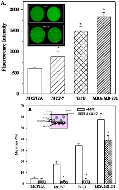
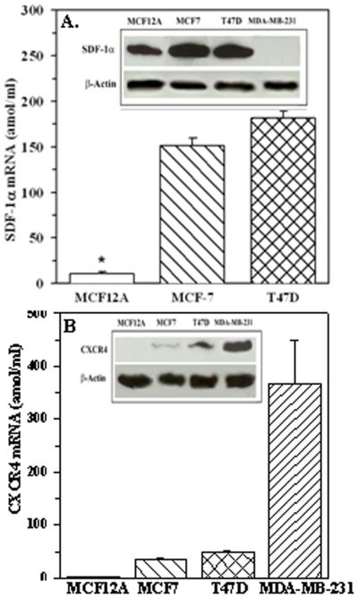
Methodology/principal findings: This study focuses on the role of mesenchymal stem cells (MSCs), Tac1, SDF-1 and CXCR4 in BCC entry into BM. The model is established in studies with low numbers of tumor cells, and focuses on cancer cells with low metastatic and invasion potential. This allowed us to recapitulate early event, and to study cancer cells with low invasive potential, even when they are part of larger numbers of highly metastatic cells. A novel migration assay showed a facilitating role of MSCs in BCC migration across BM endothelial cells. siRNA and ectopic expression studies showed a central role for Tac1 and secondary roles for SDF-1alpha and CXCR4. We also observed differences in the mechanisms between low invasive and highly metastatic cells. The in vitro studies were verified in xenogeneic mouse models that showed a preference for low invasive BCCs to BM, but comparable movement to lung and BM by highly metastatic BCCs. The expressions of Tac1 and production of SDF-1alpha were verified in primary BCCs from paired samples of BM aspirates and peripheral blood.

Conclusions/significance: MSC facilitate BCC entry into BM, partly through Tac1-mediated regulation of SDF-1alpha and CXCR4. We propose a particular population of BCC with preference for BM could be isolated for characterization. This population might be the subset that enter BM at an early time period, and could be responsible for cancer resurgence and resistance to current therapies.


Conflict of interest statement

Competing Interests: The authors have declared that no competing interests exist.

Figures

Figure 1
Figure 1. Adhesion and transmigration of BCCs.
A. Image is shown of bilayered adherence between MSCs and T47D. B. The adherence of MCF12A, MCF7, T47D and MDA-MB-231 to MSCs are presented as the mean fluorescence intensities, n = 5. Each experiment was done with MSCs from a different donor. C. Migration of BCCs across BM endothelial cells with or without MSCs placed on the lower side of the insert (Refer to inset). The results are presented as the mean percent migration±SD, n = 5. * p<0.05 vs. assays with MSCs.
Figure 2
Figure 2. Relative expression levels of CXCR4, and SDF-1α mRNA in BCCs.
SDF-1α (A) and CXCR4 (B) mRNA levels were quantitated in BCCs and presented as mean±SD, n = 5. Representative of four western blots for SDF-1α (A, inset) and CXCR4 (B, inset) with membrane extracts from four different cell lines.
Figure 3
Figure 3. Role of CXCR4 and SDF-1 in BCC migration and adherence.
A. Representative of three western blots for CXCR4 and SDF-1α with combined whole cell and membrane extracts from T47D, and CXCR4 for MDA-MB-231. Lanes 1: Untransfected; 2: mutant siRNA; 3: wild-type siRNA. B. Representative of three western blots for SDF-1α (left panel) from three cell passages, and SDF-1α and CXCR4 for MSCs, each from a different donor. C. Adherence studies with T47D and MDA-MB-231 as untransfected, CXCR4 knockdown and control with mutant siRNA, are presented as the mean fluorescence±SD, n = 5. D. Migration studies were done with the same cells and the results presented as mean % migration±SD, n = 5. *p<0.05 vs. untransfected or mutant siRNA or wild-type siRNA. E. Migration studies with T47D, and MSCs knockdown for SDF-1α or mutant siRNA, or untransfected. F. Migration studies were similarly done with CXCR4 knockdown cells. The results are presented as the mean % migration±SD, n = 5. *p<0.05 vs. untransfected or mutant siRNA.
Figure 4
Figure 4. Effects of Tac1 on adhesion and transmigration of T47D.
A. Adhesion of Tac1 knockdown T47D and MDA-MB-231, vector transfectants or untransfected cells. B. Transmigration studies with the cells described in ‘A’. Results are presented as mean±SD, n = 5. * p<0.05 vs. untransfected or vector transfectants.
Figure 5
Figure 5. Migration with Tac1 knockdown T47D and MDA-MB-231, expressed with CXCR4 and/or SDF-1α.
A. Cells were analyzed for CXCR4 by flow cytometry (n = 4). B. SDF-1α production by ELISA, mean±SD, n = 5. *p<0.05 vs. untransfected for vector-transfected T47D. C. Western blots with whole cell lysates from Tac1 knockdown cells and/or expressed for SDF-1α and/or CXCR4 (Lanes 1: Tac1 siRNA; 2: Tac1 siRNA+SDF-1α; 3: Tac1 siRNA+CXCR4; 4: Tac1 siRNA+SDF-1α+CXCR4. D. Transmigration assays with T47D and MDA-MDB-231 and their variants as for ‘A’. * p<0.05 vs. Tac1 siRNA. ** p>0.05 vs. Tac1 siRNA.
Figure 6
Figure 6. Representative of three sections obtained from femurs nude mice after 24 h of injections with T47D, untransfected, knockdown (KO) for Tac1; Tac1 knockdown with re-expressions of SDF-1α and/or CXCR4.
The femurs were treated as described for Figure S1 and the slides were triple labeled as for Figure S3 with PE-anti-CD105, FITC-anti cytokeratin and APC-anti-CD31. Arrows in the merged images depict cytokeratin (+) cells in contact with MSCs (green and red), but not in contact with CD31+ cells (blue).
Figure 7
Figure 7. Tac1 mRNA in cytokeratin-expressing cells from BM aspirates and peripheral blood of Stage III BC patients.
Pairs of BM aspirates (A) and peripheral blood (B) were taken from BC patients at the time of diagnosis. Cytokeratin positive cells were selected and then subjected to RT-PCR for Tac1 mRNA and GAPDH.

References

    1. Bigioni M, Benzo A, Irrissuto C, Maggi CA, Goso C. Role of NK-1 and NK-2 tachykinin receptor antagonism on the growth of human breast carcinoma cell line MDA-MB-231. Anticancer Drugs. 2007;16:1083–1089. - PubMed
    1. Patel HJ, Ramkissoon SH, Patel PS, Rameshwar P. Transformation of breast cells by truncated neurokinin-1 receptor is secondary to activation by preprotachykinin-A peptides. Proc Natl Acad Sci USA. 2005;102:17436–17441. - PMC - PubMed
    1. Rao G, Patel PS, Idler SP, Maloof P, Gascon P, et al. Facilitating role of preprotachykinin-I gene in the integration of breast cancer cells within the stromal compartment of the bone marrow: A model of early cancer progression. Cancer Res. 2004;64:2874–2881. - PubMed
    1. Singh D, Joshi DD, Hameed M, Qian J, Gascon P, et al. Increased expression of preprotachykinin-I and neurokinin receptors in human breast cancer cells: Implications for bone marrow metastasis. Proc Natl Acad Sci USA. 2000;97:388–393. - PMC - PubMed
    1. Greco SJ, Corcoran KE, Cho KJ, Rameshwar P. Tachykinins in the emerging immune system: Relevance to bone marrow homeostasis and maintenance of hematopoietic stem cells. Frontiers Biosci. 2004;9:1782–1793. - PubMed