Review
. 2008 Aug;130(2):247-62.
doi: 10.1007/s00418-008-0452-5. Epub 2008 Jun 25.

The renal cortical interstitium: morphological and functional aspects

Affiliations
Review

The renal cortical interstitium: morphological and functional aspects

Brigitte Kaissling et al. Histochem Cell Biol. 2008 Aug.

Abstract

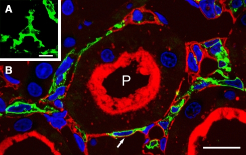
The renal interstitial compartment, situated between basement membranes of epithelia and vessels, contains two contiguous cellular networks. One network is formed by interstitial fibroblasts, the second one by dendritic cells. Both are in intimate contact with each other. Fibroblasts are interconnected by junctions and connected to basement membranes of vessels and tubules by focal adhesions. Fibroblasts constitute the "skeleton" of the kidney. In the renal cortex, fibroblasts produce erythropoietin and are distinguished from other interstitial cells by their prominent F-actin cytoskeleton, abundance of rough endoplasmic reticulum, and by ecto-5'-nucleotidase expression in their plasma membrane. The resident dendritic cells belong to the mononuclear phagocyte system and fulfil a sentinel function. They are characterized by their expression of MHC class II and CD11c. The central situation of fibroblasts suggests that signals from tubules, vessels, and inflammatory cells converge in fibroblasts and elicit an integrated response. Following tubular damage and inflammatory signals fibroblasts proliferate, change to the myofibroblast phenotype and increase their collagen production, potentially resulting in renal fibrosis. The acquisition of a profibrotic phenotype by fibroblasts in renal diseases is generally considered a main causal event in the progression of chronic renal failure. However, it might also be seen as a repair process.


Figures

Fig. 1
Fig. 1
Cortical peritubular interstitium in a mouse kidney (3 μm cryostat section). The interstitial space extends between tubules and capillaries (c). It is occupied by fibroblasts (stars) and their processes, expressing 5′NT (red) and dendritic cells (asterisks), expressing MHC class II (green). Cellular nuclei are labeled in blue; the tissue structure is shown by differential interference contrat (DIC). The brush border of proximal tubules (P) is weakly labeled by 5′NT. Bar 10 μm
Fig. 2
Fig. 2
Interstitial cells, highlighted by immunogold staining for ecto-5′-nucleotidase (a) and MHC class II (b) on consecutive cryostat sections. a 5′NT, detected by immunogold, depicts the abundance of interstitial fibroblasts (arrow); the brush border of proximal tubules (P) is stained too. b Immunogold labeling for MHC class II reveals the distribution of dendritic cells (arrow) throughout the interstitium. Bar 50 μm
Fig. 3
Fig. 3
Fibroblasts in the cortical peritubular interstitium. a Cryostat section. Fibroblasts, shown by immunogold labeling for 5′NT, bridge the interstitial space between the tubules and capillaries (c); (b) transmission electron microscopy of healty rat kidney. Stellate pericaryon of a fibroblast, connected to the basement membrane of a proximal tubule (1, magnified in insert 1), the basement membrane of a capillary (2, magnified in insert 2) and to another fibroblast process (3, magnified in insert 3). Most cell organelles are located in cellular processes but lack in the pericaryon. Note the bundles of F-actin filaments at the connection sites. Insert 4 rough endoplasmic reticulum in a fibroblast process. Barsa 10 μm, b 1 μm, inserts 0.25 μm
Fig. 4
Fig. 4
Three-dimensional view of a fibroblast in the cortical interstitium of a rat. Scanning electron microscopy, after digestion of the tubular and vascular basement membranes The pericaryon (S) shows characteristic sharp angles; the processes adjacent to the proximal tubule (PT) are extremely attenuated and perforated; the asterisk indicates a dendritic cell enclosed by fibroblast processes, arrows and arrowheads indicate processes of the dendritic cell; C capillary. Bar 10 μm. (Image from Takahashi-Iwanaga (1991) Cell Tissue Res 264:269–281)
Fig. 5
Fig. 5
Fibroblast in the cortical peritubular interstitium of a healthy mouse (transmission electron microscopy). a Stellate pericaryon with a thin cytoplasmic rim and large processes, and close juxtaposition to a lymphocyte; matrix, released by the fibroblast (asterisk); F-actin filaments beneath the plasma membrane (arrow head). b The abundant rough endoplasmatic reticulum (rER), mitochondria and Golgi apparatus are located in the large cell processes; the rER cisterns are densely studded with ribosomes and free ribosomes are frequent. c attachment to the basement membranes of tubules and capillaries frequently occurs by spine-like processes, densely filled with F-actin stress fibers (arrow heads). d Tangential sections of an endothelial cell and part of a fibroblast (F), showing the anchoring of the fibroblast to the basement membrane by spine-like processes. Barsa 4 μm, b 1 μm, c 0.5 μm, d 2 μm
Fig. 6
Fig. 6
Dendritic cells in the cortical peritubular interstitium. a Here comes the sentinel! dendritic cell (green) in the interstitial space. b 1 μm cryostat section; dendritic cells express MHC class II (green), fibroblasts express 5′NT (red), nuclei are stained in blue. Note the juxtaposition of fibroblast processes (arrow) and dendritic cells. P proximal tubule with brush border (red). Bara 1 μm, b 10 μm
Fig. 7
Fig. 7
Dendritic cells in the cortical peritubular interstium. a, b Cryostat sections, immunogold staining for MHC class II, showing dendritic cells, and differential interference contrast (DIC). The processes of the dendritic cells have extensive contact with fibroblasts (arrow). c the peripheral processes of dendritic cells are virtually devoid of large cell organelles and appear lighter than the processes of the F-actin filament-displaying fibroblast processes (arrows), here labeled with immunogold for ecto-5′-nucleotidase. d The pericaryon displays a rounded nucleus, numerous mitochondria, Golgi apparatus (arrows) and rough endoplasmic reticulum (see insert 1); the cell processes are branched and appear much lighter than those of fibroblasts (arrowheads). Insert 2 adhesion between processes of a dendritic cell and of a lymphocyte. e Birbeck granule, in a dendritic cell. Barsa, b 10 μm; c 0.1 μm; d 1 μm; insert 0.5 μm; e 0.01 μm
Fig. 8
Fig. 8
Gallery of myofibroblasts (transmission electron microscopy). Myofibroblasts show a heterogeneous morphology. a myofibroblasts after 24 h of ureter ligature with transitory features between a “quiescent” fibroblast with narrow rER (around the nucleus) and a typical myofibroblasts with inflated cisterns of rER (in the cellular process); b 2 days after ureter ligature with largely inflated cisterns; c after 3 days showing a stellate shape; d after 3 days of ureter ligature. Bar 2 μm
Fig. 9
Fig. 9
Myofibroblast in the cortical peritubular interstitium in a rat kidney after 3 days of ureter ligature. a The rounded nucleus is surrounded by a large pericaryon, containing enlarged cisterns of rough endoplasmic reticulum (rER); αSMA (arrow) is detectabe traversing the cytoplasm; note the abundant fibrillar matrix in the surrounding of the cell. b Junction between myofibroblasts processes with stress-like F-actin filaments (asterisk) are frequent; basement membrane-like material is apposed to the plasmalemm (arrows); insert bundle of αSMA filaments with dense bodies (arrows). c Inflated rER, filled with flocculent material. Barsa 2 μm; b, c and insert 0.5 μm
Fig. 10
Fig. 10
Glomerular arterioles are ensheathed by ecto-5′-nucleotidase-expressing processes (arrowheads); 5′NT (green); aa afferent arteriole, ae efferent arteriole, G Glomerulus. Barsa 50 μm; b 20 μm
Fig. 11
Fig. 11
Ecto-5′-nucleotidase in the renal cortex of a normemic (a, c) and an anemic (b, d) rat (1 μm cryostat sections; P proximal tubules; c capillaries). Fibroblasts (stars) in anemic rats reveal a markedly increased 5′NT expression and more cellular processes. Bars 10 μm
Fig. 12
Fig. 12
Renal cortex in sham-operated (a) and in ureter-ligated kidneys (b, c) (3 μm cryostat sections, red ecto-5′nucleotidase (5′NT), green alpha smooth muscle actin (αSMA), blue cell nuclei). In controls (a) the interstitium and the brush border of proximal tubules are strongly labeled by 5′NT, αSMA labels exclusively arterial vessels (a). After 2 days of ureter ligature (b) interstitial 5′NT staining decreases, whereas αSMA appears and becomes increasingly prominent throughout the cortex after 3 days (c). Interstitial αSMA-staining progressively appears also in the outer stripe of the outer medulla, seen at the bottom of the images. df Interstitial fibroblast in ureter-ligated kidney after 2 days. The weakly expressed 5′NT is distributed in a granular manner over the plasma membrane and the cytoplasm, αSMA is apparent along the plasma membrane and in the cellular processes. Barac 100 μm, df 10 μm (from Picard et al. 2008)
Fig. 13
Fig. 13
Renal cortex after 4 days of ureter ligation [3 μm cryostat sections; red ecto-5′ nucleotidase (5′NT), green alpha smooth muscle actin (αSMA), blue nuclei]. a Merge of channels, (b) 5′NT, (c) αSMA; the framed area (1) comprises interstitium with still high 5′NT and faint αSMA staining, adjacent to rather intact tubules showing open lumina, the framed area (2) comprises interstitium with strong αSMA and faint 5′NT staining, adjacent to collapsed tubules, showing signs of tubular atrophy. Barac ~100 μm; 1, 2 ~ 10 μm (from Picard et al. 2008)
Fig. 14
Fig. 14
Mitotic cells in the peritubular interstitium in rat kidneys after 1 day (a) and 2 days (b) of ureter-ligation (3 μm cryostat sections; green αSMA, red 5′NT, blue nuclei). The mitotic cell in a heavily expresses 5′NT and faintly αSMA, the adjacent cell shows strong αSMA and faint 5′NT staining. The 2 mitotic cells in b display strong staining for αSMA along the plasma membrane and in the cell processes, 5′NT staining is weak, granular and is seen over the cytoplasm. Bar 10 μm (from Picard et al. 2008)
Fig. 15
Fig. 15
Cortical peritubular fibroblast in a focal inflammation, related to injury of a distal tubule (Transmission electron microscopy). This figure exemplifies the central position of the fibroblast (F), spanned between a proximal tubule (PT) and an injured cell of a distal convoluted tubule (DT), and its juxtaposition to a capillary (c), a lymphocyte (L) and a dendritic cell (D); two thin processes (arrow heads) of the fibroblast seem to embrace the DT. Signals from the different cells may converge in the fibroblast and elicit an integrated response. Insert “kiss”-like adhesions between a fibroblast and a lymphocyte. Bars 2 μm, insert 0.5 μm
Fig. 16
Fig. 16
Focal peritubular inflammation, associated with injured distal tubules (D) in rats thiazide-treated for 72 h. a 1 μm Epon section; accumulation of interstitial cells around a DCT profile with degenerating epithelial cells, the downstream connecting tubule (CNT) segment and the surrounding interstitium is intact; bd 1 μm cryostat sections; (b) dendritic cells (MHC class II, green) and 5′NT-labeled fibroblasts (red) adjacent injured distal tubule; (c) vimentin (green) in mononuclear cells (plus) and in 5′NT-positive (red) fibroblasts (arrows); weak expression of vimentin also in the degenerating distal tubular epithelial cells; (d) some fibroblasts (arrows) show reduced 5′NT (red); MHC II-labeled dendritic cells (green). Bar 10 μm
Fig. 17
Fig. 17
Up-regulation of alpha smooth muscle actin in the interstitium surrounding injured distal tubular profiles (D) of a rat treated for 72 h with a thiazide-diuretic (5 μm cryostat section). Fibroblasts in extensive contact with the injured tubule show the strongest fluorescence for αSMA. More distant fibroblasts, which appear/are connected to the area of injury by cellular processes (arrows), show a weaker upregulation of αSMA; a arteriole. Insert close view of an interstitial αSMA-positive fibroblast. Bars 20 μm; insert 10 μm

References

    1. {'text': '', 'ref_index': 1, 'ids': [{'type': 'DOI', 'value': '10.1007/s004670050624', 'is_inner': False, 'url': 'https://doi.org/10.1007/s004670050624'}, {'type': 'PubMed', 'value': '10454789', 'is_inner': True, 'url': 'https://pubmed.ncbi.nlm.nih.gov/10454789/'}]}
    2. Alcorn D, Maric C, McCausland J (1999) Development of the renal interstitium. Pediatr Nephrol 13:347–354 - PubMed
    1. {'text': '', 'ref_index': 1, 'ids': [{'type': 'DOI', 'value': '10.1038/ki.1997.66', 'is_inner': False, 'url': 'https://doi.org/10.1038/ki.1997.66'}, {'type': 'PubMed', 'value': '9027726', 'is_inner': True, 'url': 'https://pubmed.ncbi.nlm.nih.gov/9027726/'}]}
    2. Bachmann S, Ramasubbu K (1997) Immunohistochemical colocalization of the alpha-subunit of neutrophil NADPH oxidase and ecto-5′-nucleotidase in kidney and liver. Kidney Int 51:479–482 - PubMed
    1. {'text': '', 'ref_index': 1, 'ids': [{'type': 'PubMed', 'value': '8429197', 'is_inner': True, 'url': 'https://pubmed.ncbi.nlm.nih.gov/8429197/'}]}
    2. Bachmann S, Le Hir M, Eckardt KU (1993) Co-localization of erythropoietin mRNA and ecto-5′-nucleotidase immunoreactivity in peritubular cells of rat renal cortex indicates that fibroblasts produce erythropoietin. J Histochem Cytochem 41:335–341 - PubMed
    1. {'text': '', 'ref_index': 1, 'ids': [{'type': 'DOI', 'value': '10.1159/000057607', 'is_inner': False, 'url': 'https://doi.org/10.1159/000057607'}, {'type': 'PubMed', 'value': '12021522', 'is_inner': True, 'url': 'https://pubmed.ncbi.nlm.nih.gov/12021522/'}]}
    2. Bertani T, Mazzucco G, Monga G (2002) How glomerular extracapillary proliferation might lead to loss of renal function: light microscopic and immunohistochemical investigation. Nephron 91:74–78 - PubMed
    1. {'text': '', 'ref_index': 1, 'ids': [{'type': 'DOI', 'value': '10.1681/ASN.2005070730', 'is_inner': False, 'url': 'https://doi.org/10.1681/asn.2005070730'}, {'type': 'PubMed', 'value': '17135399', 'is_inner': True, 'url': 'https://pubmed.ncbi.nlm.nih.gov/17135399/'}]}
    2. Broekema M, Harmsen MC, van Luyn MJ, Koerts JA, Petersen AH, van Kooten TG, van Goor H, Navis G, Popa ER (2007) Bone marrow-derived myofibroblasts contribute to the renal interstitial myofibroblast population and produce procollagen I after ischemia/reperfusion in rats. J Am Soc Nephrol 18:165–175 - PubMed