

Inhibition of beta-catenin signaling in articular chondrocytes results in articular cartilage destruction
- PMID: 18576323
- PMCID: PMC2667964
- DOI: 10.1002/art.23614
Inhibition of beta-catenin signaling in articular chondrocytes results in articular cartilage destruction
Abstract
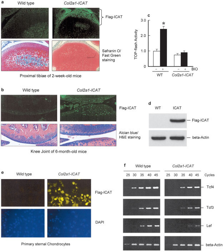
Objective: Osteoarthritis is a degenerative joint disease whose molecular mechanism is currently unknown. Wnt/beta-catenin signaling has been demonstrated to play a critical role in the development and function of articular chondrocytes. To determine the role of beta-catenin signaling in articular chondrocyte function, we generated Col2a1-ICAT-transgenic mice to inhibit beta-catenin signaling in chondrocytes.
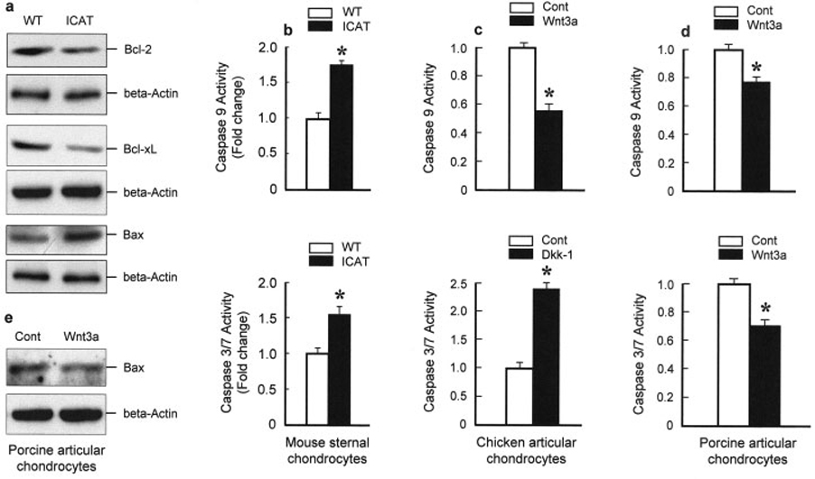
Methods: The expression of the ICAT transgene was determined by immunostaining and Western blot analysis. Histologic analyses were performed to determine changes in articular cartilage structure and morphology. Cell apoptosis was determined by TUNEL staining and the immunostaining of cleaved caspase 3 and poly(ADP-ribose) polymerase (PARP) proteins. Expression of Bcl-2, Bcl-x(L), and Bax proteins and caspase 9 and caspase 3/7 activities were examined in primary sternal chondrocytes isolated from 3-day-old neonatal Col2a1-ICAT-transgenic mice and their wild-type littermates and in primary chicken and porcine articular chondrocytes.
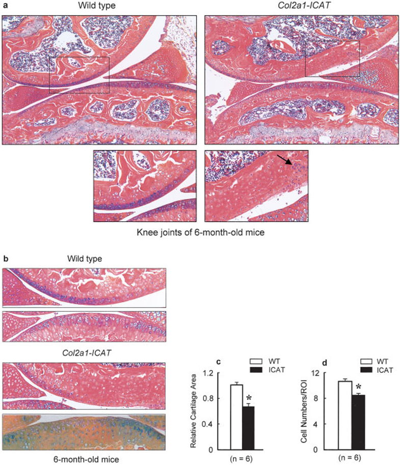
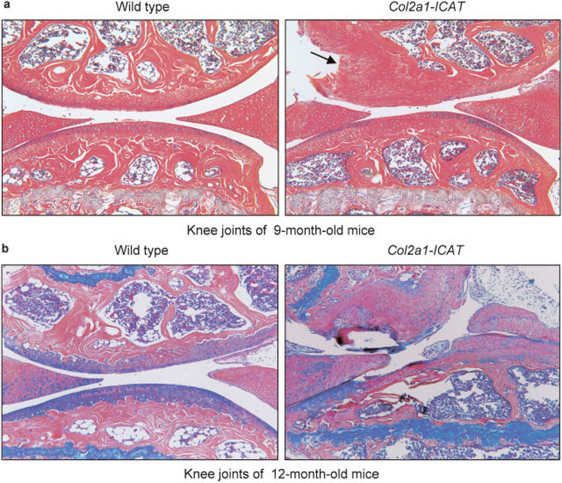
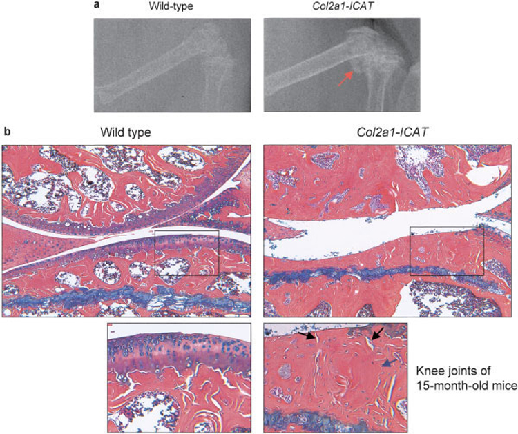
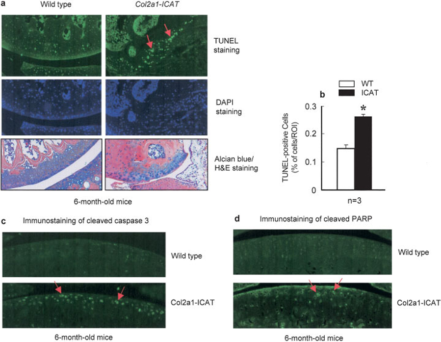
Results: Expression of the ICAT transgene was detected in articular chondrocytes of the transgenic mice. Associated with this, age-dependent articular cartilage destruction was observed in Col2a1-ICAT-transgenic mice. A significant increase in cell apoptosis in articular chondrocytes was identified by TUNEL staining and the immunostaining of cleaved caspase 3 and PARP proteins in these transgenic mice. Consistent with this, Bcl-2 and Bcl-x(L) expression were decreased and caspase 9 and caspase 3/7 activity were increased, suggesting that increased cell apoptosis may contribute significantly to the articular cartilage destruction observed in Col2a1-ICAT-transgenic mice.
Conclusion: Inhibition of beta-catenin signaling in articular chondrocytes causes increased cell apoptosis and articular cartilage destruction in Col2a1-ICAT- transgenic mice.
Figures
Similar articles
-
Chondrocyte-specific inhibition of β-catenin signaling leads to dysplasia of the caudal vertebrae in mice.Spine (Phila Pa 1976). 2013 Nov 15;38(24):2079-84. doi: 10.1097/01.brs.0000435024.57940.8d. Spine (Phila Pa 1976). 2013. PMID: 24026150 Free PMC article.
-
Inhibition of beta-catenin signaling causes defects in postnatal cartilage development.J Cell Sci. 2008 May 1;121(Pt 9):1455-65. doi: 10.1242/jcs.020362. Epub 2008 Apr 8. J Cell Sci. 2008. PMID: 18397998 Free PMC article.
-
Activation of beta-catenin signaling in articular chondrocytes leads to osteoarthritis-like phenotype in adult beta-catenin conditional activation mice.J Bone Miner Res. 2009 Jan;24(1):12-21. doi: 10.1359/jbmr.080901. J Bone Miner Res. 2009. PMID: 18767925 Free PMC article.
-
Smurf2 induces degradation of GSK-3beta and upregulates beta-catenin in chondrocytes: a potential mechanism for Smurf2-induced degeneration of articular cartilage.Exp Cell Res. 2009 Aug 15;315(14):2386-98. doi: 10.1016/j.yexcr.2009.05.019. Epub 2009 May 27. Exp Cell Res. 2009. PMID: 19481076 Free PMC article.
-
Beta-catenin, cartilage, and osteoarthritis.Ann N Y Acad Sci. 2010 Mar;1192(1):344-50. doi: 10.1111/j.1749-6632.2009.05212.x. Ann N Y Acad Sci. 2010. PMID: 20392258 Free PMC article. Review.
Cited by
-
Chondrogenesis, chondrocyte differentiation, and articular cartilage metabolism in health and osteoarthritis.Ther Adv Musculoskelet Dis. 2012 Aug;4(4):269-85. doi: 10.1177/1759720X12448454. Ther Adv Musculoskelet Dis. 2012. PMID: 22859926 Free PMC article.
-
Targets, models and challenges in osteoarthritis research.Dis Model Mech. 2015 Jan;8(1):17-30. doi: 10.1242/dmm.016881. Dis Model Mech. 2015. PMID: 25561745 Free PMC article. Review.
-
Wnt signaling: a promising target for osteoarthritis therapy.Cell Commun Signal. 2019 Aug 16;17(1):97. doi: 10.1186/s12964-019-0411-x. Cell Commun Signal. 2019. PMID: 31420042 Free PMC article. Review.
-
Profiling of Stem/Progenitor Cell Regulatory Genes of the Synovial Joint by Genome-Wide RNA-Seq Analysis.Biomed Res Int. 2018 Jun 26;2018:9327487. doi: 10.1155/2018/9327487. eCollection 2018. Biomed Res Int. 2018. PMID: 30046613 Free PMC article.
-
Wnt/β-catenin signaling contributes to articular cartilage homeostasis through lubricin induction in the superficial zone.Arthritis Res Ther. 2019 Nov 27;21(1):247. doi: 10.1186/s13075-019-2041-5. Arthritis Res Ther. 2019. PMID: 31771658 Free PMC article.
References
-
- Kuettner KE, Goldberg VM. Introduction. In: Kuettner KE, Goldberg VM, editors. Osteoarthritic disorders. Rosemont (IL): American Academy of Orthopaedic Surgeons; 1995. pp. xxi–xxv.
-
- Kuettner KE, Aydelotte MB, Thonar EJ. Articular cartilage matrix and structure: a minireview [review] J Rheumatol Suppl. 1991;27:46–48. - PubMed
-
- Day TF, Guo X, Garrett-Beal L, Yang Y. Wnt/β-catenin signaling in mesenchymal progenitors controls osteoblast and chondrocyte differentiation during vertebrate skeletogenesis. Dev Cell. 2005;8:739–750. - PubMed
Publication types
MeSH terms
Substances
Grants and funding
LinkOut - more resources
Full Text Sources
Other Literature Sources
Research Materials
