. 2008 Aug 29;283(35):24000-10.
doi: 10.1074/jbc.M803361200. Epub 2008 Jun 24.

The ubiquitin-protein ligase Nedd4-2 differentially interacts with and regulates members of the Tweety family of chloride ion channels

Affiliations

The ubiquitin-protein ligase Nedd4-2 differentially interacts with and regulates members of the Tweety family of chloride ion channels

Yaowu He et al. J Biol Chem. .

Abstract

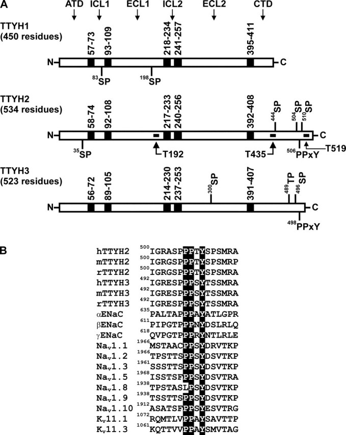
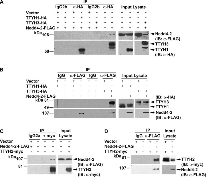
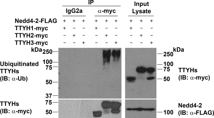
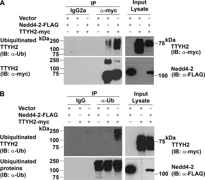
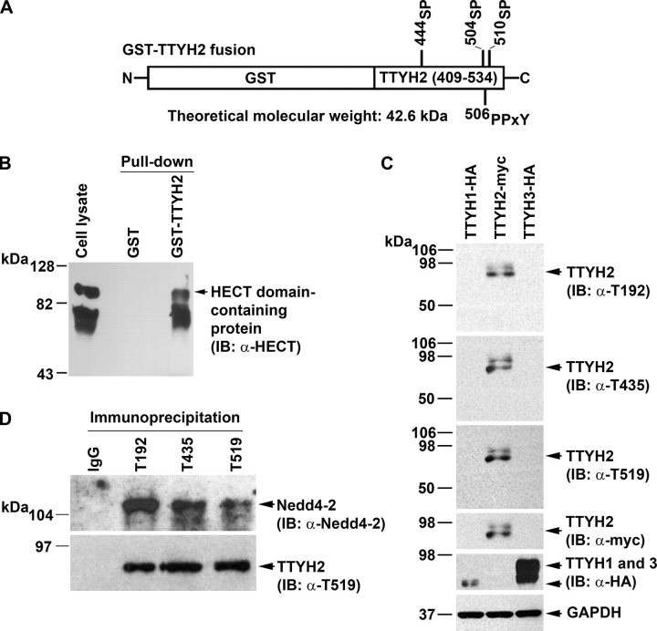
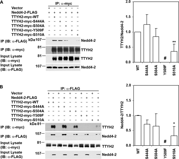
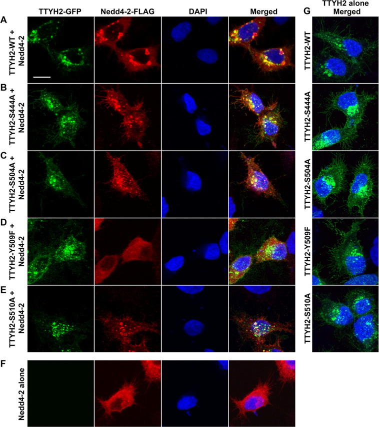
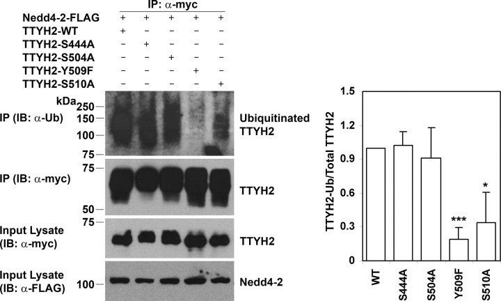
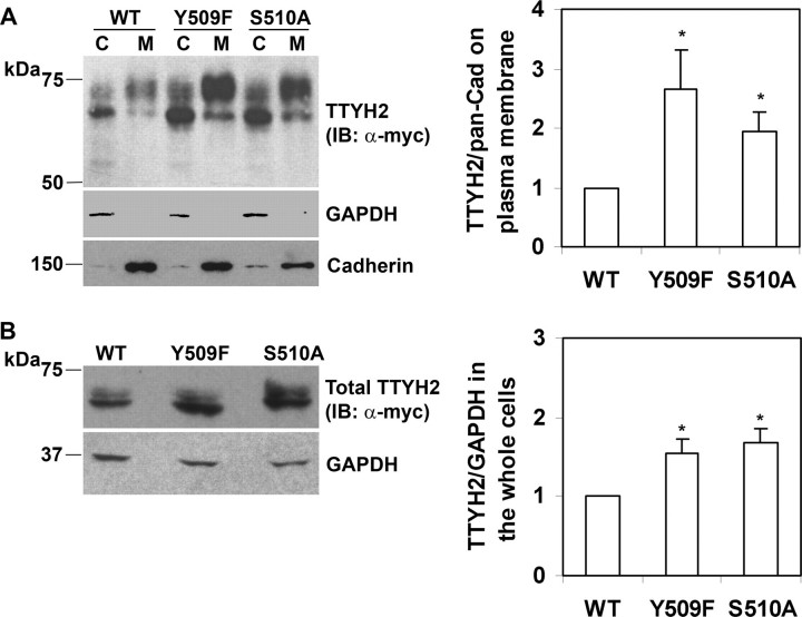
The Tweety proteins comprise a family of chloride ion channels with three members identified in humans (TTYH1-3) and orthologues in fly and murine species. In humans, increased TTYH2 expression is associated with cancer progression, whereas fly Tweety is associated with developmental processes. Structurally, Tweety proteins are characterized by five membrane-spanning domains and N-glycan modifications important for trafficking to the plasma membrane, where these proteins are oriented with the amino terminus located extracellularly and the carboxyl terminus cytoplasmically. In addition to N-glycosylation, ubiquitination mediated by the HECT type E3 ubiquitin ligase Nedd4-2 is a post-translation modification important in regulating membrane proteins. In the present study, we performed a comprehensive analysis of the ability of each of TTYH1-3 to interact with Nedd4-2 and to be ubiquitinated and regulated by this ligase. Our data indicate that Nedd4-2 binds to two family members, TTYH2 and TTYH3, which contain consensus PY ((L/P)PXY) binding sites for HECT type E3 ubiquitin ligases, but not to TTYH1, which lacks this motif. Consistently, Nedd4-2 ubiquitinates both TTYH2 and TTYH3. Importantly, we have shown that endogenous TTYH2 and Nedd4-2 are binding partners and demonstrated that the TTYH2 PY motif is essential for these interactions. We have also shown that Nedd4-2-mediated ubiquitination of TTYH2 is a critical regulator of cell surface and total cellular levels of this protein. These data, indicating that Nedd4-2 differentially interacts with and regulates TTYH1-3, will be important for understanding mechanisms controlling Tweety proteins in physiology and disease.


Figures

FIGURE 1.
FIGURE 1.
Consensus HECT type E3 ubiquitin-protein ligase binding motifs of human Tweety family members. A, TTYH1, TTYH2, and TTYH3 showing transmembrane domains (black boxes) and consensus SP, TP, and PPXY motifs. The predicted positions of transmembrane domains and the total number of residues are indicated, as are the location of peptides used to generate three anti-TTYH2 polyclonal antibodies (designated T192, T435, and T519). The orientations of the Tweety proteins at the plasma membrane are also shown. ATD, amino-terminal domain; ICL1 and ICL2, intracellular loop 1 and 2; ECL1 and ECL2, extracellular loop 1 and 2; CTD, carboxyl-terminal domain. B, alignment of PY motifs and neighboring sequences (±6 residues) of human, mouse, and rat TTYH2 and TTYH3 against human ENaC subunits α, β, and γ, human PY motif-containing voltage-gated sodium channels (Navs), and mouse voltage-gated potassium channels (Kvs). Each motif is numbered on the left to indicate its position in the amino acid sequence of the respective ion channel.
FIGURE 2.
FIGURE 2.
TTYH2 and TTYH3 but not TTYH1 interact with Nedd4-2. HEK293 cells were transfected with Nedd4-2-FLAG and either TTYH1-HA, TTYH2-Myc, or TTYH3-HA expression constructs or vector (pcDNA3.1) control. Proteins were immunoprecipitated (IP) using either monoclonal anti-HA or anti-Myc antibodies or a rabbit anti-FLAG antibody or isotype-matched (IgG2b for anti-HA; IgG2a for anti-Myc) or species-matched (rabbit IgG for anti-FLAG) control IgG. Western blot analysis (IB) was performed using anti-FLAG and anti-HA antibodies (A and B) or anti-FLAG and anti-Myc antibodies (C and D). Lysates were probed with anti-HA and anti-FLAG antibodies (A and B) or anti-FLAG and anti-Myc antibodies (C and D).
FIGURE 3.
FIGURE 3.
Comparison of Nedd4-2-mediated ubiquitination of human Tweety proteins. Lysates from HEK293 cells co-transfected with Nedd4-2-FLAG and either TTYH1-Myc, TTYH2-Myc, or TTYH3-Myc expression constructs were immunoprecipitated (IP) with a mouse anti-Myc antibody or isotype-matched (IgG2a) control IgG. To prevent degradation of ubiquitinated proteins, cells were treated for 4 h with the proteasome inhibitor MG-132 before the collection of lysates. Immunoprecipitated proteins were examined by Western blot analysis (IB) using anti-ubiquitin (Ub) and anti-Myc antibodies. Lysates were examined by Western blot analysis using rabbit anti-Myc and anti-FLAG antibodies. Ubiquitinated and total Tweety proteins (TTYHs) and Nedd4-2-FLAG are indicated.
FIGURE 4.
FIGURE 4.
Ubiquitination of TTYH2 in the presence and absence of Nedd4-2. Lysates from HEK293 cells co-transfected with the indicated constructs were subjected to immunoprecipitation with either anti-Myc or anti-ubiquitin antibodies or control IgGs. To prevent degradation of ubiquitinated proteins, cells were treated for 4 h with the proteasome inhibitor MG-132 before collection of lysates. A, proteins immunoprecipitated (IP) with either an anti-Myc antibody or control IgGs were examined by Western blot analysis (IB) using anti-ubiquitin (Ub) and anti-Myc antibodies. Lysates were examined by Western blot analysis using anti-Myc or anti-FLAG antibodies. B, proteins immunoprecipitated with either an anti-ubiquitin antibody or control IgGs were examined by Western blot analysis using anti-Myc and anti-ubiquitin antibodies. Lysates were examined by Western blot analysis using anti-Myc and anti-FLAG antibodies. Ubiquitinated and total TTYH2-Myc and Nedd4-2-FLAG are indicated.
FIGURE 5.
FIGURE 5.
Interactions between endogenous TTYH2 and Nedd4-2. A, schematic representation of a fusion of GST and the carboxyl-terminal tail of TTYH2 (residues 409-534). Consensus binding sites for HECT type E3 ubiquitin-protein ligases are indicated. B, lysates from opossum kidney cells were subjected to pull-down using either GST or GST-TTYH2. Input lysates and captured proteins were subjected to Western blot analysis using an anti-HECT domain polyclonal antibody that detects the HECT domain of Nedd4 and Nedd4-2. C, specificity of three anti-TTYH2 polyclonal antibodies (T192, T435, and T519). Chinese hamster ovary cells were transfected with expression constructs encoding either TTYH1-HA, TTYH2-Myc, or TTYH3-HA. Lysates were subjected to Western blot analysis (IB) using anti-TTYH2 T192, T435, and T519 antibodies. GAPDH was used as a loading control, and the expression of TTYH1-HA, TTYH2-Myc, and TTYH3-HA was confirmed by reprobing membranes with anti-HA or anti-Myc antibodies as appropriate. D, Western blot analysis using a specific anti-Nedd4-2 antibody of endogenous proteins immunoprecipitated from HEK293 cells using one of the anti-TTYH2 polyclonal antibodies T192, T435, or T519 or control rabbit IgG. The membrane was stripped and reprobed for immunoprecipitated endogenous TTYH2 using antibody T519 (the same pattern was obtained when the blot was reprobed with either T192 or T435 antibodies; data not shown).
FIGURE 6.
FIGURE 6.
The role of carboxyl-terminal domain SP and PY motifs in binding of TTYH2 to Nedd4-2. HEK293 cells were co-transfected with expression constructs encoding Nedd4-2-FLAG and either wild type (WT) or mutant TTYH2-Myc as indicated. Immunoprecipitates from cells co-transfected with vector (pcDNA3.1) and the Nedd4-2-FLAG expression construct were used as controls. Western blot analyses are representative of three experiments. The mean and S.D. of signal intensities, determined by densitometry analysis, from these three experiments, relative to data from TTYH2 wild type (WT), are graphed on the right of each panel. A, proteins immunoprecipitated (IP) with an anti-Myc monoclonal antibody and input lysates were subjected to Western blot analysis (IB) using either anti-FLAG or anti-Myc antibodies (left). The ratio of Nedd4-2-FLAG to TTYH2-Myc in immunoprecipitates is graphed in the right panel. #, binding completely abolished in each of the three experiments; **, p < 0.01. B, proteins immunoprecipitated with an anti-FLAG antibody and input lysates were subjected to Western blot analysis using either anti-Myc or anti-FLAG antibodies (left). The ratio of TTYH2-Myc to Nedd4-2-FLAG in immunoprecipitates is graphed in the right panel. #, binding completely abolished in each of the three experiments; *, p < 0.05.
FIGURE 7.
FIGURE 7.
Localization of Nedd4-2 and wild type and mutant TTYH2. HEK293 cells were co-transfected with expression constructs encoding Nedd4-2-FLAG and either wild type (A), S444A (B), S504A (C), Y509F (D), or S510A (E) mutant TTYH2-GFP expression constructs as indicated. In controls, cells were transfected with either Nedd4-2-FLAG (F) or TTYH2-GFP (wild type or mutant) (G) expression constructs alone. Cells were fixed and then incubated with an anti-FLAG antibody, followed by a fluorescently tagged secondary antibody. Nuclei were stained with 4′, 6-diamidino-2-phenylindole hydrochloride. Images were acquired using a Leica TCS SP5 confocal microscope and processed and displayed using Adobe Photoshop CS3.
FIGURE 8.
FIGURE 8.
The effect of loss of SP and PY motifs on ubiquitination of TTYH2 by Nedd4-2. Lysates from HEK293 cells co-transfected with Nedd4-2-FLAG and either wild type or mutant TTYH2-Myc expression constructs were subjected to immunoprecipitation using an anti-Myc antibody. To prevent degradation of ubiquitinated proteins, cells were treated for 4 h with the proteasome inhibitor MG-132 before the collection of lysates. Proteins immunoprecipitated (IP) with an anti-Myc antibody were examined by Western blot analysis (IB) using anti-ubiquitin and anti-Myc antibodies. Lysates were also examined by Western blot analysis using anti-Myc and anti-FLAG antibodies. Ubiquitinated and total TTYH2-Myc and Nedd4-2-FLAG are indicated. Western blot analyses are representative of three experiments. Signal intensities, determined by densitometry analysis, from these three experiments were used to calculate the ratio of ubiquitinated to total immunoprecipitated TTYH2-Myc. The mean and S.D. of the ratio for each experimental condition, relative to data from TTYH2 wild type (WT), are graphed in the right panel. *, p < 0.05; ***, p < 0.001.
FIGURE 9.
FIGURE 9.
Interactions between Nedd4-2 and TTYH2 impact on plasma membrane and total cellular levels of TTYH2. HEK293 cells were co-transfected with Nedd4-2-FLAG and wild type, Y509F, or S510A TTYH2-Myc expression constructs. A, plasma membrane (M) and cytoplasmic (C) fractions, separated by biotinylation of intact cells, were subjected to anti-Myc, anti-GAPDH, and anti-pancadherin Western blot analyses. The displayed blots are representative of three experiments. The mean and S.D. of the ratio of plasma membrane TTYH2 to pancadherin from these three experiments are graphed in the right panel.*, p < 0.05. B, whole cell lysates were subjected to anti-Myc and anti-GAPDH Western blot analyses. The displayed blots are representative of three experiments. The mean and S.D. of the ratio of whole cell TTYH2 to GAPDH from these three experiments, relative to data from TTYH2-WT, are graphed in the right panel.*, p < 0.05. IP, immunoprecipitation; IB, immunoblot; WT, wild type.

Similar articles

Cited by

References

    1. Sullivan, M., and Morgan, D. O. (2007) Nat. Rev. Mol. Cell Biol. 8894 -903 - PubMed
    1. Muratani, M., and Tansey, W. P. (2003) Nat. Rev. Mol. Cell Biol. 4192 -201 - PubMed
    1. Liu, Y. C. (2004) Annu. Rev. Immunol. 2281 -127 - PubMed
    1. DiAntonio, A., and Hicke, L. (2004) Annu. Rev. Neurosci. 27223 -246 - PubMed
    1. Mukhopadhyay, D., and Riezman, H. (2007) Science 315201 -205 - PubMed

Publication types

MeSH terms