. 2008 Jul;131(Pt 7):1831-44.
doi: 10.1093/brain/awn113. Epub 2008 Jun 24.

Paroxysmal exercise-induced dyskinesia and epilepsy is due to mutations in SLC2A1, encoding the glucose transporter GLUT1

Affiliations

Paroxysmal exercise-induced dyskinesia and epilepsy is due to mutations in SLC2A1, encoding the glucose transporter GLUT1

Arvid Suls et al. Brain. 2008 Jul.

Abstract

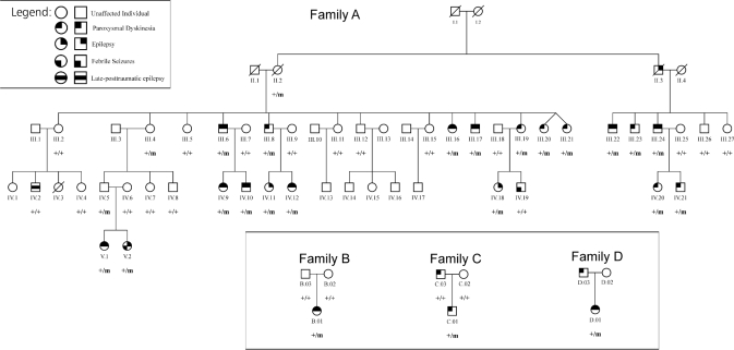
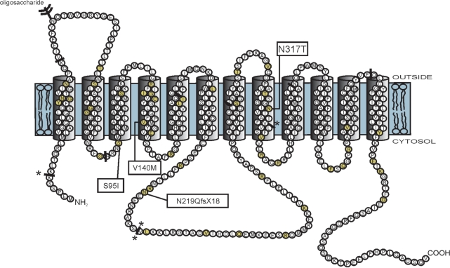
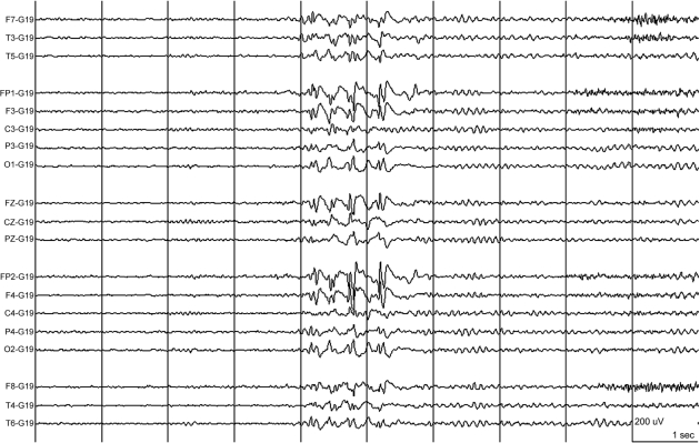
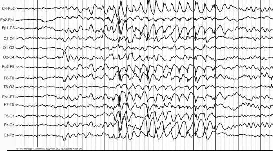
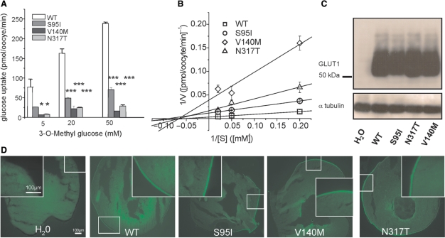
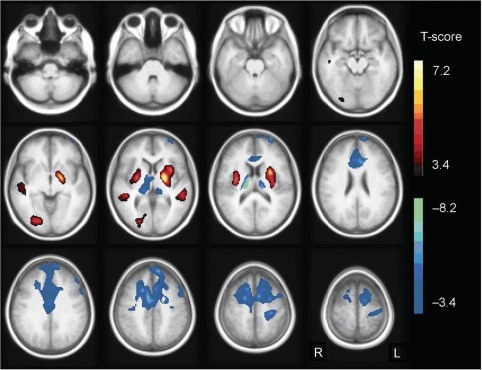
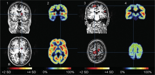
Paroxysmal exercise-induced dyskinesia (PED) can occur in isolation or in association with epilepsy, but the genetic causes and pathophysiological mechanisms are still poorly understood. We performed a clinical evaluation and genetic analysis in a five-generation family with co-occurrence of PED and epilepsy (n = 39), suggesting that this combination represents a clinical entity. Based on a whole genome linkage analysis we screened SLC2A1, encoding the glucose transporter of the blood-brain-barrier, GLUT1 and identified heterozygous missense and frameshift mutations segregating in this and three other nuclear families with a similar phenotype. PED was characterized by choreoathetosis, dystonia or both, affecting mainly the legs. Predominant epileptic seizure types were primary generalized. A median CSF/blood glucose ratio of 0.52 (normal >0.60) in the patients and a reduced glucose uptake by mutated transporters compared with the wild-type as determined in Xenopus oocytes confirmed a pathogenic role of these mutations. Functional imaging studies implicated alterations in glucose metabolism in the corticostriate pathways in the pathophysiology of PED and in the frontal lobe cortex in the pathophysiology of epileptic seizures. Three patients were successfully treated with a ketogenic diet. In conclusion, co-occurring PED and epilepsy can be due to autosomal dominant heterozygous SLC2A1 mutations, expanding the phenotypic spectrum associated with GLUT1 deficiency and providing a potential new treatment option for this clinical syndrome.


Figures

Fig. 1
Fig. 1
Pedigree of families A–D. + = normal allele; m = mutated allele. Individuals carrying a heterozygous mutation in SLC2A1 are indicated with +/m. Individuals indicated with +/+ do not carry a mutated allele. Individuals without indication were not available for screening.
Fig. 2
Fig. 2
The GLUT1 protein structure consisting of 12 transmembrane domains and intracellular amino- and carboxy-termini (Mueckler et al., 1985). GLUT1 DS (Klepper and Leiendecker, 2007) and PED/epilepsy mutations are marked on this figure (yellow colored circles). The PED/epilepsy mutations are boxed. The solid bars indicate the locations of the intron–exon boundaries in the GLUT1 gene. Splice site mutations are indicated at these solid bars with asterisk.
Fig. 3
Fig. 3
The interictal EEG of patient A. III.24 showed high voltage anterior predominant generalized spike-wave complexes on a normal background. Time base: 30 mm/s, sensitivity: see calibration: 200 µV/cm, high cut: 30.0 Hz, low cut: 0.5 Hz.
Fig. 4
Fig. 4
During hyperventilation, patient A.V.1 had a brief absence with high voltage 3 Hz generalized spike wave discharges during 6 s on EEG. Time base: 30 mm/s, sensitivity (of original recording): 300 µV/cm, high cut: 35.0 Hz, low cut: 0.5 Hz.
Fig. 5
Fig. 5
Functional studies to investigate a change in glucose uptake, protein stability or trafficking by the three point mutations in Xenopus oocytes. (A) Plotted is the glucose uptake versus 3-O-methyl-D-glucose (OMG) concentration. The uptake was significantly reduced for all three mutations (shown are representative results recorded from one batch of 3 × 10 oocytes for each glucose concentration, means ± SEM, *P < 0.05, ***P < 0.001). (B) Kinetic analysis of glucose uptakes in oocytes according to Lineweaver-Burk. The linear function 1/V (1/[S]) = 1/Vmax + Km/Vmax*1/[S] was fit to the data points, with [S] being the concentration of OMG, V the uptake velocity in pmol/oocyte/min obtained for a given [S], Vmax the maximal uptake velocity reflecting the maximal transport capacity of GLUT1 and Km the Michaelis-Menten constant representing the concentration [S] for which the half-maximal uptake velocity (V1/2) is reached. Vmax and Km were calculated from the y- and x-interceptions of the linear fits, with the y-intercept equalling 1/Vmax and the x-intercept −1/Km. The following values were obtained (Vmax is given in pmol/oocyte/min and Km in mM): WT: Vmax = 319 ± 16, Km = 19 ± 1; S95I: Vmax = 86 ± 2, Km = 11 ± 1; V140M: Vmax = 26 ± 11, Km = 15 ± 9; N317T: Vmax = 60 ± 18, Km = 15 ± 7. (C) Western blots obtained from oocytes injected with equal amounts of cRNA showed a similar amount of protein for all mutations and the WT, but no respective band for oocytes injected with H2O as a negative control; α-tubulin was used as a loading control. (D) Immunocytochemical analysis of injected oocytes using an anti-GLUT1 antibody revealed similar stainings of the surface membranes for all four clones.
Fig. 6
Fig. 6
SPM T-map of the analysis of patients versus controls. Relative hypermetabolism in the patient group compared with controls is indicated in yellow/red, hypometabolism in blue/green. Results are projected on an average spatially normalized in-house T1 image of healthy controls. R = right; L = left.
Fig. 7
Fig. 7
Subtraction ictal SPECT coregistered to MRI (SISCOM) (1, 3) during an episode of PED and interictal FDG PET (2, 4) in patient A.III.24. Top row: coronal images, bottom row: axial images. The episode of PED lasted in total around 17 min, and was interrupted by brief moments of no abnormal movements. The dyskinesia mainly involved the legs, left more than right and pelvis (see video; A.III.24 Ictal SPECT during PED, in supplementary data). The injection was given around three minutes after onset. The SISCOM (threshold: + 2 SD) showed an area of hyperperfusion in the left putamen (blue cross in 1), which coincided with interictal putaminal hypermetabolism (blue cross in 2). The largest and most hyperintense hyperperfusion cluster was in the right primary motor area of the leg (blue cross in 3), which coincided with an area of hypometabolism in the interictal FDG PET (blue cross in 4). All images are coregistered. Hyperperfusion clusters are projected on the patient's MPRAGE images. R = right; L = left.

Similar articles

Cited by

References

    1. Alderborn A, Kristofferson A, Hammerling U. Determination of single-nucleotide polymorphisms by real-time pyrophosphate DNA sequencing. Genome Res. 2000;10:1249–58. - PMC - PubMed
    1. Auburger G, Ratzlaff T, Lunkes A, Nelles HW, Leube B, Binkofski F, et al. A gene for autosomal dominant paroxysmal choreoathetosis/spasticity (CSE) maps to the vicinity of a potassium channel gene cluster on chromosome 1p, probably within 2 cM between D1S443 and D1S197. Genomics. 1996;31:90–4. - PubMed
    1. Berkovic SF. Paroxysmal movement disorders and epilepsy: links across the channel. Neurology. 2000;55:169–70. - PubMed
    1. Bhatia KP, Soland VL, Bhatt MH, Quinn NP, Marsden CD. Paroxysmal exercise-induced dystonia: eight new sporadic cases and a review of the literature. Mov Disord. 1997;12:1007–12. - PubMed
    1. Bing F, Dananchet Y, Vercueil L. [A family with exercise-induced paroxysmal dystonia and childhood absence epilepsy] Rev Neurol (Paris) 2005;161:817–22. - PubMed

Publication types

MeSH terms