. 2008 Jul 1;105(26):8989-93.
doi: 10.1073/pnas.0800388105. Epub 2008 Jun 24.

Assembly of the cnidarian camera-type eye from vertebrate-like components

Affiliations

Assembly of the cnidarian camera-type eye from vertebrate-like components

Zbynek Kozmik et al. Proc Natl Acad Sci U S A. .

Abstract

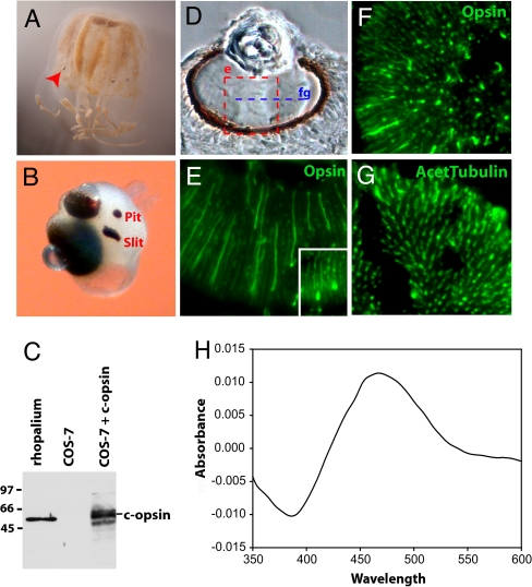
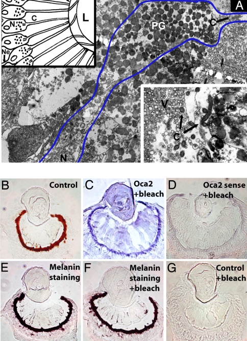
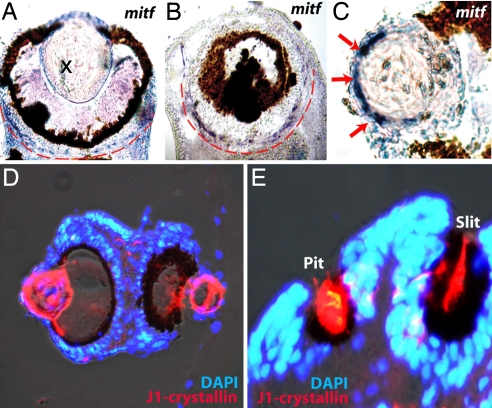
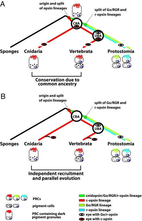
Animal eyes are morphologically diverse. Their assembly, however, always relies on the same basic principle, i.e., photoreceptors located in the vicinity of dark shielding pigment. Cnidaria as the likely sister group to the Bilateria are the earliest branching phylum with a well developed visual system. Here, we show that camera-type eyes of the cubozoan jellyfish, Tripedalia cystophora, use genetic building blocks typical of vertebrate eyes, namely, a ciliary phototransduction cascade and melanogenic pathway. Our findings indicative of parallelism provide an insight into eye evolution. Combined, the available data favor the possibility that vertebrate and cubozoan eyes arose by independent recruitment of orthologous genes during evolution.


Conflict of interest statement

The authors declare no conflict of interest.

Figures

Fig. 1.
Fig. 1.
Ciliary opsin is the functional photopigment of Tripedalia camera-type eyes. (A) Tripedalia medusae with rhopalia (arrowhead). (B) Rhopalium with two lens-containing camera-type eyes, a slit and a pit eye. Another set of slit and pit eyes is symmetrically located on the other side of the rhopalium. (C) Specificity of an anti-Tripedalia c-opsin antibody tested by Western blotting with protein extracts from rhopalia and COS-7 cells transiently transfected with Tripedalia c-opsin cDNA. (D) Bright-field section through the large camera-type eye. The area shown in E is boxed. The plane of sectioning in F and G is indicated (fg). (E–G) Immunohistochemical staining (green) of Tripedalia retina using antibodies to c-opsin (E and F) or acetylated tubulin (G). (E Inset) Immunohistochemical staining for c-opsin in the small camera-type eye. (H) Dark–light difference absorption spectrum of the reconstituted Tripedalia c-opsin photopigment. The photopigment reconstituted with 11-cis-retinal forms a functional photopigment most sensitive to blue–green light.
Fig. 2.
Fig. 2.
Melanin is the shielding pigment of Tripedalia camera-type eyes. (A) Electron micrograph of camera-type eye PRCs. Blue line borders one PRC containing shielding pigment layer (PG) as well as photosensitive cilium (C). (Upper Left Inset) Diagram of PRCs with their cilia protruding to the lens capsule. Note that all PRC nuclei are located behind the shielding pigment. (Lower Right Inset) Apical end of a PRC with cilium. C, cilium; L, lens; N, nucleus; Ne, neurite; PG, pigment granules; V, vitreous body. (B) Bright-field cryosection of camera-type eye. (C and D) In situ hybridization (blue) using oca2 antisense (C) or control sense (D) probes. (E–G) Melanin detection by Fontana–Masson staining. Tissue sections shown in C, D, F, and G were bleached to remove melanin.
Fig. 3.
Fig. 3.
Expression of mitf and J1-crystallin in Tripedalia eyes. (A–C) In situ hybridization (blue) detects mitf expression in the circle around pigment deposits (A and B) and in the lens (C, arrows). (D and E) Immunohistochemistry staining using an antibody to the major cubozoan lens crystallin, J1 (red). Nuclei of cells are visualized by DAPI staining (blue). J1-crystallin expression is localized to lenses of camera-type eyes (D) as well as to the slit and pit eyes (E).
Fig. 4.
Fig. 4.
Two scenarios for the use of ciliary phototransduction and melanogenic pathway in eye evolutionary history. A simplified view of the two evolutionary scenarios is compatible with the data in the present work. The use of similar genetic components in vertebrate and cubozoan eyes is either due to common ancestry (A) or independent parallel recruitments in cnidarian and vertebrate lineages (B). The c-opsins and Go/r-opsins arose by duplication and diversification of an ancestral opsin in the early metazoans (27). In the schemes, only the visual (i.e., the eye-specific) PRCs and opsins are considered. Different shading of pigment granules indicates possible distinct chemical composition. CBA, cnidarian–bilaterian ancestor; UBA, urbilaterian ancestor.

Similar articles

Cited by

References

    1. Fernald RD. Casting a genetic light on the evolution of eyes. Science. 2006;313:1914–1918. - PubMed
    1. Arendt D, Wittbrodt J. Reconstructing the eyes of Urbilateria. Philos Trans R Soc London Ser B. 2001;356:1545–1563. - PMC - PubMed
    1. Arendt D. Evolution of eyes and photoreceptor cell types. Int J Dev Biol. 2003;47:563–571. - PubMed
    1. Viscontini M, Hummel W, Fischer A. Isolation of pterin dimers from the eyes of Platynereis dumerilii (German) Helv Chim Acta. 1970;53:1207–1209.
    1. Shoup JR. The development of pigment granules in the eyes of wild-type and mutant Drosophila melanogaster. J Cell Biol. 1966;29:223–249. - PMC - PubMed

Publication types

MeSH terms