. 2008 Sep 1;112(5):1794-803.
doi: 10.1182/blood-2008-01-134932. Epub 2008 Jun 24.

The familial Mediterranean fever protein, pyrin, is cleaved by caspase-1 and activates NF-kappaB through its N-terminal fragment

Affiliations

The familial Mediterranean fever protein, pyrin, is cleaved by caspase-1 and activates NF-kappaB through its N-terminal fragment

Jae Jin Chae et al. Blood. .

Abstract

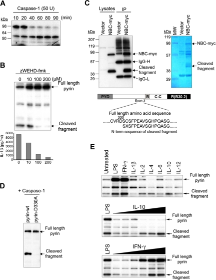
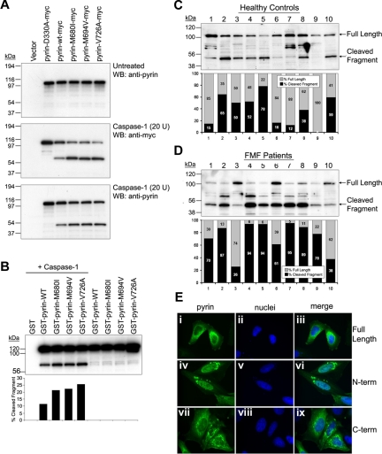
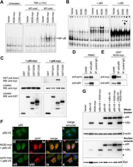
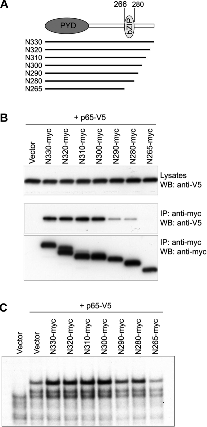
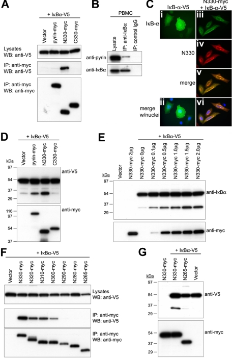
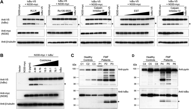
Familial Mediterranean fever (FMF) is an autoinflammatory disease caused by mutations in MEFV, which encodes a 781-amino acid protein denoted pyrin. We have previously shown that pyrin regulates caspase-1 activation and IL-1beta production through interaction of its N-terminal PYD motif with the ASC adapter protein, and also modulates IL-1beta production by interaction of its C-terminal B30.2 domain with the catalytic domains of caspase-1. We now asked whether pyrin might itself be a caspase-1 substrate, and found that pyrin is cleaved by caspase-1 at Asp330, a site remote from the B30.2 domain. Pyrin variants harboring FMF-associated B30.2 mutations were cleaved more efficiently than wild-type pyrin. The N-terminal cleaved fragment interacted with the p65 subunit of NF-kappaB and with IkappaB-alpha through its 15-aa bZIP basic domain and adjacent sequences, respectively, and translocated to the nucleus. The interaction of the N-terminal fragment with p65 enhanced entrance of p65 into the nucleus. The interaction of N-terminal pyrin with IkappaB-alpha induced calpain-mediated degradation of IkappaB-alpha, thus potentiating NF-kappaB activation. Absolute and relative quantities of cleaved pyrin and IkappaB-alpha degradation products were substantially increased in leukocytes from FMF patients compared with healthy controls. Our data support a new pyrin/caspase-1 pathway for NF-kappaB activation.


Figures

Figure 1
Figure 1
Cleavage of pyrin by caspase-1. (A) 35S-labeled pyrin was incubated with 50 units recombinant caspase-1 at 37°C for various periods as indicated, subjected to SDS-PAGE, and visualized by autoradiography. (B) Pyrin was cotransfected into Cos-7 cells with caspase-1 and pro–IL-1β. Cells were treated with increasing amounts of z-WEHD-fmk as indicated. After 24 hours, cell lysates underwent SDS-PAGE and Western blotting with a polyclonal Ab developed against the N-terminal 374 aa of pyrin (top). Cell culture supernatants were also collected and assayed for IL-1β by ELISA (bottom). (C) PT67 cells were cotransfected with pNBC-myc (construct expressing B30.2 domain–deleted pyrin) and with caspase-1. After 24 hours, cell lysates were immunoprecipitated using antimyc Ab, and subjected to SDS-PAGE followed by Western blotting with antimyc Ab (top left panel) or Coomassie blue staining (top right panel). The result of N-terminal Edman sequencing of the eluted, Coomassie-stained band corresponding to the C-terminal cleaved fragment is shown under the schematic diagram of pyrin. (D) WT or D330A pyrin was cotransfected with caspase-1 into PT67 cells. Lysates were subjected to SDS-PAGE and Western blotting with the same antipyrin Ab as in panel B. (E) PBMCs from healthy controls were cultured in RPMI supplemented with 10% fetal bovine serum in 6-well culture plates, treated with LPS (1 μg/mL) from E coli 0127:B7 (Sigma-Aldrich), IFN-γ (100 ng/mL), IL-1β (3.75 ng/mL), IL-2 (1.6 ng/mL), IL-4 (12.5 ng/mL), IL-6 (8.5 ng/mL), IL-10 (100 ng/mL) or IL-12 (10 ng/mL; BD Biosciences, San Jose, CA). After 24 hours, cells were lysed and 12 μg total protein was subjected to SDS-PAGE followed by Western blot with the same antipyrin Ab as in panel B (top). PBMCs were also stimulated with LPS or various amounts (from 10 pg/mL to 100 ng/mL) of IL-10 (middle) or IFN-γ (bottom) for 24 hours. Cells were lysed and analyzed as in top panel.
Figure 2
Figure 2
Cleavage of FMF-associated mutant pyrins by caspase-1 and cellular localization of cleaved fragments. (A) Myc-tagged D330A, WT, or FMF-associated mutant pyrin was transfected into PT67 cells. After 24 hours, cell lysates (10 μg total protein) underwent Western blotting with antipyrin Ab (top). The same amounts of lysates were incubated with 20 units of recombinant caspase-1 for 10 minutes at room temperature. The incubated cell lysates were subjected to Western blotting with antimyc, detecting the C-terminal cleavage fragment (middle), and antipyrin Abs, detecting the smaller N-terminal cleavage fragment (bottom). (B) GST-fused WT or mutant pyrin was cotransfected into PT67 cells with (lanes 1-5) or without (lanes 6-9) caspase-1. Cell lysates were analyzed by Western blots using antipyrin Ab (top panel). The cleaved and uncleaved pyrin fragments were quantified by densitometry and represented as percentage of total pyrin (bottom panel). (C,D) Lysates of PBMCs from 10 healthy individuals (C) and 10 FMF patients (D) were subjected to SDS-PAGE, and Western blotting with antipyrin Ab. Exposure times were the same for panels C and D. The full-length and cleaved pyrin were quantified by densitometry and represented as percentage of total pyrin. (E) HeLa cells were transfected with myc-tagged full-length pyrin (i-iii), N-terminal cleaved pyrin (iv-vi), or C-terminal cleaved pyrin (vii-ix). After 24 hours, cells were fixed with 4% paraformaldehyde in PBS, and stained with AlexaFluor 488–conjugated antimyc Ab (green), counterstained with DAPI (blue), and visualized on a Leica DMR microscope. Pyrin variants are shown in the first column (i, iv, and vii); nuclei are in the second column (ii, v, and viii); merged images are in the third column (iii, vi, and ix).
Figure 3
Figure 3
N-terminal cleaved pyrin activates NF-κB and interacts with p65. (A) NF-κB DNA-binding activity was measured by EMSA in nuclear lysates prepared from HeLa cells that had been transfected with myc-tagged empty vector, N330, or C330, and incubated with or without TNF-α (12.5 ng/mL) for 1 hour. As indicated, either unlabeled excess NF-κB wt (WT-cold) or mutant (MT-cold) oligonucleotides were added to the EMSA reactions as a competitor to assay binding specificity. (B) HeLa cells were cotransfected with the indicated pyrin constructs and either p65 or p50. From the nuclear lysates, NF-κB DNA-binding activity was measured by EMSA as in panel A. (C) Myc-tagged p50 (left panels) or p65 (right panels) was cotransfected into PT67 cells with GST vector, GST-fused pyrin, GST-N330, or GST-C330. Lysates were subjected to GST pull-down assay followed by Western blotting with antimyc Ab (top panels). Expression of transfected constructs is shown in the bottom 2 panels (cell lysates) probed with antimyc or anti-GST Abs. (D) Lysates from PBMCs of a healthy donor were immunoprecipitated with anti-p65 Ab or control IgG. Cell lysates and eluted proteins were analyzed by Western blotting with antipyrin (top) or anti-p65 Abs (bottom). Data are from a representative experiment from 3 separate subjects. (E) U937 cells were transduced with retroviral constructs expressing N330-myc or with empty vector. After G418 selection, cell lysates were immunoprecipitated with anti-p65 Ab or control IgG, and analyzed as in panel D. (F) HeLa cells were transfected with V5-tagged p65 (p65-V5) alone (i-ii), or cotransfected with p65-V5 and N330-myc (iii-vi), or with p65-V5 and myc-tagged full-length pyrin (vii-xi). After 24 hours, p65 and pyrin variants were stained with AlexaFluor 488–conjugated anti-V5 Ab (green) and AlexaFluor 568–conjugated antimyc Ab (red), respectively. Cells were counterstained with DAPI (blue) and visualized on a Leica DMR microscope. Expressed p65 is shown in the first column (i, iii, and vii); pyrin variants are shown in the second column (iv and viii); merged images for p65 and pyrin variants are in the third column (v and ix); merged images for p65, pyrin variants, and nuclei are in the fourth column (ii, vi, and x). (G) IκB-α–V5, p65-V5, and N330-myc were transfected individually, or cotransfected as indicated into HeLa cells. After 24 hours, total cell lysates (top 2 panels) and nuclear extracts (bottom 2 panels) were prepared and subjected to SDS-PAGE for Western blotting with anti-V5 Ab (top panel and third panel), anti–β-actin Ab (for loading control of whole-cell extracts), or anti-Ran Ab (for loading control of nuclear extracts). Asterisk denotes degraded 30-kDa IκB-α fragment that is explained in “N330 interacts with p65 and enhances the nuclear localization of p65.”
Figure 4
Figure 4
Interaction of the bZIP basic domain of N330 with p65. (A) Sequential 10-aa deleted N-terminal fragments from aa's 330 to 266 are indicated by lines under the schematic of N330 in which the PYRIN domain (PYD) and bZIP basic domain (bZIP) reside. The first and last aa numbers of bZIP basic domain are indicated on the top. (B) PT67 cells were cotransfected with p65-V5 and the 10-aa deleted N-terminal fragments depicted in panel A. Lysates were immunoprecipitated with antimyc Ab followed by Western blotting with anti-V5 (middle) and antimyc (bottom) Abs. Expression of p65-V5 in whole lysates is shown by anti-V5 Ab (top). (C) HeLa cells were cotransfected with p65-V5 and the 10-aa deleted N-terminal fragments depicted in panel A. From the nuclear lysates, NF-κB DNA-binding activity was measured by EMSA as in Figure 3A.
Figure 5
Figure 5
Interaction of N330 with IκB-α and the N330-induced proteolysis of IκB-α. (A) V5-tagged IκB-α was cotransfected into PT67 cells with myc-tagged pyrin, N330, or C330 or empty vector. Lysates were immunoprecipitated with antimyc Ab and analyzed as in Figure 4B. (B) Lysates from PBMCs of a healthy donor were immunoprecipitated with anti–IκB-α Ab or control IgG. Cell lysates and eluted proteins were analyzed by Western blotting with antipyrin (top) or anti–IκB-α Abs (bottom). Data are from a representative experiment from 3 separate donors. (C) HeLa cells were transfected with V5-tagged IκB-α (IκB-α–V5) alone (i,ii), or cotransfected with IκB-α–V5 and N330-myc (iii-vi). After 24 hours, IκB-α and N330 were stained with AlexaFluor 488–conjugated anti-V5 Ab (green) and AlexaFluor 568–conjugated antimyc Ab (red), respectively. Cells were counterstained with DAPI (blue) and visualized on a Leica DMR microscope. Expressed IκB-α is shown in panels i, ii (merged with nuclei), and iii, and N330 is in panel iv. Merged images for IκB-α and N330 without and with nuclei are in panels v and vi, respectively. (D) HeLa cells were cotransfected with IκB-α–V5 and myc-tagged pyrin variants or empty vector. Cell lysates were analyzed by Western blotting with anti-V5 (top) or antimyc Abs (bottom). (E) HeLa cells were cotransfected with IκB-α–V5 and increasing amount of N330-myc, and also transfected with empty vector or N330-myc alone. Cell lysates were analyzed by Western blotting with anti–IκB-α (top) or antimyc Abs (bottom). (F) PT67 cells were cotransfected with IκB-α–V5 and the 10-aa deleted N-terminal fragments depicted in Figure 4A. Lysates were immunoprecipitated with antimyc Ab and analyzed as in Figure 4B. (G) HeLa cells were cotransfected with IκB-α–V5 and N330-myc, N265-myc, or empty vector, and also transfected with N330-myc alone. Cell lysates were analyzed by Western blotting with anti-V5 (top) or antimyc Abs (bottom).
Figure 6
Figure 6
Effects of calpain inhibitors, colchicine, and FMF mutations on N330-induced IκB-α degradation. (A) HeLa cells were cotransfected with IκB-α–V5 and N330-myc, and treated with various inhibitors, ALLN (0.3-1.25 μg/mL, proteosome, calpain I and II inhibitor), Ro106-9920 (0.39-3.125 μM, ubiquitination inhibitor), calpain inhibitor III (0.625-5 μg/mL, calpain I and II inhibitor), EST (3.125-100 mM, calpain I inhibitor), or calpain inhibitor IV (1.25-5 μg/mL, calpain II inhibitor). Cells were also transfected with N330-myc or IκB-α–V5 alone as controls. Cell lysates were analyzed by Western blotting with anti-V5 Ab for IκB-α (top), antimyc Ab for N330 (middle), or anti–β-tubulin Ab for loading controls (bottom). The approximately 30-kDa IκB-α fragment is denoted with an asterisk. (B) HeLa cells were cotransfected or transfected as in panel A. Cotransfected cells were treated with various amounts of colchicine (5-80 pg/mL). Cell lysates were analyzed as in panel A. (C) PBMCs from 3 FMF patients who had at least one mutation in the B30.2 domain, as well as from 3 healthy controls, were lysed immediately after purification. Cell lysates were subjected to Western blotting with antipyrin (top panel) or anti–IκB-α Abs (bottom panel). Asterisks denote the approximately 30-kDa IκB-α fragment. (D) PBMCs from 3 FMF patients who have 2 mutations on B30.2 domains of both alleles, as well as from 3 healthy controls, were treated with IFN-γ for 24 hours. Cells were lysed and analyzed as in panel C.

References

    1. Ancient missense mutations in a new member of the RoRet gene family are likely to cause familial Mediterranean fever: The International FMF Consortium. Cell. 1997;90:797–807. - PubMed
    1. A candidate gene for familial Mediterranean fever: The French FMF Consortium. Nat Genet. 1997;17:25–31. - PubMed
    1. Centola M, Wood G, Frucht DM, et al. The gene for familial Mediterranean fever, MEFV, is expressed in early leukocyte development and is regulated in response to inflammatory mediators. Blood. 2000;95:3223–3231. - PubMed
    1. Diaz A, Hu C, Kastner DL, et al. Lipopolysaccharide-induced expression of multiple alternatively spliced MEFV transcripts in human synovial fibroblasts: a prominent splice isoform lacks the C-terminal domain that is highly mutated in familial Mediterranean fever. Arthritis Rheum. 2004;50:3679–3689. - PubMed
    1. Touitou I. The spectrum of Familial Mediterranean Fever (FMF) mutations. Eur J Hum Genet. 2001;9:473–483. - PubMed

Publication types

MeSH terms