. 2008 Jul 8;99(1):110-7.
doi: 10.1038/sj.bjc.6604457. Epub 2008 Jun 24.

Inducible re-expression of p16 in an orthotopic mouse model of pancreatic cancer inhibits lymphangiogenesis and lymphatic metastasis

Affiliations

Inducible re-expression of p16 in an orthotopic mouse model of pancreatic cancer inhibits lymphangiogenesis and lymphatic metastasis

P Schulz et al. Br J Cancer. .

Abstract

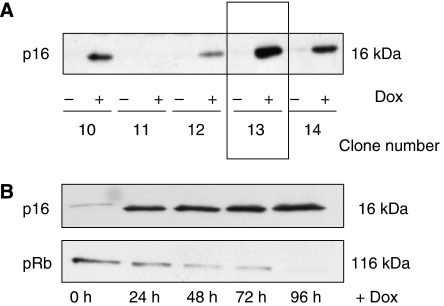
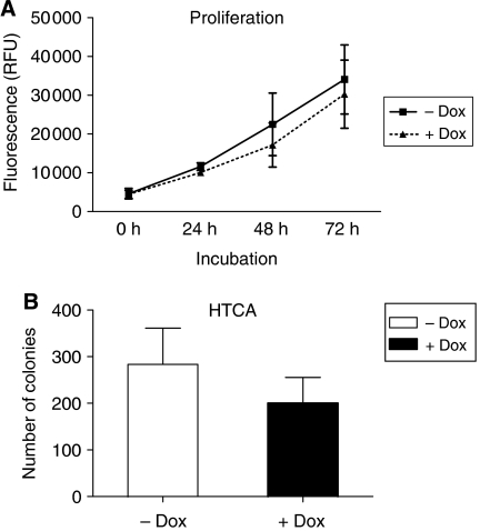
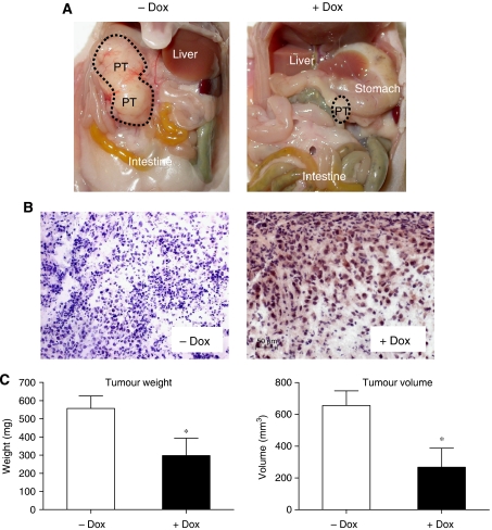
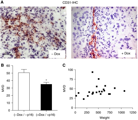
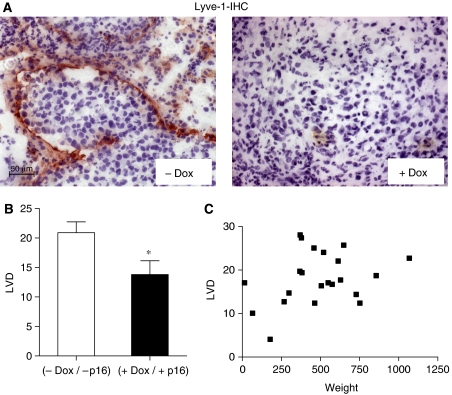
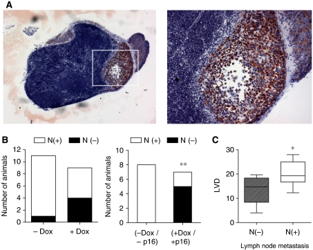
Functional inactivation of the tumour suppressor protein p16(INK4a) constitutes a key event in the multistep process of pancreatic ductal cell transformation. However, the significance of p16 inactivation for complex and tissue-specific aspects of pancreatic cancer progression, such as angiogenesis and metastasis, is less understood. Here, we inducibly re-expressed p16 in vivo in an orthotopic model of pancreatic cancer and examined the impact on these clinically relevant aspects of pancreatic cancer tumour biology. Consistent with previous work in subcutaneous xenograft models, we found p16 capable of reducing primary tumour growth. In addition, p16 restitution resulted in a marked reduction of tumour angiogenesis, largely accounted for by a p16-dependent inhibition of lymphangiogenesis. In excellent agreement with the antilymphangiogenic effect, re-expression of p16 almost completely prevented lymph node metastases of MiaPaca-2 pancreatic tumours. To our knowledge, this is the first report that experimentally links the tumour suppressor p16 to the process of lymphangiogenesis.


Figures

Figure 1
Figure 1
Generation MiaPaCa-2-TREx–p16 cells with doxycyclin-inducible expression of p16. (A) The plasmids pcDNA6/TR and pcDNA4/TO-p16 were sequentially transfected into p16-deficient MiaPaCa-2 cells and clones with inducible expression of p16 were selected (MiaPaca-2-TREx-p16). For each of the zeozine-resistant MiaPaCa-2-TREx-p16 clone, cells were grown for 48 h in the presence (+Dox) or absence (−Dox) of 1 μg ml−1 doxycycline. The expression of p16 protein was analysed by western blotting. The clones with the highest p16 induction and lowest basal expression were further analysed, for example clone 13. (B) Time course of doxycycline-induced p16 expression. Cells were cultured for 96 h in the presence or absence of doxycycline and each 24 h, lysates were prepared for western blot analysis of p16 (upper panel) and pRB (lower panel). Equal amounts of protein (20 μg) were separated on sodium dodecyl sulphate polyacrylamide gel electrophoresis.
Figure 2
Figure 2
Effects of doxycycline-induced p16 expression on growth of MiaPaCa-2-TREx cells in vitro. (A) Time-dependent proliferation of MiaPaCa-2-TREx-p16 cells in the presence and absence of Dox as determined based on Alamar Blue assay. Every 24 h alamar blue dye was applied to the media of the cells and fluorescence was determined as an indirect measurement of cell numbers. (B) Effects of p16 expression on anchorage-independent growth of MiaPaCa-2-TREx-p16/13 cells. Anchorage-independent growth was evaluated based on soft agar colony formation. 103 cells were seeded out in a methylcellulose/agar mixture and incubated in the presence (+Dox) or absence (−Dox) of doxycycline for 10 days. Vital colonies were counted. Values shown are the mean±s.e.m. for each group (+/−Dox) of three independent experiments.
Figure 3
Figure 3
Induction of p16 expression reduced primary tumour growth in orthotopic MiaPaCa-2 pancreatic carcinomas. Pancreatic MiaPaCa-2-TREx-p16 cells were grown orthotopically in the pancreas of mice treated with or without Dox as indicated. At the end of the treatment (8 weeks) tumour volumes and weights were calculated. (A) Open situ of representative mice from the control (−Dox) and treatment group (+Dox). (B) Confirmation of p16 induction in vivo. Primary tumours were analysed for p16 expression by immunohistochemistry using an antibody against p16. An example of p16 expression in a small tumour in the treatment group is shown on the right panel, with detection of p16 protein in the nucleus and in the cytoplasm of tumour cells. In contrast, no signal was obtained in control tumours (−Dox) (images at × 20 magnification). (C) Summary of primary tumour weights (left) and volumes (right) from Dox-treated animals. Data represent mean±s.e.m. for each group. *P<0.05.
Figure 4
Figure 4
Induction of p16 expression reduced angiogenesis in orthotopic MiaPaCa-2 pancreatic carcinomas. Cryosections of the tumours from control (−Dox) and treatment group (+Dox) were stained with the endothelial marker CD31. To determine microvessel density (MVD) CD31-expressing vessels were quantitated from hotspot areas, as described in Materials and methods. (A) Shown are representative CD31 stainings from a (−Dox) and a (+Dox) tumour (B) and quantitative evaluation of the results, *P<0.05. (images at × 20 magnification) (C) Relationship of MVD and tumour weight. MVD values from both groups (+/−Dox) were plotted against the respective tumour weight to assess a potential correlation of both parameters. The distribution shows no correlation.
Figure 5
Figure 5
Induction of p16 expression reduced lymphangiogenesis in orthotopic MiaPaCa-2 pancreatic carcinomas. Cryosections of the tumours were stained with Lyve-1, an endothelial cell marker, which specifically detects lymphatic endothelia. (A) Illustration of Lyve-1 immunohistochemical detection in representative tumours from control (−Dox) and treatment group (+Dox) (images at × 20 magnification). (B) Quantitative evaluation of lymphatic vessel density (LVD) for each group (+/−Dox). *P<0.05. (C) Missing correlation of LVD and tumour weight.
Figure 6
Figure 6
Induction of p16 expression inhibits lymphatic spread of MiaPaCa-2 cells to the liver hilus lymph nodes. Metastatic spread of MiaPaCa-2-TREx-p16 cells was evaluated based on inspection of lung and abdominal organs at the time of autopsy and microscopic assessment of lymph node metastasis. (A) Representative picture of a lymph node with cytokeratin-positive human tumour cells (stained red) surrounded by lymphocytes (images at × 10 magnification and × 20 (image inset). (B) Quantitative analysis of lymph node metastasis. Data represent the number of animals with presence of lymph node metastasis in the liver hilus lymph nodes (left: comparison between treatment (+Dox) and control group (−Dox). Right: subgroup analysis including only those animals with histologically confirmed expression or absence of p16 protein (−Dox/p16 vs +Dox/+p16) (**P<0.05) whereas in (C) the correlation between LVD and lymph node metastasis is shown (*P<0.05).

Similar articles

Cited by

References

    1. Aguirre AJ, Bardeesy N, Sinha M, Lopez L, Tuveson DA, Horner J, Redston MS, DePinho RA (2003) Activated K-ras and Ink4a/Arf deficiency cooperate to produce metastatic pancreatic ductal adenocarcinoma. Genes Dev 17: 3112–3126 - PMC - PubMed
    1. Ahmed SA, Gogal Jr RM, Walsh JE (1994) A new rapid and simple non-radioactive assay to monitor and determine the proliferation of lymphocytes: an alternative to [3H]thymidine incorporation assay. J Immunol Methods 170: 211–224 - PubMed
    1. Alves F, Contag S, Missbach M, Kaspareit J, Nebendahl K, Borchers U, Heidrich B, Streich R, Hiddemann W (2001) An orthotopic model of ductal adenocarcinoma of the pancreas in severe combined immunodeficient mice representing all steps of the metastatic cascade. Pancreas 23: 227–235 - PubMed
    1. Bardeesy N, Aguirre AJ, Chu GC, Cheng KH, Lopez LV, Hezel AF, Feng B, Brennan C, Weissleder R, Mahmood U, Hanahan D, Redston MS, Chin L, DePinho RA (2006) Both p16(Ink4a) and the p19(Arf)-p53 pathway constrain progression of pancreatic adenocarcinoma in the mouse. Proc Natl Acad Sci USA 103: 5947–5952 - PMC - PubMed
    1. Blouw B, Song H, Tihan T, Bosze J, Ferrara N, Gerber HP, Johnson RS, Bergers G (2003) The hypoxic response of tumors is dependent on their microenvironment. Cancer Cell 4: 133–146 - PubMed

Publication types