. 2008 Sep;16(9):1594-601.
doi: 10.1038/mt.2008.136. Epub 2008 Jun 24.

Double oxygen-sensing vector system for robust hypoxia/ischemia-regulated gene induction in cardiac muscle in vitro and in vivo

Affiliations

Double oxygen-sensing vector system for robust hypoxia/ischemia-regulated gene induction in cardiac muscle in vitro and in vivo

Ekaterina V Fomicheva et al. Mol Ther. 2008 Sep.

Abstract

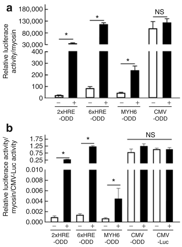
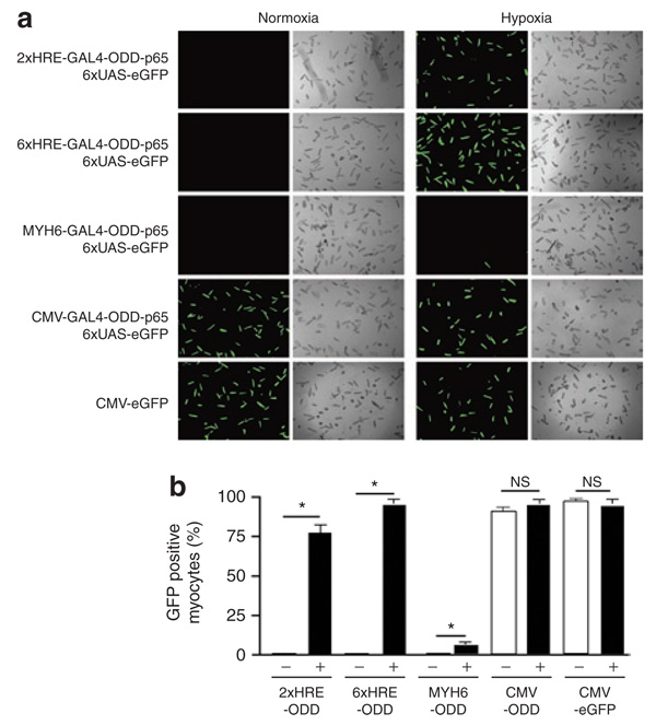
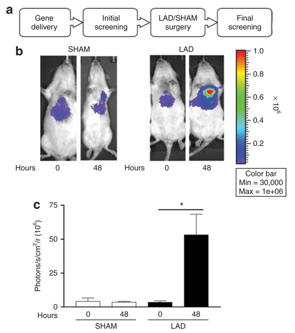
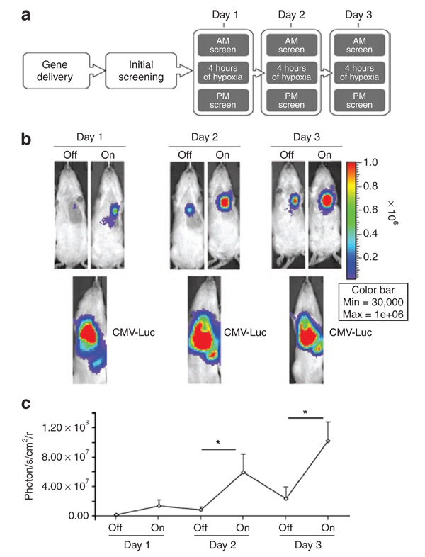
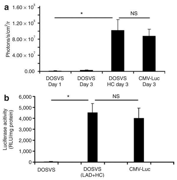
High-fidelity genetically encoded bio-sensors that respond to changes in cellular environmental milieu in disease offer great potential in a range of patho-physiological settings. Here a unique hypoxia-regulated vector-based system with double oxygen-sensing transcriptional elements was developed for rapid and robust hypoxia-regulated gene expression in the heart. Hypoxia-responsive cis elements were used in tandem with a single proline-modified oxygen-dependent degradation (ODD) domain of hypoxia-inducible factor-1alpha to form a double oxygen-sensing vector system (DOSVS). In adult cardiac myocytes in vitro, the DOSVS demonstrated a low background expression not different from baseline control in normoxia, and with 100% efficiency, robust, 1,000-fold induction upon hypoxia. In the heart in vivo, hypoxic and ischemic challenges elicited rapid 700-fold induction in living animals, exceeding that obtained by a high-fidelity constitutive cytomegalovirus (CMV) viral promoter. DOSVS also showed high temporal resolution in the heart in response to cyclical bouts of hypoxia in vivo. We propose that DOSVS will be valuable for a range of applications, including bio-sensing and therapeutic gene expression in the heart and other organ systems that are confronted by chronic or episodic hypoxic/ischemic stresses in vivo.


Figures

Figure 1
Figure 1. Schematics of viral constructs for double oxygen–sensing vector system
Six recombinant adenoviruses were generated for the double oxygen–sensing virus system. The four sensor vectors (top) have the GAL4-ODD-p65-coding sequence driven by different promoters: cytomegalovirus (CMV), Myh6, 2xHRE-mp, or 6xHRE-mp. Derivation of components: GAL4-yeast DNA-binding domain, ODD-oxygen-dependent degradation domain of hypoxia-inducible factor-1α, p65/RelA-the human pol II–activated domain from nuclear factor κB. The two effector vectors (bottom) consisted of six UAS-GAL4 binding sites upstream of a minimal viral promoter (E1BTATA) that was able to amplify the hypoxia signal by binding GAL4-ODD-p65 protein to activate transcription of enhanced green fluorescent protein (eGFP) or luciferase reporter genes. HRE, hypoxia-responsive element; LTR, left-terminal repeat; RTR, right-terminal repeat.
Figure 2
Figure 2. Hypoxia-mediated amplification of transcriptional activity in double oxygen–sensing vector system (DOSVS)
Rat cardiac myocytes were transduced with one of four different sensor viruses (Figure 1) containing unique promoters: 2xHRE-mp, 6xHRE-mp, Myh6, and cytomegalovirus (CMV) fused with GAL4-ODD-p65. The same myocytes were also transduced with the 6UAS-luciferase effector virus. The CMV-luciferase virus was used as a positive control of maximal adenoviral gene transfer efficiency. (a) At 48 hours after transduction, promoter activity of each pair of viruses was assessed by luciferase assay. (b) The transcriptional activity of each set of DOSVS was also normalized to CMV-luciferase activity. In each graph, “+” is hypoxic and “−” is normoxic. Values are mean ± SEM. HRE, hypoxia-responsive element; NS, not significant; ODD, oxygen-dependent degradation domain.
Figure 3
Figure 3. Hypoxia-mediated amplification of enhanced green fluorescent protein (eGFP) expression by double oxygen–sensing vector system
Rat cardiac myocytes were transduced with identical sensor vectors as in Figure 1, containing unique promoters: 2xHRE-mp, 6xHRE-mp, Myh6, and cytomegalovirus (CMV) fused with GAL4- ODD-p65, and the 6UAS-eGFP effector virus was used as the reporter. (a) At 48 hours after transduction, we scored eGFP positive myocytes for each pair of viruses. (b) The percentage of GFP-positive myocytes within a cover slip was estimated by dividing GFP-positive by total myocytes. The CMV-eGFP virus served as a positive control for adenoviral transduction efficiency (a,b). In each graph, “+” is hypoxic and “−” is normoxic. Values are mean ± SEM. HRE, hypoxia-responsive element; NS, not significant; ODD, oxygen-dependent degradation domain.
Figure 4
Figure 4. Fidelity of double oxygen–sensing vector system (DOSVS) in an experimental model of myocardial infarction in vivo
(a) The rats were imaged for in vivo luciferase expression using the Xenogen imaging system at 24 (initial screening) and 72 hours (final screening) after intramyocardial delivery of DOSVS. Four representative animals are shown at 24 and 72 hours after injection. (b) SHAM rats (n = 7) had the same surgical procedure but without left anterior descending artery (LAD) ligation. LAD rats (n = 11) were subjected to LAD surgery with 48 hours of recovery time. (c) Graph of imaging data showing the average luciferase expression of SHAM and LAD at time 0 of initial screening and at time 48 hours of final screening. LAD rats had significantly higher luciferase expression compared to SHAM animals; (P < 0.02). Values are mean ± SEM.
Figure 5
Figure 5. Dynamic temporal response of double oxygen–sensing vector system (DOSVS) during episodic hypoxic challenge in vivo
(a) After intramyocardial delivery of DOSVS, luciferase expression was monitored in real time in living animals during multiple hypoxic challenge events. (b,c) The same amount CMV-Luc virus was used as a positive control for adenoviral transduction efficiency. DOSVS activity could be switched “On” and “Off” by subjecting the rats (n = 8) alternately between hypoxic or normoxic conditions. At days 2 and 3, the switch to hypoxia led to significant increases in bioluminescence (*P < 0.001). In each case, the basal level of bioluminescence of DOSVS in normoxia was lower then the preceding hypoxia event. By contrast, the level of expression driven by virus encoding CMV-Luc reporter which was high over all time points (lower panels) (b). Summary of increased bioluminescence by episodic hypoxia (c). Values are mean ± SEM. CMV, cytomegalovirus.
Figure 6
Figure 6. In vivo and ex vivo analysis of luciferase expression of double oxygen–sensing vector system (DOSVS) compared to expression of CMV-Luc
(a) Luciferase activity measured in rat hearts injected with CMV-Luc viruses was compared to values from hearts injected with DOSVS, with and without hypoxic challenge (HC). The induced group (N = 8) with DOSVS showed a significantly greater signal (P < 0.01) at day 3 as compared with preinduction levels at day 1 (N = 6). Also, the induced DOSVS group shows a significantly greater (P < 0.01) signal as compared with the uninduced group at day 3 (N = 6). The signal levels in the group induced with DOSVS and in the group with CMV-Luc were similar. Values are mean ± SEM. (b) Ex vivo luciferase signal was measured in isolated hearts using a luminometer. Data are presented as relative light units (RLU) per mg of protein and shown as mean ± SEM using three rats per group. Hearts from rats which underwent LAD surgery or HC treatment expressed at the highest level compared to hearts from rats injected with DOSVS without any treatment (P < 0.01). The luciferase signal from hearts injected with CMV-Luc virus was similar compared to signals from induced (LAD or HC) hearts with DOSVS. CMV, cytomegalovirus; NS, not significant.

References

    1. Yenari MA, Sapolsky RM. Gene therapy in neurological disease. Methods Mol Med. 2005;104:75–88. - PubMed
    1. Woo SL, Skarlatos SI, Joyce MM, Croxton TL, Qasba P. Critical resources for gene therapy in Heart, Lung, and Blood Diseases Working Group. Mol Ther. 2006;13:641–643. - PubMed
    1. Williams ML, Koch WJ. Viral-based myocardial gene therapy approaches to alter cardiac function. Annu Rev Physiol. 2004;66:49–75. - PubMed
    1. Pachori AS, Melo LG, Dzau VJ. Gene therapy: role in myocardial protection. Handb Exp Pharmacol. 2006:335–350. - PubMed
    1. Hermonat PL, Mehta JL. Potential of gene therapy for myocardial ischemia. Curr Opin Cardiol. 2004;19:517–523. - PubMed

Publication types

MeSH terms

LinkOut - more resources