. 2008 Jun 24;6(6):e149.
doi: 10.1371/journal.pbio.0060149.

Crystal structure of the FeS cluster-containing nucleotide excision repair helicase XPD

Affiliations

Crystal structure of the FeS cluster-containing nucleotide excision repair helicase XPD

Stefanie C Wolski et al. PLoS Biol. .

Abstract

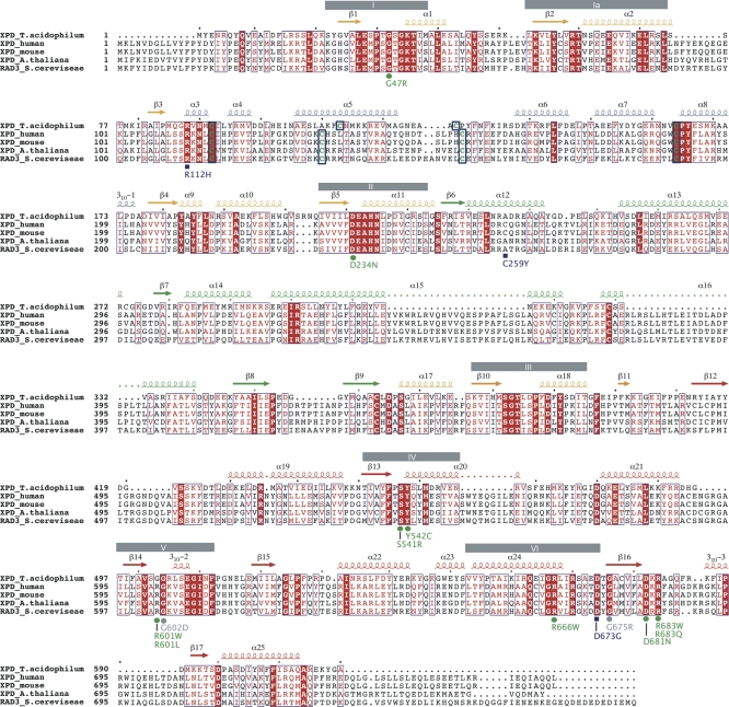
DNA damage recognition by the nucleotide excision repair pathway requires an initial step identifying helical distortions in the DNA and a proofreading step verifying the presence of a lesion. This proofreading step is accomplished in eukaryotes by the TFIIH complex. The critical damage recognition component of TFIIH is the XPD protein, a DNA helicase that unwinds DNA and identifies the damage. Here, we describe the crystal structure of an archaeal XPD protein with high sequence identity to the human XPD protein that reveals how the structural helicase framework is combined with additional elements for strand separation and DNA scanning. Two RecA-like helicase domains are complemented by a 4Fe4S cluster domain, which has been implicated in damage recognition, and an alpha-helical domain. The first helicase domain together with the helical and 4Fe4S-cluster-containing domains form a central hole with a diameter sufficient in size to allow passage of a single stranded DNA. Based on our results, we suggest a model of how DNA is bound to the XPD protein, and can rationalize several of the mutations in the human XPD gene that lead to one of three severe diseases, xeroderma pigmentosum, Cockayne syndrome, and trichothiodystrophy.


Conflict of interest statement

Competing interests. The authors have declared that no competing interests exist.

Figures

Figure 1
Figure 1. Overall Structure of XPD and the FeS Cluster
(A) Front view of XPD, with domain 1 in yellow, domain 2 in cyan, domain 3 in green, and domain 4 in red. The FeS cluster is shown in all-bonds representation. (B) Side view of XPD color-coded as in (A). (C) SIGMAA weighted 2F o-F c electron density map contoured at one times the standard deviation around the 4Fe4S cluster and the surrounding protein in grey, and at four times the standard deviation in blue. The cluster and the coordinating cysteines are shown in all-bonds representation. Secondary structure elements are labeled in grey. (D) Residues in close proximity to the 4Fe4S cluster. The cluster is shown as in (C), and residues Arg88, Tyr166, Lys117, Phe131, and Glu167 are shown in all-bonds representation, with α-helices and β-strands being labeled.
Figure 2
Figure 2. Sequence Alignment
Sequence alignment of five different XPD proteins. From top to bottom: XPD from T. acidophilum, human, mouse, Arabidopsis thaliana, and Saccharomyces cerevisiae. Secondary structure elements are indicated above the sequence and are color coded according to their domains; arrows indicate β-strands and coils α-helices. Helicase motifs are shown as gray boxes. Mutations leading to xeroderma pigmentosum, Cockayne syndrome, or trichothiodystrophy are indicated below the sequence and are colored in blue, green, and grey, referring to their occurrence in trichothiodystrophy, xeroderma pigmentosum, or xeroderma pigmentosum/Cockayne syndrome patients, respectively. Conserved residues are boxed, and strictly conserved residues are shown in white with a red background. The four cysteines that coordinate the 4Fe4S cluster are highlighted in green surrounded by thick blue boxes.
Figure 3
Figure 3. Electrostatic Surface Potential of XPD
Four different views of the electrostatic surface potential of XPD. The surface potential has been calculated with Pymol/APBS at an ionic strength of 150 mM and is contoured at ±10 kBT. The first view is the same as shown in Figure 1A; the arrows show the transition from one view to the other with the rotation indicated by the arrow. Putative DNA binding sites are marked by additional arrows.
Figure 4
Figure 4. Structural Similarity to MutY
Side-by-side presentation of the 4Fe4S clusters in MutY and XPD after superposition (A). Two arginines (Arg149 and Arg 153) are located in close proximity to the 4Fe4S cluster and point towards the DNA in MutY (B). In XPD, Arg88 occupies a similar position as observed for the arginines in MutY. The helices surrounding the cluster have been labeled.
Figure 5
Figure 5. The Wedge
A view towards helices α22 and α23 in domain 3. The color code from Figure 1A was maintained, and a transparent surface was added to provide a view of the groove where one DNA strand binds. All residues that may play a role in double strand separation are indicated and labeled. Helices α22 and α23 have been labeled and indicated by arrows.
Figure 6
Figure 6. Hypothetical XPD-DNA Model
(A) Overall view of XPD in complex with ssDNA. The model was obtained by superposition with the NS3 helicase in complex with ssDNA [26]. Residual difference map peaks at 2.5 times the standard deviation are shown in blue, and peaks used for backbone phosphate positioning are numbered. Further extension of the ssDNA towards the hole of XPD was achieved by the addition of B-form DNA. The DNA is shown with its backbone in orange, and the bases are shown as blue spokes. (B) Close-up view from (A). (C) Surface representation of the XPD–DNA model. The DNA is shown as in (A), and the surface is colored according to sequence conservation, with green indicating strictly conserved, yellow highly conserved, and gray residues that are not conserved. The DNA in this view emerges from the hole and fits nicely into the highly conserved pocket (indicated by the red arrow), which could potentially play a role in damage recognition or which couples DNA binding to the FeS cluster. (D) Top view of the XPD-DNA model. For clarity, the top part of XPD has been removed to allow a view into the highly conserved groove, which leads the ssDNA towards the hole in the donut-shaped molecule.
Figure 7
Figure 7. Mutants Leading to Xeroderma Pigmentosum, Cockayne Syndrome, or Trichothiodystrophy
XPD is shown in ribbon presentation with the same orientation and color code for the different domains as in Figure 1A. Point mutations leading to either xeroderma pigmentosum, Cockayne syndrome, or trichothiodystrophy are shown in a space-filling representation and are labeled according to their residue number for T. acidophilum XPD in black and, according to the disease they cause in humans, green (XP), blue (TTD), and grey (XP/CS). Secondary structure elements mentioned in the text are labeled.

Similar articles

Cited by

References

    1. Friedberg EC, Walker GC, Siede W, Wood RD, Schultz RA, et al. DNA repair and mutagenesis. Washington (D.C.): ASM Press; 2006. 1118
    1. Goosen N, Moolenaar GF. Role of ATP hydrolysis by UvrA and UvrB during nucleotide excision repair. Res Microbiol. 2001;152:401–409. - PubMed
    1. Sancar A. DNA excision repair. Annu Rev Biochem. 1996;65:43–81. - PubMed
    1. Van Houten B. Nucleotide excision repair in Escherichia coli. Microbiol Rev. 1990;54:18–51. - PMC - PubMed
    1. Gillet LC, Scharer OD. Molecular mechanisms of mammalian global genome nucleotide excision repair. Chem Rev. 2006;106:253–276. - PubMed

Publication types