. 2008 Jun 24;6(6):e152.
doi: 10.1371/journal.pbio.0060152.

The B cell antigen receptor and overexpression of MYC can cooperate in the genesis of B cell lymphomas

Affiliations

The B cell antigen receptor and overexpression of MYC can cooperate in the genesis of B cell lymphomas

Yosef Refaeli et al. PLoS Biol. .

Abstract

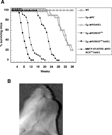
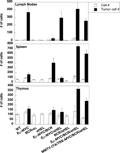
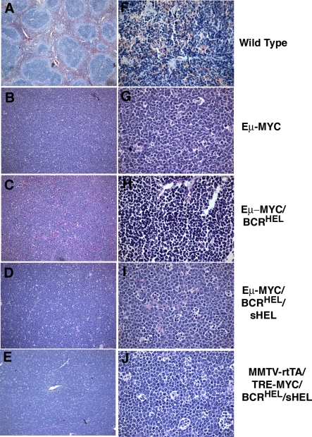
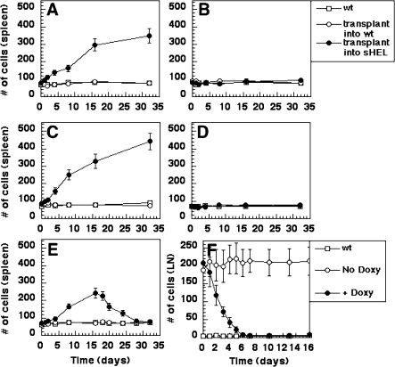
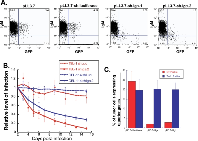
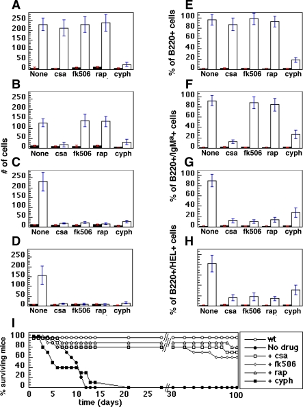
A variety of circumstantial evidence from humans has implicated the B cell antigen receptor (BCR) in the genesis of B cell lymphomas. We generated mouse models designed to test this possibility directly, and we found that both the constitutive and antigen-stimulated state of a clonal BCR affected the rate and outcome of lymphomagenesis initiated by the proto-oncogene MYC. The tumors that arose in the presence of constitutive BCR differed from those initiated by MYC alone and resembled chronic B cell lymphocytic leukemia/lymphoma (B-CLL), whereas those that arose in response to antigen stimulation resembled large B-cell lymphomas, particularly Burkitt lymphoma (BL). We linked the genesis of the BL-like tumors to antigen stimulus in three ways. First, in reconstruction experiments, stimulation of B cells by an autoantigen in the presence of overexpressed MYC gave rise to BL-like tumors that were, in turn, dependent on both MYC and the antigen for survival and proliferation. Second, genetic disruption of the pathway that mediates signaling from the BCR promptly killed cells of the BL-like tumors as well as the tumors resembling B-CLL. And third, growth of the murine BL could be inhibited by any of three distinctive immunosuppressants, in accord with the dependence of the tumors on antigen-induced signaling. Together, our results provide direct evidence that antigenic stimulation can participate in lymphomagenesis, point to a potential role for the constitutive BCR as well, and sustain the view that the constitutive BCR gives rise to signals different from those elicited by antigen. The mouse models described here should be useful in exploring further the pathogenesis of lymphomas, and in preclinical testing of new therapeutics.


Conflict of interest statement

Competing interests. The authors have declared that no competing interests exist.

Figures

Figure 1
Figure 1. A Clonal B cell Antigen Receptor Cooperates with MYC in the Development of BCLs
(A) Survival. Strains of mice in groups of 50 were observed over a period of 36 wk. Deceased mice were examined by necropsy. Death was uniformly attributable to lymphoid tumors. The difference among the mortality curves for the Eμ-MYC/BCRHEL/sHEL mice and that of MMTV-rtTA/TRE-MYC/BCRHEL/sHEL transgenic mice to each other had a significance value p = 0.05. The difference between the mortality curves for those two sets of mice and the other mice represented in the graph was p = 0.005. In addition, the statistical significance of the difference between the Eμ-MYC/BCRHEL transgenic mice and any other groups of mice presented in the graph is p < 0.01. (B) Jaw tumor in 16-wk-old MMTV-rtTA/TRE-MYC/BCRHEL/sHEL mouse.
Figure 2
Figure 2. Lymphomagenesis in Transgenic Mice
Single-cell suspensions were generated from lymph nodes (six nodes for each mouse – a pair of inguinal, axillary and brachial lymph nodes), spleens, thymii and jaw-tumors. The bar graphs represent the total number of cells (x10−6) obtained for the indicated organs. Counts represent the mean derived from 10 independent mice ± the standard deviation for those values. Healthy animals were euthanized at 21 d of age, Eμ-MYC mice at 200–240 d, Eμ-MYC/BCRHEL mice at 112–130 d, Eμ-MYC/BCRHEL/sHEL mice at 26–30 d, and MMTV-rtTA/TRE-MYC/sHEL/BCRHEL mice at 71–86 d. All tumors contained homogeneous populations of cells with distinctive surface phenotypes: B220+/IgM- cells for Eμ-MYC tumors, B220+/IgMa+ cells for both Eμ-MYC/BCRHEL and Eμ-MYC/BCRHEL/sHEL tumors, and B220+/BCRHEL cells for MMTV-rtTA/TRE-MYC/BCRHEL/sHEL tumors. Open bars represent normal mice. Filled bars represent tumor-bearing mice.
Figure 3
Figure 3. Histological Analysis of Tumors
Tissues were sectioned, stained with hematoxylin and eosin, and microscopic images obtained as described in Methods. Magnification was 10X for (A–E), 100X for (F–J). (A and F) Spleen from a normal wild-type mouse. (B and G) Lymph node tumor from an Eμ-MYC mouse. (C and H) Spleen tumor from an Eμ-MYC/BCRHEL mouse. (D and I) Spleen tumor from an Eμ-MYC/BCRHEL/sHEL mouse. (E and J) Jaw tumor from an MMTV-rtTA/TRE-MYC/BCRHEL/sHEL mouse.
Figure 4
Figure 4. Establishment and Maintenance of Murine BL by Antigenic Stimulus and MYC Overexpression
(A and B) Primary transplants. Spleen and lymph node cells were harvested from an MMTV-rtTA/TRE-MYC/BCRHEL mouse at 4 wk of age. Cells from spleen and lymph nodes were pooled at a 1:1 ratio, and 106 cells were introduced into either syngeneic wild-type mice (empty circles) or sHEL transgenic mice (filled circles) by intravenous injection. Cohorts of mice were either kept on regular food (A), or on doxycycline-containing food (B). Tissues were collected at indicated time points from spleens and analyzed for total number of cells. Samples taken from wild-type mice were analyzed at the same times (empty squares). (C and D) Secondary transplants. Cells were collected from tumors of spleens and lymph nodes represented in (A), 16 d after their initiation by transplantation. Cells from spleen and lymph nodes were pooled at a 1:1 ratio, and 105 cells were introduced into either wild-type recipients (empty circles) or sHEL transgenic mice (filled circles) by intravenous injection. The empty squares represent wild-type, unmanipulated mice that were analyzed in parallel with the experimental groups. Cohorts of mice were either kept on regular food (C), or on doxycycline-containing food (D). Cells were collected from spleens at the indicated times after the transplantation and analyzed as in (A and B). (E and F) BCLs regress after MYC overexpression is extinguished. (E) A cohort of mice similar to those described in (A) was allowed to develop externally visible lymphadenopathy. 16 d later, those mice were switched to a doxycycline-containing diet. The empty circles represent wild-type recipient mice that received transplants of MMTV-rtTA/TRE-MYC/BCRHEL cells, the filled circles represent sHEL transgenic mice that received transplants of those cells, the empty squares represent wild-type, unmanipulated mice that were analyzed in these experiments in parallel with the experimental mice. Cells were collected from spleens at the indicated times after the transplantation and analyzed as in (A and B). (F) MMTV-rtTA/TRE-MYC/BCRHEL/sHEL mice were allowed to develop tumors spontaneously, as a result of transgene function. Approximately 40 d later, mice with externally apparent lymphadenopathy were given doxycycline containing food (day 0 in figure). Cells were collected from lymph nodes at the indicated times after exposure of the mice to doxycycline and analyzed as in (A and B). The empty circles represent MMTV-rtTA/TRE-MYC/BCRHEL mice that were never exposed to docycycline, the filled circles represent MMTV-rtTA/TRE-MYC/BCRHEL mice that were given doxycycline-containing food after they developed externally apparent lymphadenopathy, the empty squares represent wild-type, unmanipulated mice that were analyzed in parallel with the experimental mice.
Figure 5
Figure 5. The Maintenance of Tumors Derived from Eμ-MYC/BCRHEL and Eμ-MYC/BCRHEL/sHEL Mice Depends on the Continued Expression of Igα or Igβ
(A) Cell lines were generated from either Eμ-MYC/BCRHEL tumors and designated as DBL114, or from Eμ-MYC/BCRHEL/sHEL tumors and designated TBL-1. These cell lines uniformly express B220 and IgM on their surface. To determine whether the shRNA sequences targeting Igα were able to knock down their target protein, we measured the levels of IgM expressed on the surface of DBL-114 cells that were transduced with lentiviral constructs that encode a reporter gene (GFP). The expression of IgM on the surface is co-modulated with Igα expression, hence the loss of Igα should reduce the levels of surface IgM. The panels represent the flow cytometric profile of the DBL-114 cells that had been transduced with the parental virus (pLL3.7), or a variant that encodes an shRNA specific for firefly luciferase, as a negative control, or two variants of pLL3.7 that encode different shRNAs specific for Igα. The GFP-positive cells in the panels that contained shRNAs specific for Igα showed a loss of surface IgM expression. This is not the case for the GFP-negative fraction of the same cell populations. Similar results were obtained in TBL-1 cells (unpublished data). (B) The shRNA-mediated knock-down of Igα or Igβ in cells obtained from either Eμ-MYC/BCRHEL or Eμ-MYC/BCRHEL/sHEL tumors confers a competitive disadvantage on those cells in vitro, compared to their nontransduced counterparts. Single-cell suspensions were generated from the respective tumors, and used for lentiviral transductions. The cells were maintained in cultured and assayed for GFP expression, by flow cytometry every 24 h. The data for the GFP+ fraction in the population of cells harboring a lentivirus encoding and shRNA was divided by the fraction of GFP+ cells in the population of cells that was transduced with the parental vector, in order to standardize the values and examine the rates of change from the starting level of GFP+ cells, as previously reported [104]. The cells that were transduced with lentiviruses encoding shRNAs specific for either Igα or Igβ exhibited a significant competitive disadvantage when compared to the cells harboring lentiviruses encoding shRNAs specific for firefly luciferase. All wells were set up in triplicates. The graphs represent data from one experiment, representative of eight independent experiments. (C) In vivo validation of the effects of Igα-specific shRNAs on the maintenance of Eμ-MYC/BCRHEL tumors. Cells were obtained from Eμ-MYC/BCRHEL tumors, and transduced in vitro with pLL3.77-sh.luciferase (uses thy1.1 as a reporter gene) or pLL3.7-sh.Igα.1 (uses GFP as a reporter gene). The different cell populations were then mixed in order to generate mixtures of cells that contained an approximately equal fraction of cells that harbored the control lentivirus (pLL3.77.sh.luciferase) and the experimental lentivirus (pLL3.7.sh.Igα). The mixtures of cells were transplanted into cohorts of Rag-1–/– mice. The mice were observed daily until they exhibited externally evident signs of lymphoma, and the organs were harvested. The graphs represent the fraction of cells in the tumorous lymph nodes that retained expression of either thy1.1 (for the control lentivirus), or GFP (for the Igα-specific lentivirus. These results confirm the requirement for Igα expression in the maintenance of the murine BCLs.
Figure 6
Figure 6. Suppression of Tumor Growth by Pharmacological Agents
Tumor cells were harvested from lymph nodes and spleens and transplanted as described in Methods. The recipient mice were held until tumors became clinically apparent. Tumor recipient (open bars) and wild type (filled bars) mice then received daily injections of the indicated drugs for 7 d of either cyclosporine A (csa), FK506, rapamycin (rap), or cyclophosphamide (cyph). For (A–H), the mice were euthanized 24 h after the last injection of drug, and lymph nodes were harvested for analysis of either total number of cells (A–D) (expressed in single units representing 106 cells each) or surface markers of donor cells (E–H). For (I), the mice were observed over a span of 100 da and deaths recorded, as shown. (A and E) Eμ-MYC tumors. (B and F) Eμ-MYC/BCRHEL tumors. (C and G) Eμ-MYC/BCRHEL/sHEL tumors. (D and H) MMTV-rtTA/TRE-MYC/BCRHEL/sHEL tumors. (I) Survival of animals bearing Eμ-MYC/BCRHEL/sHEL tumors. The statistical significance of the differences observed in the kinetics of mortality between the tumor-bearing mice that were either untreated, or treated transiently with cyclophosphamide is 0.01. The statistical significance of the difference in the mortality curves observed between those two groups and the tumor-bearing mice treated with either of the three immunosuppressant drugs is p < 0.001.

References

    1. Jaffe E, Harris NL, Stein H, Vardiman JW. World Health Organization classification of tumors. In: Kleihuis P, Sobin L, editors. Pathology and genetics of tumors of hematopoietic and lymphoid tissues. Lyon (France): Interantional Agency for Research on Cancer; 2001.
    1. Leoncini L, Delsol G, Gascoyne RD, Harris NL, Pileri SA, et al. Aggressive B-cell lymphomas: a review based on the workshop of the XI Meeting of the European Association for Haematopathology. Histopathology. 2005;46:241–255. - PubMed
    1. Küppers R. Mechanisms of B-cell lymphoma pathogenesis. Nat Rev Cancer. 2005;5:251–262. - PubMed
    1. Lam KP, Kuhn R, Rajewsky K. In vivo ablation of surface immunoglobulin on mature B cells by inducible gene targeting results in rapid cell death. Cell. 1997;90:1073–1083. - PubMed
    1. Neuberger MS. Antigen receptor signaling gives lymphocytes a long life. Cell. 1997;90:971–973. - PubMed

Publication types