Review
. 2008 Summer;9(2):148-57.
doi: 10.1089/ham.2007.1079.

Mechanisms of hemoglobin adaptation to high altitude hypoxia

Affiliations
Review

Mechanisms of hemoglobin adaptation to high altitude hypoxia

Jay F Storz et al. High Alt Med Biol. 2008 Summer.

Abstract

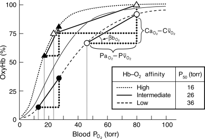
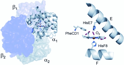
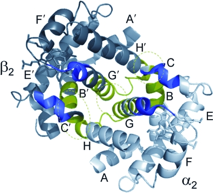
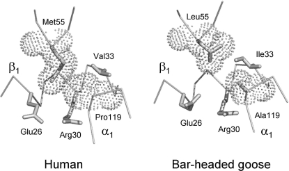
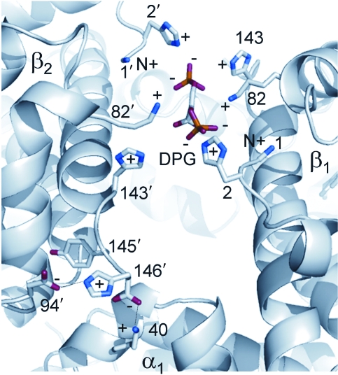
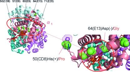
Evidence from a number of vertebrate taxa suggests that modifications of hemoglobin (Hb) function may often play a key role in mediating an adaptive response to high altitude hypoxia. The respiratory functions of Hb are a product of the protein's intrinsic O(2)-binding affinity and its interactions with allosteric effectors such as protons, chloride ions, CO(2), and organic phosphates. Here we review several case studies involving high altitude vertebrates where it has been possible to identify specific mechanisms of Hb adaptation to hypoxia. In addition to comparative studies of Hbs from diverse animal species, functional studies of human Hb mutants also suggest that there is ample scope for evolutionary adjustments in Hb-O(2) affinity through alterations of the equilibrium constants of O(2) binding to deoxy- and oxyHb or through changes in the allosteric equilibrium constants for the transition between the deoxy- and oxyHb quaternary structures. It may be the case that certain evolutionary paths are followed more often than others simply because they are subject to less stringent pleiotropic constraints.


Figures

FIG. 1.
FIG. 1.
O2 equilibrium curves showing the theoretical influence of a change in Hb–O2 affinity on blood O2 transport under conditions of moderate hypoxia (open symbols) and severe hypoxia (filled symbols). Each curve is a plot of the O2 saturation of Hb (vertical axis) versus blood formula image (horizontal axis), with paired values for arterial and venous blood connected by solid lines. For each pair of arterial and venous points, the formula image for venous blood (formula image) is marked by a vertical gray line that extends to the horizontal axis. The sigmoid O2 equilibrium curves are shown for high, intermediate, and low Hb–O2 affinities; formula image of blood at which the O2 saturation of Hb is at 50%. Each change in Hb–O2 affinity produces a shift in formula image, but the formula image of arterial blood (formula image) is assumed to remain constant. In this graph, formula image is the difference in O2 concentration between arterial and mixed venous blood, and formula image is the corresponding arterial–venous difference in formula image. formula image, the blood O2 capacitance coefficient, is defined as the ratio formula image (= the slope of the line connecting the arterial and venous points on the O2 equilibrium curve). The interrelationships of these parameters are summarized by the following Fick's equation: formula image, where formula image is the total O2 consumption and Vb is the total cardiac bloodflow. The product formula image is the circulatory conductance of O2 in the bloodstream. Note that under conditions of moderate hypoxia the right-shifted curve produces the greatest formula image and results in a less severe drop in formula image, the overall index of tissue oxygenation. By contrast, under severe hypoxia, the left-shifted curve produces the greatest values of formula image and formula image. When the kinetics of O2 transfer across the alveolar gas–blood barrier become a limiting step (diffusion limitation), a left-shifted O2 equilibrium curve may also be advantageous under conditions of less severe hypoxia (Bencowitz et al., ; Bouverot, 1985).
FIG. 2.
FIG. 2.
Three-dimensional structure of the Hb tetramer (left) and a detailed view of the heme–ligand complex (right). The distal histidine (E7) and proximal histidine (F8) residues are shown above and below the heme plane, respectively.
FIG. 3.
FIG. 3.
The α2β2 dimer (one-half of a functional Hb tetramer) shown in a side view. The intradimer α2β2 packing contacts are shown in green and the residues participating in interdimer (α1β2 and α2β2)sliding contacts are shown in purple.
FIG. 4.
FIG. 4.
Dot representation of van der Waals radii at an intradimer α1β1 contact in human Hb (left) and bar-headed goose Hb (right). Note that in the bar-headed goose Hb the replacement of Ala for Pro at the α119(H2) residue position results in a loss of atomic contact between the α1 and β1 subunits. The disruption of this interchain van der Waals contact destabilizes the T-state deoxyHb quaternary structure and therefore results in an increased Hb–O2 affinity, because the allosteric equilibrium is shifted in favor of the R-state oxyHb structure.
FIG. 5.
FIG. 5.
Binding of 2,3-DPG in the central cavity between the β1 and β2 chains of deoxyHb. Also shown (lower-left corner) is an intrachain salt bridge that is formed in deoxyHb between the imidazole ring of the N-terminal β146(HC3)His and the negatively charged β94(FG1)Asp. This bond increases the affinity of FG1 Asp for protons and therefore contributes to the Bohr effect. The protons are released upon transition to R-state oxyHb.
FIG. 6.
FIG. 6.
Homology-based structural model of deer mouse oxyHb showing the location of five amino acid replacement polymorphisms that span the E-helix domain of the α-chain subunit. Two charge-changing substitutions at α50(CD8) and α64(E13) are highlighted in the inset figure.

References

    1. Arnone A. Mechanism of action of hemoglobin. Ann. Rev. Med. 1974;25:123–130. - PubMed
    1. Baldwin J. Chothia C. Haemoglobin: the structural changes related to ligand binding and its allosteric mechanism. J. Mol. Biol. 1979;129:175–220. - PubMed
    1. Bettati S. Mozzarelli A. Perutz M.F. Allosteric mechanism of hemoglobin: rupture of salt bridges raises oxygen affinity of the T-structure. J. Mol. Biol. 1983;281:581–585. - PubMed
    1. Bencowitz H.Z. Wagner P.D. West J.B. Effect of change in P50 on exercise tolerance at high-altitude: a theoretical study. J. Appl. Physiol. 1982;53:1487–1495. - PubMed
    1. Bouverot P. Springer-Verlag; Berlin: 1985. Adaptation to Altitude-Hypoxia in Vertebrates.

Publication types

LinkOut - more resources