. 2008 Jun 25:5:26.
doi: 10.1186/1742-2094-5-26.

Characterization of early and terminal complement proteins associated with polymorphonuclear leukocytes in vitro and in vivo after spinal cord injury

Affiliations

Characterization of early and terminal complement proteins associated with polymorphonuclear leukocytes in vitro and in vivo after spinal cord injury

Hal X Nguyen et al. J Neuroinflammation. .

Abstract

Background: The complement system has been suggested to affect injury or disease of the central nervous system (CNS) by regulating numerous physiological events and pathways. The activation of complement following traumatic CNS injury can also result in the formation and deposition of C5b-9 membrane attack complex (C5b-9/MAC), causing cell lysis or sublytic effects on vital CNS cells. Although complement proteins derived from serum/blood-brain barrier breakdown can contribute to injury or disease, infiltrating immune cells may represent an important local source of complement after injury. As the first immune cells to infiltrate the CNS within hours post-injury, polymorphonuclear leukocytes (PMNs) may affect injury through mechanisms associated with complement-mediated events. However, the expression/association of both early and terminal complement proteins by PMNs has not been fully characterized in vitro, and has not observed previously in vivo after traumatic spinal cord injury (SCI).

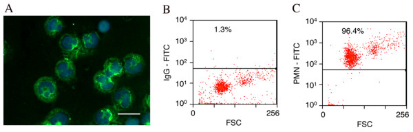
Method: We investigated the expression of complement mRNAs using rt-PCR and the presence of complement proteins associated with PMNs using immunofluroescence and quantitative flow cytometry.

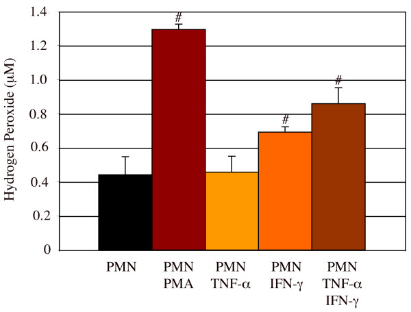
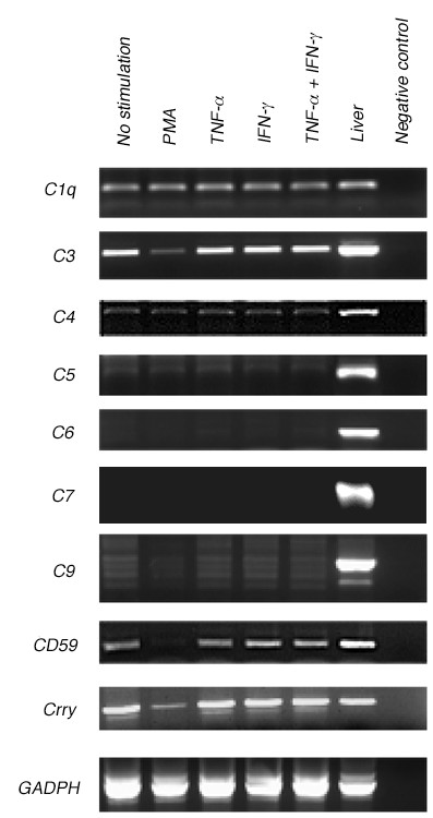
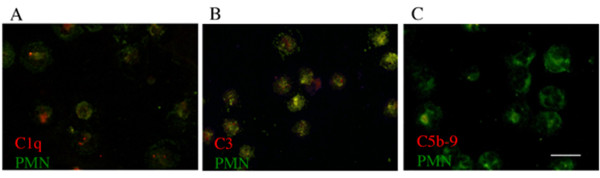
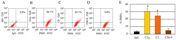
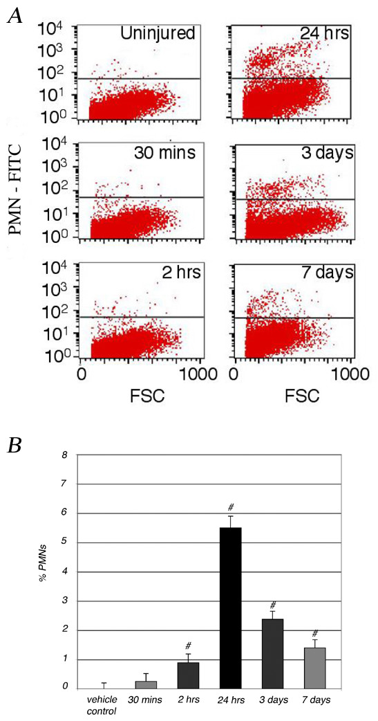
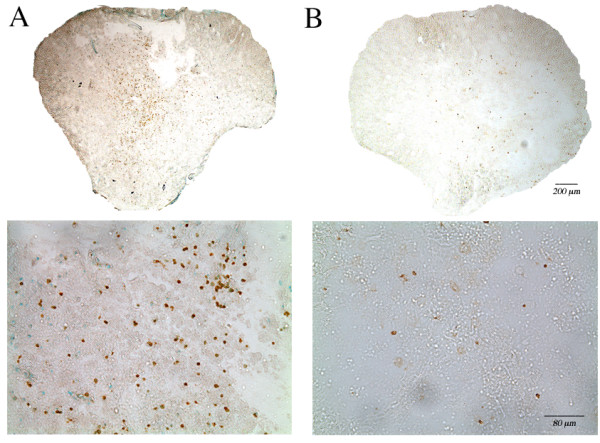
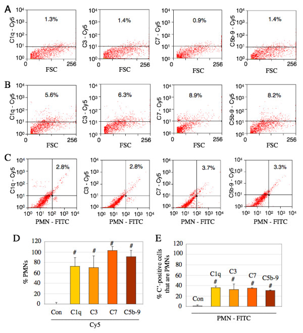
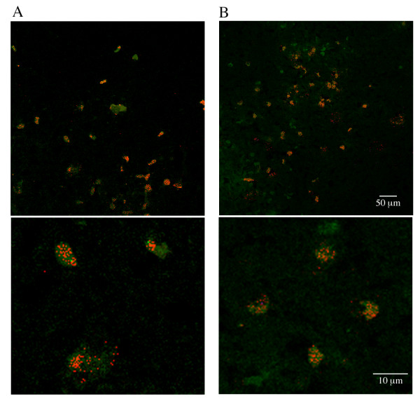
Results: Stimulated or unstimulated PMNs expressed mRNAs encoding for C1q, C3, and C4, but not C5, C6, C7 or C9 in culture. Complement protein C1q or C3 was also detected in less than 30% of cultured PMNs. In contrast, over 70% of PMNs that infiltrated the injured spinal cord were associated with C1q, C3, C7 and C5b-9/MAC 3 days post-SCI. The localization/association of C7 or C5b-9/MAC with infiltrating PMNs in the injured spinal cord suggests the incorporation or internalization of C7 or C5b-9/MAC bound cellular debris by infiltrating PMNs because C7 and C5b-9/MAC were mostly localized to granular vesicles within PMNs at the spinal cord epicenter region. Furthermore, PMN presence in the injured spinal cord was observed for many weeks post-SCI, suggesting that this infiltrating cell population could chronically affect complement-mediated events and SCI pathogenesis after trauma.

Conclusion: Data presented here provide the first characterization of early and terminal complement proteins associated with PMNs in vitro and in vivo after SCI. Data also suggest a role for PMNs in the local internalization or deliverance of complement and complement activation in the post-SCI environment.


Figures

Figure 1
Figure 1
Rat peritoneal neutrophils (PMNs) were identified by immunofluorescence (A, labeled with Hoechst (blue) and an anti-PMN anti-body (green)) and flow cytometry (C). All flow cytometric gates were set using control IgG isotype labeled cells (B). Over 95% of cells isolated were PMNs; scale bar = 10 μm.
Figure 2
Figure 2
Rat peritoneal PMNs were activated by PMA, TNF-α, or IFN-γ to release hydrogen peroxide. PMNs (5.0 × 10⁶/ml) cultured with or without PMA (0.64 μM), TNF-α (2000 U/ml), or IFN-γ (2000 U/ml) stimulation released high levels of hydrogen peroxide. # = significantly different (P < 0.05) from PMN only. n = 6. Number of experiment replication = 3.
Figure 3
Figure 3
Rat peritoneal PMNs express early proteins (C1q, C3, and C4) but not terminal proteins (C5, C6, C7, and C9) of the complement system, shown by rt-PCR. mRNAs were collected from rat peritoneal PMNs (5 × 10⁶/ml) after 24 hours in culture with no stimulant or with PMA (0.64 μM), TNF-α (2000 U/ml), IFN-γ (2000 U/ml), or a combination of TNF-α and IFN-γ. Positive control mRNAs for all complement proteins and inhibitors (CD59 and Crry) of complement activation were isolated from rat liver, while negative controls were absent of mRNAs.
Figure 4
Figure 4
Rat peritoneal PMNs are associated with the early proteins of the complement system but not C5b-9/MAC in vitro, shown by immunofluorescence. PMNs were labeled with an anti-PMN anti-body (green) and an anti-C1q (A), anti-C3 (B), or anti-C5b-9/MAC (C) antibody (red). Scale bar = 10 μm.
Figure 5
Figure 5
Rat peritoneal PMNs are associated with the early proteins of the complement system but not C5b-9/MAC in vitro, shown by flow cytometry. Cells (5 × 10⁶/ml) labeled with a FITC-conjugated anti-PMN antibody and a Cy5-conjugated antibody for C1q (B), C3 (C), or C5b-9 (D), or appropriate IgG controls (A) showed that nearly all cells were PMNs (B, C, & D), but only some cells were associated with C1q (B & E) and C3 (C & E); however, these cells were not associated with C5b-9/MAC (D & E). All flow cytometric gates were set using control IgG isotype labeled cells. # = significantly differ from IgG control. n = 3. Number of experiment replication = 3.
Figure 6
Figure 6
Time course of PMN infiltration after a moderate SCI at T9, demonstrated by flow cytometry in the injured spinal cord. Cells isolated from spinal cord (T8-T10) were labeled with a FITC-conjugated anti-PMN antibody before analyzed on a FACS Calibur flow cytometer. Spinal cord tissues were collected from uninjured control or injured animal 30 minutes, 2 hours, 24 hours, 3 days, or 7 days after SCI. The number of PMNs detected in the injured or uninjured spinal cord was visualized by dot plots (A), and represented as the percent of cells in the spinal cord relative to uninjured control (B). All flow cytometric gates were set using control IgG isotype labeled cells. # = significantly (P < 0.05) different from uninjured control. n = 5. Number of experiment replication = 2.
Figure 7
Figure 7
PMN infiltration was observed at the spinal cord epicenter region after SCI. Cross sections of rat spinal cord adjacent to the epicenter region were immunolabeled for PMNs 3 days (A) or 42 days (B) after a moderate contusion to the spinal cord (T9).
Figure 8
Figure 8
Infiltrating PMNs in the injured spinal cord are associated with the early and terminal complement proteins 3 days post-SCI. Cells isolated from spinal cord (T8-T10) of control (A) or injured rats (B & C) were labeled with a FITC-conjugated anti-PMN antibody and a Cy5-conjugated antibody for C1q, C3, C7 or C5b-9, or appropriate IgG controls before analyzed on a FACS Calibur flow cytometer. After 3 days following SCI (B), the number of cells positive for C1q, C3, C7, or C5b-9 was elevated compared to that from control (A). However, only some of the complement-positive cells were PMNs (C & E) and that most PMNs that infiltrated the injured spinal cord were also positive for C1q, C3, C7, and C5b-9 (C & D). All flow cytometric gates were set using control IgG isotype labeled cells. # = significantly (P < 0.05) different from uninjured control (Con). n = 5. Number of experiment replication = 2.
Figure 9
Figure 9
PMNs are associated with C7 (A) and C5b-9 (B) at the injury epicenter region 3 days post-SCI. Cross sections of rat spinal cord epicenter region were immunolabeled for PMNs (green) and C7 (red) or C5b-9 (red) 3 days after a moderate contusion injury to the spinal cord (T9). Lower panels are higher magnifications of selected areas taken from upper panels.

Similar articles

Cited by

References

    1. Anderson AJ. Mechanisms and pathways of inflammatory responses in CNS trauma: spinal cord injury. J Spinal Cord Med. 2002;25:70–79. - PubMed
    1. Tenner AJ. Complement in Alzheimer's disease: opportunities for modulating protective and pathogenic events. Neurobiol Aging. 2001;22:849–861. doi: 10.1016/S0197-4580(01)00301-3. - DOI - PubMed
    1. Rus H, Cudrici C, Niculescu F. The role of the complement system in innate immunity. Immunol Res. 2005;33:103–112. doi: 10.1385/IR:33:2:103. - DOI - PubMed
    1. Bohana-Kashtan O, Ziporen L, Donin N, Kraus S, Fishelson Z. Cell signals transduced by complement. Mol Immunol. 2004;41:583–597. doi: 10.1016/j.molimm.2004.04.007. - DOI - PubMed
    1. Anderson AJ, Robert S, Huang W, Young W, Cotman CW. Activation of complement pathways after contusion-induced spinal cord injury. J Neurotrauma. 2004;21:1831–1846. doi: 10.1089/neu.2004.21.1831. - DOI - PubMed

Publication types